Abstract
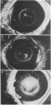
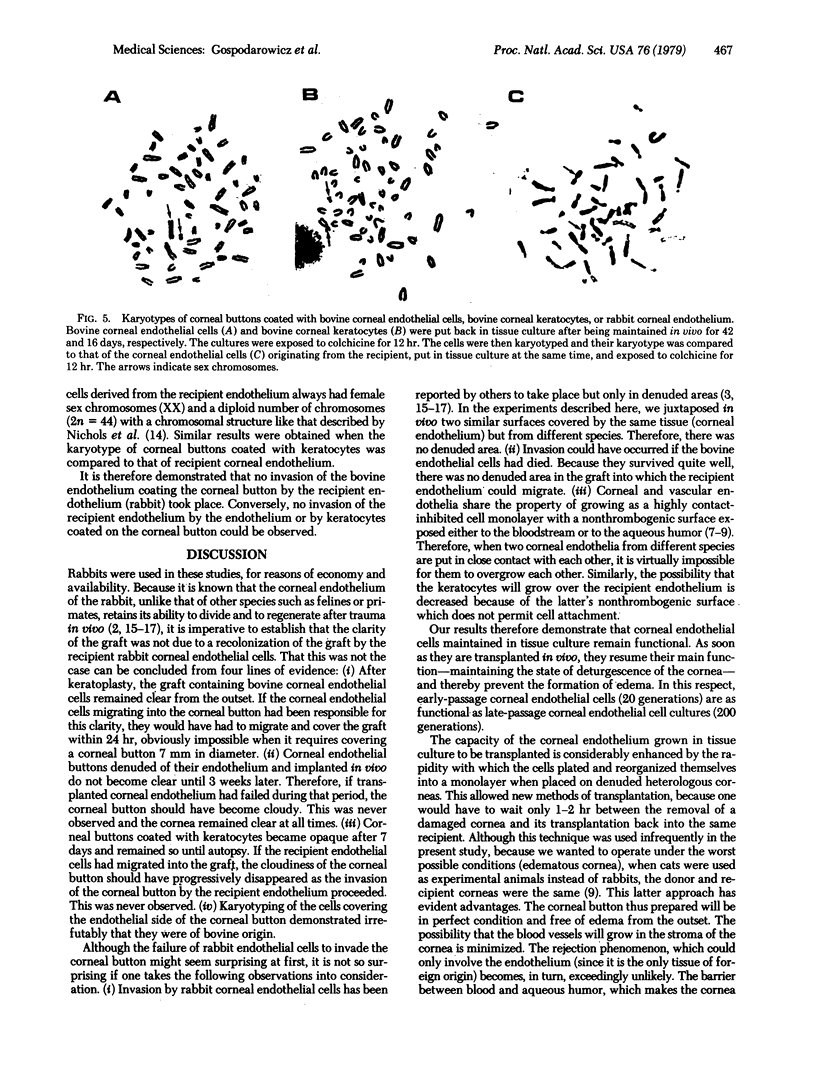
Rabbit corneas denuded of their endothelium were coated with bovine corneal endothelial cells (from steers) previously maintained in tissue culture for short (20 generations) or prolonged (200 generations) periods. When grafted back into female rabbits, the corneal buttons remained clear and showed no edema. In contrast, denuded corneas coated with bovine keratocytes and grafted into rabbits became opaque and edematous within 7 days and remained so thereafter. Bovine corneal endothelial cells of the grafted corneas, which had remained clear for over 100 days, proliferated actively when put back into tissue culture. The corneal endothelial cells of the graft were characteristic of the male (XY). The chromosome number of the endothelium of the recipient rabbit was 2n = 44 with sex chromosomes characteristic of the female (XX). Results of the karyotype analysis show that there was no invasion of the corneal button by the recipient endothelium and, conversely, no invasion of the recipient endothelium by the endothelium on the corneal button. These results demonstrate that cultured corneal endothelial cells remain functional in vitro and can replace a damaged or nonfunctional endothelium in vivo.
Full text
PDF
Images in this article
Selected References
These references are in PubMed. This may not be the complete list of references from this article.
- Birdwell C. R., Gospodarowicz D., Nicolson G. L. Identification, localization, and role of fibronectin in cultured bovine endothelial cells. Proc Natl Acad Sci U S A. 1978 Jul;75(7):3273–3277. doi: 10.1073/pnas.75.7.3273. [DOI] [PMC free article] [PubMed] [Google Scholar]
- Gospodarowicz D., Greenburg G., Birdwell C. R. Determination of cellular shape by the extracellular matrix and its correlation with the control of cellular growth. Cancer Res. 1978 Nov;38(11 Pt 2):4155–4171. [PubMed] [Google Scholar]
- Gospodarowicz D., Mescher A. L., Birdwell C. R. Stimulation of corneal endothelial cell proliferations in vitro by fibroblast and epidermal growth factors. Exp Eye Res. 1977 Jul;25(1):75–89. doi: 10.1016/0014-4835(77)90248-2. [DOI] [PubMed] [Google Scholar]
- Howard B. V., Macarak E. J., Gunson D., Kefalides N. A. Characterization of the collagen synthesized by endothelial cells in culture. Proc Natl Acad Sci U S A. 1976 Jul;73(7):2361–2364. doi: 10.1073/pnas.73.7.2361. [DOI] [PMC free article] [PubMed] [Google Scholar]
- Kanai A. [The regeneration of corneal endothelial cell]. Nippon Ganka Gakkai Zasshi. 1972 Oct;76(10):1163–1175. [PubMed] [Google Scholar]
- Khodadoust A. A., Green K. Physiological function of regenerating endothelium. Invest Ophthalmol. 1976 Feb;15(2):96–101. [PubMed] [Google Scholar]
- Nichols W. W., Levan A., Hansen-Melander E., Melander Y. The idiogram of the rabbit. Hereditas. 1965;53(1):63–76. doi: 10.1111/j.1601-5223.1965.tb01980.x. [DOI] [PubMed] [Google Scholar]
- Van Horn D. L., Hyndiuk R. A. Endothelial wound repair in primate cornea. Exp Eye Res. 1975 Aug;21(2):113–124. doi: 10.1016/0014-4835(75)90076-7. [DOI] [PubMed] [Google Scholar]
- Van Horn D. L., Sendele D. D., Seideman S., Buco P. J. Regenerative capacity of the corneal endothelium in rabbit and cat. Invest Ophthalmol Vis Sci. 1977 Jul;16(7):597–613. [PubMed] [Google Scholar]


