

Abstract
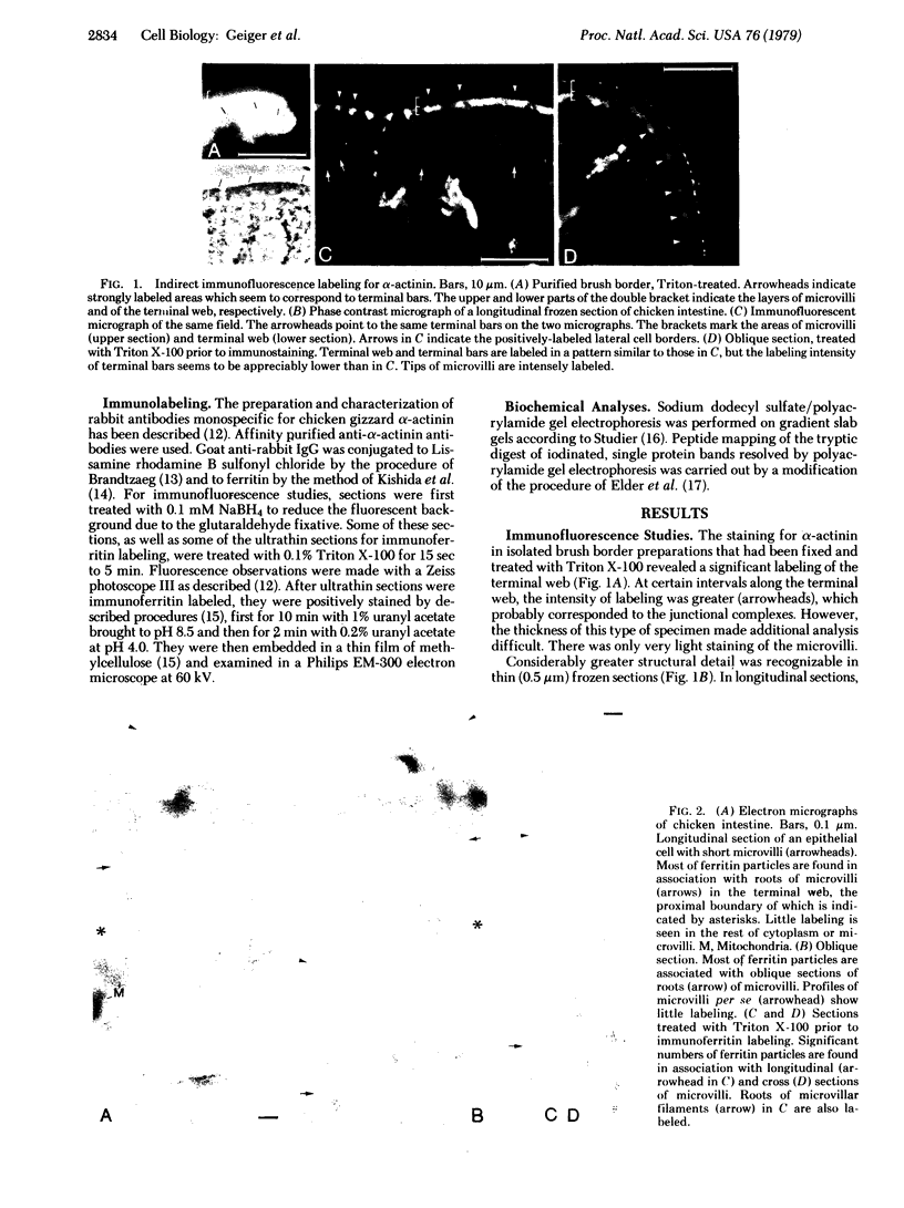
alpha-Actinin was localized in chicken intestinal epithelial cells by immunofluorescence and immunoferritin labeling of thin frozen sections. Most of the label of the brush border was confined to the terminal web area. The label there was concentrated mainly along the "roots" of the microvilli core microfilaments and in the vicinity of the zonula adherens. In the latter structure, the narrow electron-dense zones adjacent to the cell membranes, however, were not significantly labeled. This suggests that alpha-actinin does not mediate directly the association of the transverse terminal web microfilaments to the membrane at the zonula adherens. Sparse ferritin labeling was found near the tight junction, whereas the staining associated with the spot desmosome was negligible. The microvilli were not significantly labeled by either immunofluorescence or immunoferritin staining unless the sections were previously treated with detergent. Moreover, alpha-actinin (or a structurally related protein) was not detected in preparations of purified microvillar vesicles, suggesting the possibility that the alpha-actinin staining in the microvilli may be an artificial due to its translocation by the detergent from the terminal web onto the microvilli. The possible roles of alpha-actinin in the organization and function of the brush border are discussed.
Full text
PDF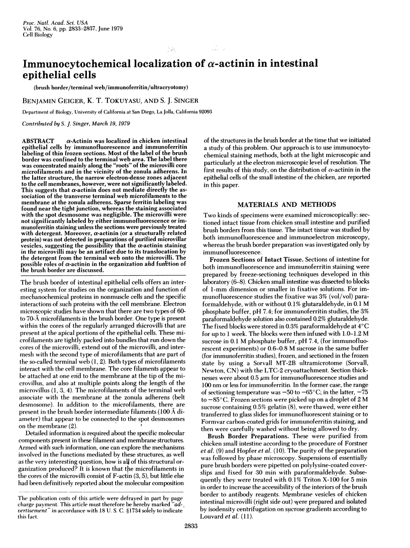


Images in this article


Selected References
These references are in PubMed. This may not be the complete list of references from this article.
- Brandtzaeg P. Conjugates of immunoglobulin G with different fluorochromes. I. Characterization by anionic-exchange chromatography. Scand J Immunol. 1973;2(3):273–290. doi: 10.1111/j.1365-3083.1973.tb02037.x. [DOI] [PubMed] [Google Scholar]
- Bretscher A., Weber K. Localization of actin and microfilament-associated proteins in the microvilli and terminal web of the intestinal brush border by immunofluorescence microscopy. J Cell Biol. 1978 Dec;79(3):839–845. doi: 10.1083/jcb.79.3.839. [DOI] [PMC free article] [PubMed] [Google Scholar]
- Bretscher A., Weber K. Purification of microvilli and an analysis of the protein components of the microfilament core bundle. Exp Cell Res. 1978 Oct 15;116(2):397–407. doi: 10.1016/0014-4827(78)90463-9. [DOI] [PubMed] [Google Scholar]
- Craig S. W., Pardo J. V. alpha-Actinin localization in the junctional complex of intestinal epithelial cells. J Cell Biol. 1979 Jan;80(1):203–210. doi: 10.1083/jcb.80.1.203. [DOI] [PMC free article] [PubMed] [Google Scholar]
- Elder J. H., Pickett R. A., 2nd, Hampton J., Lerner R. A. Radioiodination of proteins in single polyacrylamide gel slices. Tryptic peptide analysis of all the major members of complex multicomponent systems using microgram quantities of total protein. J Biol Chem. 1977 Sep 25;252(18):6510–6515. [PubMed] [Google Scholar]
- Forstner G. G., Tanaka K., Isselbacher K. J. Lipid composition of the isolated rat intestinal microvillus membrane. Biochem J. 1968 Aug;109(1):51–59. doi: 10.1042/bj1090051. [DOI] [PMC free article] [PubMed] [Google Scholar]
- Geiger B., Singer S. J. The participation of alpha-actinin in the capping of cell membrane components. Cell. 1979 Jan;16(1):213–222. doi: 10.1016/0092-8674(79)90202-2. [DOI] [PubMed] [Google Scholar]
- Holmes G. R., Goll D. E., Suzuki A. Effect of -actinin on actin viscosity. Biochim Biophys Acta. 1971 Nov 2;253(1):240–253. doi: 10.1016/0005-2728(71)90250-7. [DOI] [PubMed] [Google Scholar]
- Hopfer U., Nelson K., Perrotto J., Isselbacher K. J. Glucose transport in isolated brush border membrane from rat small intestine. J Biol Chem. 1973 Jan 10;248(1):25–32. [PubMed] [Google Scholar]
- Hull B. E., Staehelin L. A. The terminal web. A reevaluation of its structure and function. J Cell Biol. 1979 Apr;81(1):67–82. doi: 10.1083/jcb.81.1.67. [DOI] [PMC free article] [PubMed] [Google Scholar]
- Kishida Y., Olsen B. R., Berg R. A., Prockop D. J. Two improved methods for preparing ferritin-protein conjugates for electron microscopy. J Cell Biol. 1975 Feb;64(2):331–339. doi: 10.1083/jcb.64.2.331. [DOI] [PMC free article] [PubMed] [Google Scholar]
- Lazarides E., Burridge K. Alpha-actinin: immunofluorescent localization of a muscle structural protein in nonmuscle cells. Cell. 1975 Nov;6(3):289–298. doi: 10.1016/0092-8674(75)90180-4. [DOI] [PubMed] [Google Scholar]
- Louvard D., Maroux S., Baratti J., Desnuelle P., Mutaftschiev S. On the preparation and some properties of closed membrane vesicles from hog duodenal and jejunal brush border. Biochim Biophys Acta. 1973 Feb 16;291(3):747–763. doi: 10.1016/0005-2736(73)90478-1. [DOI] [PubMed] [Google Scholar]
- Masaki T., Endo M., Ebashi S. Localization of 6S component of a alpha-actinin at Z-band. J Biochem. 1967 Nov;62(5):630–632. doi: 10.1093/oxfordjournals.jbchem.a128717. [DOI] [PubMed] [Google Scholar]
- McNutt N. S. A thin-section and freeze-fracture study of microfilament-membrane attachments in choroid plexus and intestinal microvilli. J Cell Biol. 1978 Dec;79(3):774–787. doi: 10.1083/jcb.79.3.774. [DOI] [PMC free article] [PubMed] [Google Scholar]
- Mooseker M. S., Tilney L. G. Organization of an actin filament-membrane complex. Filament polarity and membrane attachment in the microvilli of intestinal epithelial cells. J Cell Biol. 1975 Dec;67(3):725–743. doi: 10.1083/jcb.67.3.725. [DOI] [PMC free article] [PubMed] [Google Scholar]
- Mukherjee T. M., Staehelin L. A. The fine-structural organization of the brush border of intestinal epithelial cells. J Cell Sci. 1971 May;8(3):573–599. doi: 10.1242/jcs.8.3.573. [DOI] [PubMed] [Google Scholar]
- Painter R. G., Tokuyasu K. T., Singer S. J. Immunoferritin localization of intracellular antigens: the use of ultracryotomy to obtain ultrathin sections suitable for direct immunoferritin staining. Proc Natl Acad Sci U S A. 1973 Jun;70(6):1649–1653. doi: 10.1073/pnas.70.6.1649. [DOI] [PMC free article] [PubMed] [Google Scholar]
- Podlubnaya Z. A., Tskhovrebova L. A., Zaalishtsbvili M. M., Stefanenko G. A. Electron microscopic study of alpha-actinin. J Mol Biol. 1975 Feb 25;92(2):357–359. doi: 10.1016/0022-2836(75)90234-x. [DOI] [PubMed] [Google Scholar]
- Tokuyasu K. T. A study of positive staining of ultrathin frozen sections. J Ultrastruct Res. 1978 Jun;63(3):287–307. doi: 10.1016/s0022-5320(78)80053-7. [DOI] [PubMed] [Google Scholar]
- Tokuyasu K. T. A technique for ultracryotomy of cell suspensions and tissues. J Cell Biol. 1973 May;57(2):551–565. doi: 10.1083/jcb.57.2.551. [DOI] [PMC free article] [PubMed] [Google Scholar]
- Tokuyasu K. T., Singer S. J. Improved procedures for immunoferritin labeling of ultrathin frozen sections. J Cell Biol. 1976 Dec;71(3):894–906. doi: 10.1083/jcb.71.3.894. [DOI] [PMC free article] [PubMed] [Google Scholar]