Abstract
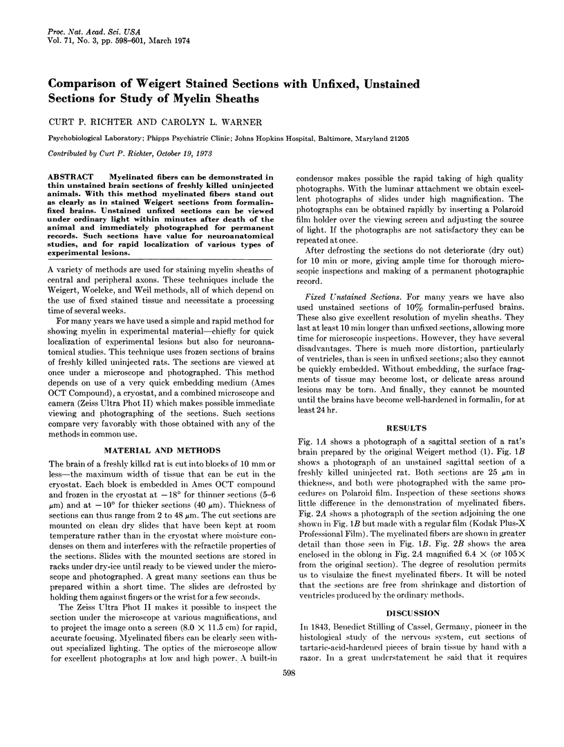
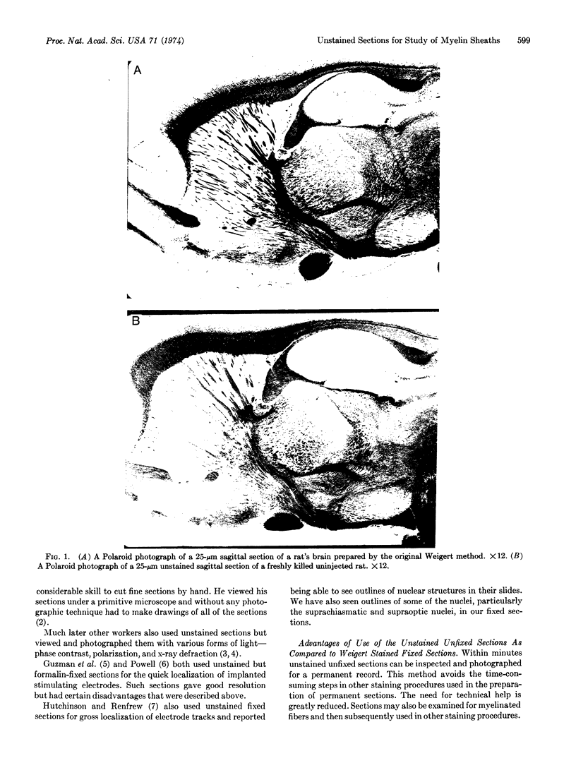
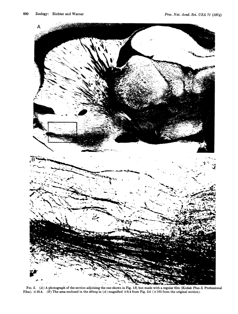
Myelinated fibers can be demonstrated in thin unstained brain sections of freshly killed uninjected animals. With this method myelinated fibers stand out as clearly as in stained Weigert sections from formalin-fixed brains. Unstained unfixed sections can be viewed under ordinary light within minutes after death of the animal and immediately photographed for permanent records. Such sections have value for neuroanatomical studies, and for rapid localization of various types of experimental lesions.
Full text
PDF


Images in this article
Selected References
These references are in PubMed. This may not be the complete list of references from this article.
- Hutchinson R. R., Renfrew J. W. A simple histological technique for localizing electrode tracks and lesions within the brain. J Exp Anal Behav. 1967 May;10(3):277–280. doi: 10.1901/jeab.1967.10-277. [DOI] [PMC free article] [PubMed] [Google Scholar]
- POWELL E. W. A RAPID METHOD OF INTRACRANIAL ELECTRODE LOCALIZATION USING UNSTAINED FROZEN SECTIONS. Electroencephalogr Clin Neurophysiol. 1964 Oct;17:432–434. doi: 10.1016/0013-4694(64)90168-3. [DOI] [PubMed] [Google Scholar]
- SHANKLIN W. M., ISSIDORIDES M., SALAM M. Histochemistry of the cerebral cortex from a case of amaurotic family idiocy. J Neuropathol Exp Neurol. 1962 Apr;21:284–293. doi: 10.1097/00005072-196204000-00009. [DOI] [PubMed] [Google Scholar]


