Abstract
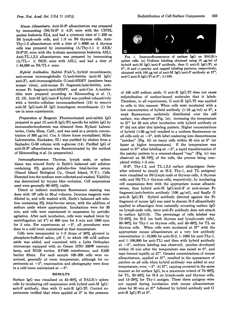
Redistribution of surface immunoglobulins, H-2b, Thy-1.2, and TL.1,2,3 alloantigens, and concanavalin A receptors on mouse lymphoid cells induced by hybrid rabbit F(ab′)2 antibody (anti-mouse immunoglobulin/anti-visual marker or anti-concanavalin A/anti-visual marker) was studied by immunofluorescence. When used directly to label surface immunoglobulin, and indirectly to label alloantigens and concanavalin A receptors, hybrid antibodies induced similar displacement of all surface components from a uniform distribution into “patches” and “caps” at 37°. One hybrid antibody preparation, antimouse immunoglobulin/anti-ferritin, contained negligible amounts of bivalent anti-mouse immunoglobulin antibody, and was therefore “monovalent” for the antimouse immunoglobulin specificity. This observation suggests that factors other than multivalent crosslinking are responsible for hybrid antibody-induced redistribution of cell-surface components. Cap formation induced by hybrid antibody was enhanced markedly by attachment of the visual marker, either ferritin or southern bean mosaic virus, at 37°. At -5°, hybrid antibody does not displace uniformly distributed H-2b alloantigen-alloantibody complexes, but patches of label develop when ferritin attaches to the hybrid antibody. These results explain the patchy distribution of cell-surface components, which is a temperature-independent characteristic of labeling with hybrid antibodies and visual markers for electron microscopy.
Keywords: cell surface, immunofluorescence, cap formation, ferritin
Full text
PDF


Images in this article
Selected References
These references are in PubMed. This may not be the complete list of references from this article.
- Agrawal B. B., Goldstein I. J. Protein-carbohydrate interaction. VI. Isolation of concanavalin A by specific adsorption on cross-linked dextran gels. Biochim Biophys Acta. 1967 Oct 23;147(2):262–271. [PubMed] [Google Scholar]
- Aoki T., Hämmerling U., De Harven E., Boyse E. A., Old L. J. Antigenic structure of cell surfaces. An immunoferritin study of the occurrence and topography of H-2' theta, and TL alloantigens on mouse cells. J Exp Med. 1969 Nov 1;130(5):979–1001. doi: 10.1084/jem.130.5.979. [DOI] [PMC free article] [PubMed] [Google Scholar]
- Davis W. C., Alspaugh M. A., Stimpfling J. H., Walford R. L. Cellular surface distribution of transplantation antigens: discrepancy between direct and indirect labeling techniques. Tissue Antigens. 1971;1(2):89–93. doi: 10.1111/j.1399-0039.1971.tb00083.x. [DOI] [PubMed] [Google Scholar]
- Davis W. C. H-2 antigen on cell membranes: an explanation for the alteration of distribution by indirect labeling techniques. Science. 1972 Mar 3;175(4025):1006–1008. doi: 10.1126/science.175.4025.1006. [DOI] [PubMed] [Google Scholar]
- Edelman G. M., Yahara I., Wang J. L. Receptor mobility and receptor-cytoplasmic interactions in lymphocytes. Proc Natl Acad Sci U S A. 1973 May;70(5):1442–1446. doi: 10.1073/pnas.70.5.1442. [DOI] [PMC free article] [PubMed] [Google Scholar]
- Frye L. D., Edidin M. The rapid intermixing of cell surface antigens after formation of mouse-human heterokaryons. J Cell Sci. 1970 Sep;7(2):319–335. doi: 10.1242/jcs.7.2.319. [DOI] [PubMed] [Google Scholar]
- Gingell D. Membrane permeability change by aggregation of mobile glycoprotein units. J Theor Biol. 1973 Mar;38(3):677–679. doi: 10.1016/0022-5193(73)90266-x. [DOI] [PubMed] [Google Scholar]
- Hämmerling U., Aoki T., Wood H. A., Old L. J., Boyse E. A., de Harvin E. New visual markers of antibody for electron microscopy. Nature. 1969 Sep 13;223(5211):1158–1159. doi: 10.1038/2231158a0. [DOI] [PubMed] [Google Scholar]
- Hämmerling U., Aoki T., de Harven E., Boyse E. A., Old L. J. Use of hybrid antibody with anti-gamma-G and anti-ferritin specificities in locating cell surface antigens by electron microscopy. J Exp Med. 1968 Dec 1;128(6):1461–1473. doi: 10.1084/jem.128.6.1461. [DOI] [PMC free article] [PubMed] [Google Scholar]
- Hämmerling U., Rajewsky K. Evidence for surface-associated immunoglobulin on T and B lymphocytes. Eur J Immunol. 1971 Dec;1(6):447–452. doi: 10.1002/eji.1830010608. [DOI] [PubMed] [Google Scholar]
- Loor F., Forni L., Pernis B. The dynamic state of the lymphocyte membrane. Factors affecting the distribution and turnover of surface immunoglobulins. Eur J Immunol. 1972 Jun;2(3):203–212. doi: 10.1002/eji.1830020304. [DOI] [PubMed] [Google Scholar]
- Neauport-Sautes C., Lilly F., Silvestre D., Kourilsky F. M. Independence of H-2K and H-2D antigenic determinants on the surface of mouse lymphocytes. J Exp Med. 1973 Feb 1;137(2):511–526. doi: 10.1084/jem.137.2.511. [DOI] [PMC free article] [PubMed] [Google Scholar]
- Pinto da Silva P. Translational mobility of the membrane intercalated particles of human erythrocyte ghosts. pH-dependent, reversible aggregation. J Cell Biol. 1972 Jun;53(3):777–787. doi: 10.1083/jcb.53.3.777. [DOI] [PMC free article] [PubMed] [Google Scholar]
- Singer S. J., Nicolson G. L. The fluid mosaic model of the structure of cell membranes. Science. 1972 Feb 18;175(4023):720–731. doi: 10.1126/science.175.4023.720. [DOI] [PubMed] [Google Scholar]
- Stackpole C. W., Aoki T., Boyse E. A., Old L. J., Lumley-Frank J., De Harven E. Cell surface antigens: serial sectioning of single cells as an approach to topographical analysis. Science. 1971 Apr 30;172(3982):472–474. doi: 10.1126/science.172.3982.472. [DOI] [PubMed] [Google Scholar]
- Tillack T. W., Scott R. E., Marchesi V. T. The structure of erythrocyte membranes studied by freeze-etching. II. Localization of receptors for phytohemagglutinin and influenza virus to the intramembranous particles. J Exp Med. 1972 Jun 1;135(6):1209–1227. doi: 10.1084/jem.135.6.1209. [DOI] [PMC free article] [PubMed] [Google Scholar]
- Unanue E. R., Perkins W. D., Karnovsky M. J. Ligand-induced movement of lymphocyte membrane macromolecules. I. Analysis by immunofluorescence and ultrastructural radioautography. J Exp Med. 1972 Oct 1;136(4):885–906. doi: 10.1084/jem.136.4.885. [DOI] [PMC free article] [PubMed] [Google Scholar]
- Yahara I., Edelman G. M. Restriction of the mobility of lymphocyte immunoglobulin receptors by concanavalin A. Proc Natl Acad Sci U S A. 1972 Mar;69(3):608–612. doi: 10.1073/pnas.69.3.608. [DOI] [PMC free article] [PubMed] [Google Scholar]
- de Petris S., Raff M. C. Distribution of immunoglobulin on the surface of mouse lymphoid cells as determined by immunoferritin electron microscopy. Antibody-induced, temperature-dependent redistribution and its implications for membrane structure. Eur J Immunol. 1972 Dec;2(6):523–535. doi: 10.1002/eji.1830020611. [DOI] [PubMed] [Google Scholar]


