

Abstract
The fertilization reaction of echinoderm eggs (Lytechinus pictus, a sea urchin, and Dendraster excentricus, a sand dollar) was followed with intracellular electrodes. Membrane potential and K+ activity were recorded.
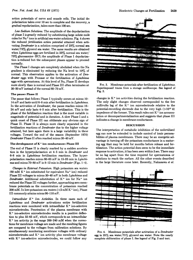
The unfertilized egg of Lytechinus has a membrane potential of -8 mV, inside negative. Within 5 sec after the addition of sperm, a fertilization action potential develops, going to +10 mV, inside positive. The time from the initial depolarization to a return to the original -8 mV is 120-150 sec. The repolarization continues until a potential of -10 to -14 mV is reached, at which point it pauses for 3-4 min. At 6-8 min after fertilization, a further and relatively rapid hyperpolarization begins, going to -60 to -65 mV by 15-25 min after fertilization and remaining constant at these values.
The membrane potential of the unfertilized egg appears to depend on a general permeability to anions. The fertilization action potential seems to reflect a prolonged influx of sodium. The final depolarization to -60 mV is attributable to the development of potassium conductance.
Simultaneous measurements with a K+ ion-selective electrode gives constant readings of about 240 mM K+ in the unfertilized eggs throughout the fertilization process.
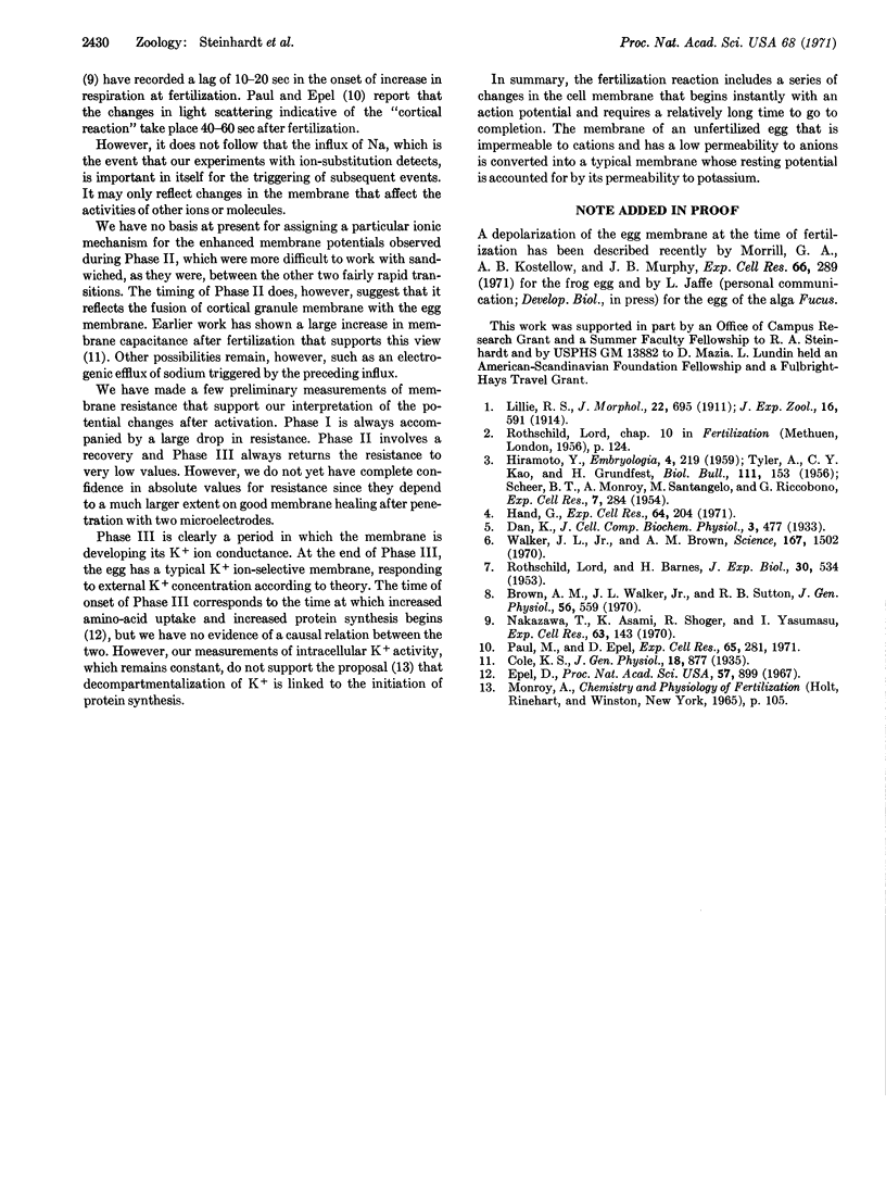
Similar results were obtained with Dendraster eggs. The resting potential of the unfertilized eggs was -7 mV; the action potential on activation attained +18 mV; the repolarization paused at -16 to -24 mV and the final potential attained was -70 mV. The electrical changes after fertilization with spermatozoa or activation with Pronase were identical.
Keywords: fertilization, sea-urchin egg, membrane potential, sodium permeability, intracellular potassium, potassium permeability
Full text
PDF

Images in this article


Selected References
These references are in PubMed. This may not be the complete list of references from this article.
- Brown A. M., Sutton R. B., Walker J. L., Jr Increased chloride conductance as the proximate cause of hydrogen ion concentration effects in Aplysia neurons. J Gen Physiol. 1970 Nov;56(5):559–582. doi: 10.1085/jgp.56.5.559. [DOI] [PMC free article] [PubMed] [Google Scholar]
- Cole K. S. ELECTRIC IMPEDANCE OF HIPPONOE EGGS. J Gen Physiol. 1935 Jul 20;18(6):877–887. doi: 10.1085/jgp.18.6.877. [DOI] [PMC free article] [PubMed] [Google Scholar]
- Epel D. Protein synthesis in sea urchin eggs: a "late" response to fertilization. Proc Natl Acad Sci U S A. 1967 Apr;57(4):899–906. doi: 10.1073/pnas.57.4.899. [DOI] [PMC free article] [PubMed] [Google Scholar]
- Hand G. S., Jr Stimulation of protein synthesis in unfertilized sea urchin and sand dollar eggs treated with trypsin. Exp Cell Res. 1971 Jan;64(1):204–208. doi: 10.1016/0014-4827(71)90212-6. [DOI] [PubMed] [Google Scholar]
- Morrill G. A., Kostellow A. B., Murphy J. B. Sequential forms of ATPase activity correlated with changes in cation binding and membrane potential from meiosis to first clevage in R. pipiens. Exp Cell Res. 1971 Jun;66(2):289–298. doi: 10.1016/0014-4827(71)90680-x. [DOI] [PubMed] [Google Scholar]
- Nakazawa T., Asami K., Shoger R., Fujiwara A., Yasumasu I. Ca2+ uptake H+ ejection and respiration in sea urchin eggs on fertilization. Exp Cell Res. 1970 Nov;63(1):143–146. doi: 10.1016/0014-4827(70)90342-3. [DOI] [PubMed] [Google Scholar]
- Paul M., Epel D. Fertilization-associated light-scattering changes in eggs of the sea urchin Strongylocentrotus purpuratus. Exp Cell Res. 1971 Apr;65(2):281–288. doi: 10.1016/0014-4827(71)90003-6. [DOI] [PubMed] [Google Scholar]
- SCHEER B. T., MONROY A., SANTANGELO M., RICCOBONO G. Action potentials in sea urchin eggs at fertilization. Exp Cell Res. 1954 Aug;7(1):284–287. doi: 10.1016/0014-4827(54)90065-8. [DOI] [PubMed] [Google Scholar]
- Walker J. L., Jr, Brown A. M. Unified account of the variable effects of carbon dioxide on nerve cells. Science. 1970 Mar 13;167(3924):1502–1504. doi: 10.1126/science.167.3924.1502. [DOI] [PubMed] [Google Scholar]