Abstract
AIRmax (maximal inflammation) and AIRmin (minimal inflammation) mice show distinct susceptibilities to pristane-induced arthritis (PIA). The Slc11a1 gene, which regulates macrophage and neutrophil activity, is involved in this infirmity. AIRmaxSS mice homozygous for the non-functional Slc11a1 S (gly169asp) allele obtained by genotype-assisted crosses from AIRmax and AIRmin mice are more susceptible than mice homozygous for the Slc11a1 resistant (R) allele. The present work sought to identify the quantitative trait loci (QTL) regulating PIA and to examine the interactions of these QTL with Slc11a1 alleles in modulating PIA. Mice were given two ip injections of 0.5 mL pristane at 60 day intervals, and the incidence and severity of PIA was scored up to 160 days. Genome-wide linkage studies were performed to search for arthritis QTL in an F2 (AIRmax × AIRmin, n = 290) population. Significant arthritis QTL (LODscore>4) were detected on chromosomes 5 and 8, and suggestive QTL on chromosomes 7, 17 and 19. Global gene expression analyses performed on Affymetrix mouse 1.0 ST bioarrays (27k genes) using RNA from arthritic or control mice paws showed 419 differentially expressed genes between AIRmax and AIRmin mice and demonstrated significantly (P<0.001) over-represented genes related to inflammatory responses and chemotaxis. Up-regulation of the chemokine genes Cxcl1, Cxcl9, Cxcl5, Cxcl13 on chromosome 5 was higher in AIRmaxSS than in the other lines. Macrophage scavenger receptor 1 and hemeoxigenase (decycling) 1 genes on chromosome 8 were also expressed at higher levels in AIRmaxSS mice. Our results show that the gene expression profiles of the two arthritis QTL (on chromosomes 5 and 8) correlate with Slc11a1 alleles, resulting in enhanced AIRmaxSS mice susceptibility to PIA.
Introduction
Mouse lines phenotype-selected for the maximum (AIRmax) or minimum (AIRmin) acute inflammatory reactivity (AIR) were used to study the impact of the genetic control of nonspecific immunity on susceptibility to autoimmune [1], neoplastic [2], and infectious diseases [3]. AIRmax and AIRmin mice were developed through bidirectional selection, starting from a highly polymorphic population (F0) derived from the intercrossing of eight inbred mouse strains (A, DBA2, P, SWR, CBA, SJL, BALB/c and C57BL/6). The selection phenotypes chosen were localized leukocyte influx and exudated plasma proteins 24 hr after the subcutaneous injection of polyacrylamide beads (Biogel), a non-antigenic, insoluble, and chemically inert substance [4]. The progressive divergence of the AIRmax and AIRmin lines during successive generations of selective breeding reached 20- and 2.5-fold differences in leukocyte infiltration and exudated protein concentrations respectively. These differences resulted from the accumulation of alleles in quantitative trai loci (QTL) endowed with opposite and additive effects on the inflammatory response. We can consider AIRmax and AIRmin as outbred stock mice derived from eight inbred lines and submitted to extensive bidirectional selective process for strong and weak acute inflammation phenotypes. Inbreeding was avoided for the selective breeding, as such AIRmax and AIRmin mice maintain a heterogeneous genetic background but are homozygosus in acute inflammation modifier loci. Analysis of the selective processes indicated that the AIR phenotype is regulated by at least 11 QTL [2].
The acute inflammation response to Biogel, as well as susceptibility to pristane induced-arthritis [5], to S. enterica serotype Typhimurium infection, and to the LPS of the bacteria were all modified in these mice; and linkage analysis with microsatellite markers mapped QTL in chromosomes 1, 6, and 11 which are relevant to these phenotypes [6].
Alterations in bone marrow granulopoiesis in response to hematopoietic factors and the production of chemotactic factors by infiltrated or local resident cells both contribute to phenotypic differences between the two lines. Convergent phenotypes in AIRmax mice were observed that were characterized by high neutrophil production in bone marrow, a high number of neutrophils in the blood, high concentrations of chemotatic agents, and increased resistance of infiltrating neutrophils to spontaneous apoptosis [7].
Tissue repair was also investigated in these two lines, revealing that AIRmax mice present a high regeneration capacity in comparison to AIRmin mice. Inflammatory QTL on chromosomes 1 (Slc11a1 gene region) and 14 were found to regulate tissue regrowth in this model [8]. Additionally, the same chromosome 1 QTL seems to regulate leukocyte and protein influx during acute inflammation, as well as arthritis incidence and severity [5].
Rheumatoid arthritis (RA) affects approximately 0.5 to 1% of the world population [9] and is modulated by immune and inflammatory processes, as well as by class II MHC (in particular the locus HLA-DRB1) [10], non-MHC genes [11] and environmental factors. Among RA mouse models, pristane-induced arthritis (PIA) [12] resembles the human condition by its chronic inflammatory nature, similar histopathology and dependency of specific immunity, in particular CD4+ T cells [13]. Another feature of PIA in the mouse is the requirement of microbiota stimulation [14].
Pevious studies have shown that AIRmax mice are extremely susceptible to PIA, whereas AIRmin mice are resistant [1]. The incidence and severity of PIA in AIRmax mice is similar to that of inbred DBA/1 mice [15], but is more intense than those seen in BALB/c [12] or CBA Ighb [16] mice. Fifteen to 25% of the susceptible inbred individuals in lines such as BALB/c [12] and CBA Ighb [17] developed arthritis 200 days after pristane injection. Specific pathogen-free CBA Ighb mice do not develop this disease, but will develop arthritis upon transfer to a conventional environment in the same way as CBA Ighb mice raised conventionally since birth – indicating the involvement of environmental factors in PIA [14]. Susceptibility to PIA is CD4+ T cell (Th) dependent [13] and has been associated with increased agalactosylIgG levels mediated by IL6 production [18], while protection against PIA is mediated by Th2-associated cytokines produced after hsp65 pre-immunization [19].
In contrast to the immune response profile observed in inbred mice, Th2-type response high IgG1 anti-hsp65 levels and high numbers of IL4, IL6, and TNFa secreting splenic cells were observed in susceptible AIRmax mice, following pristane treatment. On the other side, Th1-dependent IgG2a was the predominant isotype in resistant AIRmin mice and increased IFNg producing cells in spleen was evident in these mice only [1].
SOLUTE CARRIER FACTOR 11 A 1 (SLC11A1) gene polymorphism has been implicated in human RA susceptibility [20]. In the mouse, Slc11a1 codes for a transport protein expressed at the membrane of macrophage phagosomes [21] and it is associated with the transport of essential ions. This gene has been described in mice as a major modulator of susceptibility to infectious diseases (first named Natural resistance associate macrophage protein 1 (Nramp1) gene) [22], and is expressed in macrophages and neutrophils [23]. Slc11a1 is pleiotropic, interfering with macrophage activation, oxidative and nitrosamine bursts [24], TNFα, IFNγ, and IL-1 production [25], and the expression of MHC class II molecules [26]. The mutation corresponding to the Slc11a1 S (susceptibility) allele determines a gly169asp substitution resulting in a non-functional protein [27] that promotes ion accumulations inside the phagosome that favor pathogen replication [28].
In the selection process for AIRmax and AIRmin mouse lines, the Slc11a1 S allele frequency was 25% in the founder population (F0), but shifted to 60% in AIRmin and to 9% in AIRmax after 30 generations of selective breeding for inflammatory response, suggesting that these frequency changes were, in fact, the result of genetic selection [3]. In order to determine if the accumulation of Slc11a1 alleles in the AIR lines was due to selection of inflammation QTL and not a chance event, Slc11a1 homozygous mice for the susceptibility (S) or resistant (R) alleles with either AIRmax or AIRmin genetic backgrounds were produced through genotype-assisted mating. The resulting sublines were designated AIRmaxRR, AIRmaxSS, AIRminRR, and AIRminSS. The interaction of the Slc11a1 S allele with high inflammatory background QTL in AIRmax mice was evident through the down-modulation of Biogel-induced early acute inflammation and resistance to Salmonella Typhimurium infection [6], as well as by the aggravation of pristane-induced arthritis [5].
We recently identified several acute inflammatory QTL modulating neutrophil migration, interleukin 1 beta production, and protein concentration in the exudate using the F2 intercross of AIRmax and AIRmin mice [29], [30]. Additionally, our group detected the first QTL modulating PIA in mice, the Prtia1 locus, located on chromosome 3, using mice selected for high and low antibody production [31]. The goal of the present work was therefore to discover new PIA QTL using the AIRmax and AIRmin mouse models, and to study the molecular basis underlying Slc11a1 R and S alleles effect in arthritis development through gene expression profiling within these QTL.
Materials and Methods
Mice
AIRmax and AIRmin lines (Ibut:AIRH and Ibut:AIRL formal stock designations at ILAR, Institute for Laboratory Animal Research, National Research Council), AIRmaxRR, AIRmaxSS, AIRminRR, and AIRminSS sublines, and F2 intercrosses were developed and maintained at the animal facilities of the Laboratory of Immunogenetics of the Butantan Institute. Male and female 8 to 12- week-old mice were used in the experiments. The F2 population was obtained by intercrossing AIRmax with AIRmin mice as described [29]. All procedures were approved by the Institutional Animal Care and Use Committee of the Butantan Institute.
Pristane-induced Arthritis (PIA)
Mice received two intraperitoneal injections with 0.5 ml of the non-immunogenic mineral oil pristine (2,6,10,14-tetramethylpentadecane, Sigma Chemical Co., San Diego, USA) at 60-day intervals. Arthritis development was examined twice weekly for 160 days, by recording arthritis incidence and maximum severity scores for each paw. The severity scores, evaluated for each paw, were: 0–no signs of arthritis; 1–mild swelling of the toes or ankle joint; 2–moderate swelling; 3–severe swelling and/or ankylosis. The maximum score possible for any animal was 12. Phenotypes were assessed twice weekly by two independent observers and the animals were considered arthritic when the mean score assigned by the two observers was ≥2 [31].
Genome-wide SNP Genotyping
Genomic DNA was extracted from tail tips using the E.Z.N.A.® Tissue DNA Kit (Omega Bio-Tek) and quantified using the Quant-iT™ PicoGreen®dsDNA Assay Kit (Invitrogen, Carlsbad, CA). SNPs were genotyped in F2 intercross mice utilizing the Bead-Array Platform (Illumina Inc. San Diego, CA), using the 1449-SNP loci mouse linkage panel as described [29]. Searches for QTL affecting the arthritis severity phenotype under study were carried out through genome-wide linkage analyses between genotypes and phenotypes by interval mapping using GridQTL version 3.1.0 [32] that uses a linear model to fit phenotype data according to genotypes. Additive and dominant effects at the QTL were included along with other explanatory variables of sex and family. The significance thresholds of phenotype-genotype associations were estimated by genome-wide permutation analysis.
Affymetrix Microarray Analysis
Total RNA from the footpads of mice on days 0 and 160 after the second pristane injections were isolated using the RNAspin mini kit (GE Healthcare, Buckinghamshire, UK). The concentrations of the extracted RNA were checked using Nano-Drop (Thermo scientific), and their integrity was tested using Agilent 2100 Bioanalyzer (Agilent Technologies, Santa Clara, CA). The Affymetrix (Santa Clara, CA) Mouse Gene 1.0 ST Array was used, which is a whole transcript-based array that interrogates 28,853 well-annotated genes. Staining, hybridization, washing, and scanning of the array were performed following the manufacturers’ protocols at the AFIP-UNIFESP Molecular Facility, Federal University of Sao Paulo. Four experimental samples were run independently, providing replicates for each experimental group. Gene expression data was stored in a.cel format and subsequently exported to.txt files to be analyzed by Multi-viewer array software [33]. The data discussed in this publication have been deposited in NCBI’s Gene Expression Omnibus [34] and are accessible through GEO Series accession number GSE51516 (http://www.ncbi.nlm.nih.gov/geo/query/acc.cgi?acc=GSE51516).
Real-time Quantitative PCR
Microarray results were validated by quantitative real-time PCR using gene-specific primers. Real-time PCR amplification mixtures contained 0.5 µl template cDNA, 12.5 µl SYBRGreen PCR Master Mix (Invitrogen, Carlsbad, CA, USA), and 0.3 µM specific PCR primers. Reactions were carried out in a Chromo 4 thermocycler (Bio-Rad, Hercules, CA). Targets and housekeeping genes were amplified with the primers described in Table 1. Relative differences were calculated according to the delta-delta Ct method [35]. Microarray data was correlated with qPCR results using Pearson’s analyses.
Table 1. qPCR versus Microarray results correlations and primers used.
| Genes | Pearson (r) | Statistics | Foward | Reverse |
| Il6 | 0,91 | p = 0,001 | 5′-GTTCTCTGGGAAATCGTGGA-3′ | 5′-TGTACTCCAGGTAGCTATGG-3′ |
| Il12a | 0,89 | p = 0,001 | 5′-GATCATGAAGACATCACACGG-3′ | 5′-AGAATGATCTGCTGATGGTGG-3′ |
| Il10 | 0,84 | p = 0,01 | 5′-TCAAACAAAGGACCAGCTGGACAACATACTG-3′ | 5′-CTGTCTAGGTCCTGGAGTCCAGCAGACTCAA-3′ |
| Il1b | 0,81 | p = 0,01 | 5′-CAGTTCTGCCATTGACCATC-3′ | 5′-TCTCACTGAAACTCAGCCGT-3′ |
| Tnfa | 0,82 | p = 0,01 | 5′-TCTCATCAGTTCTATGGCCC-3′ | 5′-GGGAGTAGACAAGGTACAAC-3′ |
| Csf2 | 0,80 | p = 0,01 | 5′-TGAACCTCCTGGATGACATG-3′ | 5′-GTGTTTCACAGTCCGTTTCC-3′ |
| Cxcl1 | 0.89 | p = 0,001 | 5′- TGAAGCTCCCTTGGTTCAGA-3′ | 5′- AGGTGCCATCAGAGCAGTCT-3′ |
| Msr1 | 0.91 | p = 0,001 | 5′- GGGGAGTGTAGGCGGATCAACCCC-3′ | 5′- CGGCCCTCATGGGCTCCACTA-3′ |
| Hmox1 | 0.87 | p = 0,001 | 5′- ACCAGAGTCCCTCACAGATGGCG-3′ | 5′- GCAGGGGCAGTATCTTGCACCA-3′ |
| β2microglubulin | 0.97 | p = 0,001 | 5′TGACCGGCTTGTATGCTATC-3′ | 5′-CAGTGTGAGCCAGGATATAG-3′ |
Relative differences were calculated according to the delta-delta Ct method. Microarray data was correlated with qPCR results using Pearson’s analyses and the same four samples on each line.
Statistical Analyses
The gene expression observed in each array was log-transformed to approximate a Gaussian distribution and then standardized over the array to adjust for systematic differences in their expressions. Differentially expressed genes were detected using Significance of Analysis of Microarray (SAM) software (Two class unpaired, FDR ≤5%) that examined the data from four biological replicates on each line [36]. We analyzed all of the significant differentially-expressed genes for the over-represented biological themes using Expression Analysis Systematic Explorer (EASE) software [37]. This program automates the process of biological theme determination using Gene Ontology (GO) classification. EASE calculates over-representation with respect to the total number of genes assayed and annotated within each system, allowing for side-by-side comparisons of categories from categorization systems with varying levels of annotation. EASE will thus rapidly convert lists of genes into an ordered table of robust biological themes. Calculating statistics on thousands of gene categories can, however, lead to a few seemingly significant probabilities simply due to chance. To address this multiple comparison issue, we used a Bonferroni-type probability correction.
Results
QTL Detection
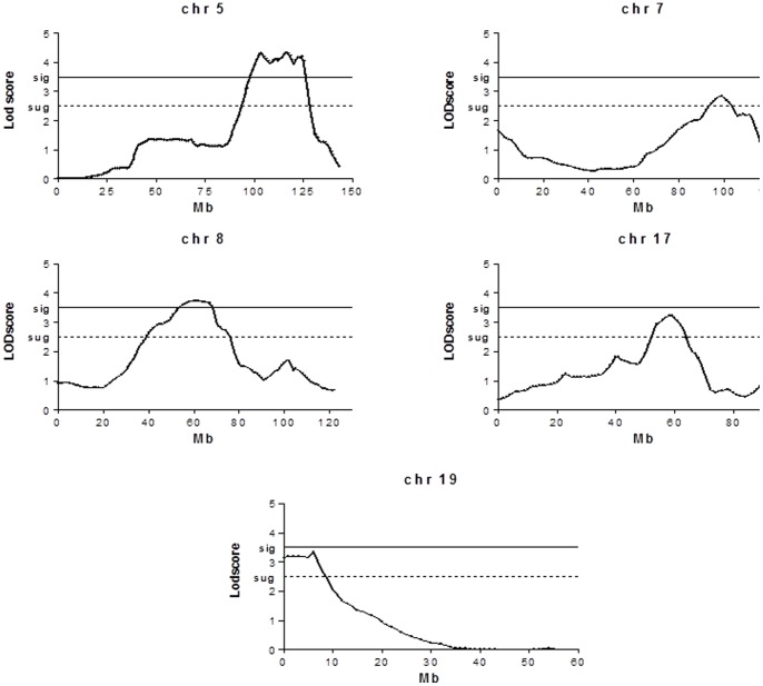
Genome-wide scanning was performed in 290 F2 intercross mice with the PIA severity scores phenotype. Figure 1 shows PIA severity quantitative trait loci (arthritis QTL) presenting high linkage significance (LODscore>4) on chromosomes 5 and 8, and several suggestive QTL on chromosomes 7, 17 and 19. The 1 LOD score confidence interval (CI) on Chromosome 5 maps from 95 to 125 Mb while on chromosome 8 the CI ranges from 40 to 85 Mb, considering LOD scores of 4.177 and 3.865 respectively. The three other suggestive QTL showed wide CI, with LOD scores around 3.
Figure 1. Mapping of the QTL regulating PIA severity phenotype in mice selected for acute inflammation.
SNPs were genotyped in F2 intercrosses employing the Bead-Array Platform (Illumina Inc. San Diego, CA) and using the 1449-SNP loci mouse linkage panel as described. Searches for QTL affecting the arthritis severity phenotype under study were carried out using genome-wide linkage analysis between genotypes and phenotypes by interval mapping, using GridQTL version 3.1.0.
Global Gene Expression and qPCR Analysis
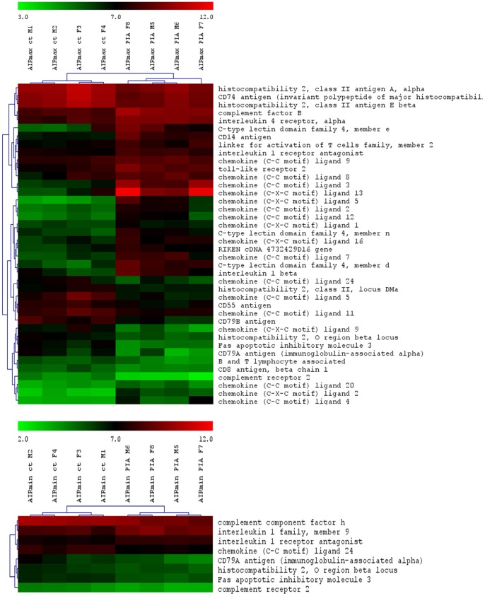
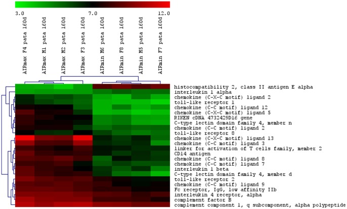
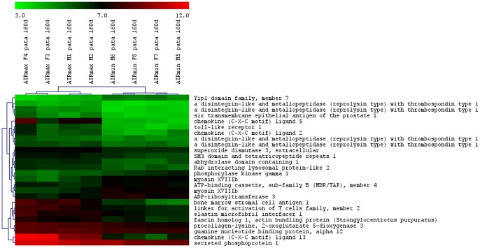
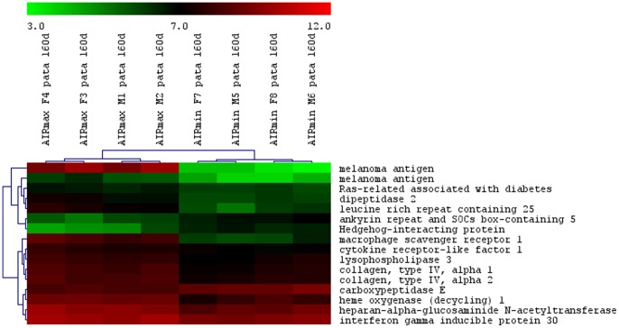
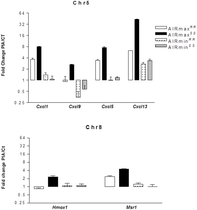
The total numbers of the up- and down-modulated inflammatory response genes are distinct in each line, being higher in AIRmax than in AIRmin mice (Figure 2). These genes are expressed in the swelled footpad of the mice, where there is a large infiltrate of inflammatory neutrophils and macrophages. Global gene expression analysis indicated 419 differentially expressed genes between AIRmax and AIRmin mice. EASE analysis demonstrated significantly over-represented genes (P<0.001) related to inflammatory responses and chemotaxis (Figure 3). Figures 4 and 5 show genes differentially expressed on the confidence intervals of the PIA QTLs at chromosomes 5 and 8 respectively. Several genes related to inflammation, cell adhesion, and chemotaxis could be observed on chromosome 5, while tissue antigens, cell differentiation, hemeoxigenase and scavenger receptor genes were observed on chromosome 8. AIRmax and AIRmin Slc11a1 sublines showed distinct gene expression profiles, reinforcing these results. Higher up-regulation of the chemokine genes Cxcl1, Cxcl9, Cxcl5, Cxcl13 on chromosome 5 were observed in AIRmaxSS than in the other lines. The macrophage scavenger receptor 1(Msr1) and hemeoxigenase (decycling) 1(Hmox1) genes on chromosome 8, were also more actively expressed in AIRmaxSSmice (Figure 6). High correlations among qPCR and Microarray results can be seen in Table 1, indicating the validity of these experiments. These results revealed two significant arthritis QTL (on chromosomes 5 and 8) interacting with the Slc11a1 gene to create a gene expression profile that contributes to the enhanced susceptibility of AIRmaxSS mice to PIA.
Figure 2. Up- and down-modulated inflammatory and chemokine genes in AIRmax and AIRmin mice.
Mouse Gene 1.0(SAM) software (Two class unpaired, FDR ≤5%) that examined the data from four biological replicates on each line.
Figure 3. Differentially expressed inflammatory and chemokine genes in AIRmax and AIRmin mice.
Mouse Gene 1.0(SAM) software (Two class unpaired, FDR ≤5%) that examined the data from four biological replicates on each line.
Figure 4. Differentially expressed genes on chromosome 5 between AIRmax and AIRmin mice.
Mouse Gene 1.0(SAM) software (Two class unpaired, FDR ≤5%) that examined the data from four biological replicates on each line.
Figure 5. Differentially expressed genes on chromosome 8 in AIRmax and AIRmin mice.
Mouse Gene 1.0(SAM) software (Two class unpaired, FDR ≤5%) that examined the data from four biological replicates on each line.
Figure 6. Signal intensities of differentially-expressed genes from AIRmax and AIRmin mouse sublines.
Mouse Gene 1.0(SAM) software (Two class unpaired, FDR ≤5%) that examined the data from four biological replicates on each line.
Discussion
The present work mapped two new PIA QTL (Prtia 2 and Prtia3), on chromosomes 5 and 8, respectively. Three suggestive QTL were detected on chromosomes 7, 17 and 19. The first PIA loci detected in mice was Prtia 1 (by our group) on chromosome 3 using animals selected for high and low antibody production [31]. We also described the involvement of Slc11a1 alleles in PIA using the present AIRmax and AIRmin mouse model [5].
We have recently mapped loci that regulate the intensity of the acute inflammatory response on chromosomes 5, 7, 8 and 17, which overlap the newly detected PIA QTL, suggesting common regulations [29], [30]. Co-located chromosome 5 QTL controlling arthritis severity and humoral responses during B. burgdorferi infection were identified in the F2 intercross of C3H/HeNCr and C57BL/6NCr mice [38], confirming the involvement of the chemokine Cxcl9 gene in this model [39]. QTL on chromosomes 5 and 8 were also detected in two other arthritis models (collagen and proteoglycan induced arthritis) [40], suggesting the importance of these related gene clusters to arthritis susceptibility.
Regarding the suggestive QTL detected in this work, the loci on chromosomes 7, 17 and 19 overlap regions detected for acute inflammation where map inflammasome/integrins, MHC and ion transport related genes, respectively (Table 2) [30]. AIRmax and AIRmin mice present distinct MHC haplotypes: AIRmax H2b and AIRmin H2kd [1]. H2Ea gene expression was low in AIRmax mice (Figure 3), in agreement with a report describing a correlation of the H2b haplotype with low H2Ea gene expression [41].
Table 2. Differentially-expressed major candidate genes detected in microarray related to PIA QTL.
| Genes | Chromosomes | AIRmax/ AIRmin ratio | GO Biological process |
| Slc11a1 | 1 | 2.98 | Transport |
| Cxcl1 | 5 | 3.78 | Chemokine |
| Cxcl9 | 5 | 2.67 | Chemokine |
| Cxcl5 | 5 | 12.11 | Chemokine |
| Cxcl13 | 5 | 27.50 | Chemokine |
| Saa3 | 7 | 21.39 | Inflammation |
| Il21r | 7 | 2.82 | Inflammation |
| Bcl3 | 7 | 2.03 | Apoptosis |
| Msr1 | 8 | 5.02 | Scavenger |
| Hmox1 | 8 | 2.05 | Angiogenesis/apoptosis |
| H2ea | 17 | 0.03 | Antigen Presentation |
| Slc15a3 | 19 | 2.55 | Transport |
Differentially expressed genes in AIRmax and AIRmin mice. Mouse Gene 1.0 ST Array was used. Differentially expressed genes were detected using Significance of Analysis of Microarray (SAM) software (Two class unpaired, FDR ≤5%) that examined the data from four biological replicates on each line.
The total number of up- and down-regulated genes in each line was distinct, as can be seen in Figure 2. More genes were modulated in AIRmax than in AIRmin mice, although we observed an over-representation of genes related to inflammatory reactions and chemotaxis (gene ontology) biological themes in both lines. This same gene expression profile was observed in bone marrow cells from these lines after Biogel stimulus [42], indicating the generalized action of selective pressure during the phenotype selection process.
Ibrahim and collaborators investigated the gene expression profiles of inflamed paws in DBA1 inbred mice using a similar approach for collagen-induced arthritis [43]. In that work, inflammation resulted in increased gene expression of matrix metalloproteinases, and immune-related extra-cellular matrix and cell-adhesion molecules, as well as molecules involved in cell division and transcription, in a manner very similar to our model. However, the total number of differentially-expressed genes involved in the inbred mouse model (223) was lower than in our model (419), suggesting that the heterogeneous background of AIRmax and AIRmin mice permitted a larger genome involvement in this phenotype.
The high acute inflammation observed in AIRmax mice appears to result from the accumulation of three convergent elements during their selection: 1) higher numbers of neutrophils in their bone marrow, as a consequence of an elevated response to granulopoietic cytokines; 2) high concentrations of chemotatic factors in their exudates; and, 3) strong resistance of their neutrophils to spontaneous apoptosis [7]. Consistent with these alterations, AIRmax mice are more susceptible to arthritis [1], LPS shock [6], and colon carcinogenesis [44]. However, this high-responder line demonstrated extreme resistance to bacterial infections [3] as well as to lung [45] and skin carcinogenesis [2]. Neutrophils are the first cells to be recruited to a damaged site [46], and CXC chemokines (including CXCL2 and CXCL1) are the most critical inflammatory mediators for this recruitment [47]. Among the differentially-expressed genes observed in the present work, inflammatory and chemokine genes on chromosome 5 and macrophage scavenger receptor 1 (Msr1) and hemeoxigenase 1 (Hmox1) genes on chromosome 8 appear to be the major candidates.
Chemokines are involved in leukocyte recruitment to inflammatory sites, such as to synovial tissue in rheumatoid arthritis (RA). However, they may also be homeostatic as these functions often overlap [48]. Chemokines have essential roles in the recruitment and activation of leucocyte subsets within tissue microenvironments, and stromal cells actively contribute to these networks. It has been demonstrated that inappropriate constitutive chemokine expression contributes to the persistence of inflammation by actively blocking its resolution [49]. This was likewise observed in lung carcinogenesis in our model, as transcriptome analysis revealed that the genes involved in transendotelial migration and chemokine-cell adhesion were differently-expressed in normal lungs of susceptible AIRmin and resistant AIRmax mice [50], suggesting important roles for these phenotypes in chronic diseases.
Macrophages play a central role in the pathogenesis of rheumatoid arthritis (RA), which is marked by an imbalance of inflammatory and anti-inflammatory macrophages in RA synovium. Although the polarization and heterogeneity of macrophages in RA have not been fully elucidated, the identities of macrophages in RA can potentially be defined by their products, including co-stimulatory molecules, scavenger receptors, different cytokines/chemokines and receptors, and transcription factors. Efforts have been made to understand polarization, apoptosis regulation, and the novel signaling pathways in macrophages [51]. Serum from rheumatoid arthritis patients (but not from healthy subjects) increased mRNA expression of the Msr1 gene in macrophages. Human arterial endothelial cells are inhibited by both anti-IL-6 receptor antibodies (α-IL-6R Ab) and TNF-α receptors (p75)-Fc (TNFR-Fc), suggesting an important role for this receptor in chronic inflammatory disease [52].
Heme oxygenase-1 has potent antioxidant and anti-inflammatory functions, although the underlying mechanisms are not yet well understood [53]. The disruption of Hmox1 in macrophages in vivo and the release of nonmetabolized heme cause tissue inflammation [54]. Additionally, expression of the Slc11a1 gene has been associated with a twofold increase in Hmox1 transcriptional levels in macrophages undergoing erythrophagocytosis [55]. Although Hmox1 seems to alter macrophage activity, no involvement with arthritis is described in the literature. In the present work, AIRmaxSS mice homozygous for the Slc11a1 S allele showed higher Hmox1 and Msr1 gene expression than AIRmaxRR mice, suggesting that Slc11a1 interacts with these two genes (figure 4). Similarly, chemokine genes are also up-regulated in AIRmaxSS mice, evidencing a specific gene expression profile of this line that favors chronic inflammation and consequent susceptibility to arthritis. In this way, Slc11a1 S allele should alter the macrophage activity, promoting modulation of chemokines, heme oxygenase, apoptosis, transport, scavenger receptor and inflammatory genes expressed by these cells (Table 2).
The interaction of the Slc11a1 S allele with high inflammatory background QTL in AIRmax mice modulated the Biogel-induced early acute inflammation, infection resistance [6], and pristane induced-arthritis [5]. AIRmaxSS also showed faster ear-wound closure than AIRmaxRR mice, suggesting that the Slc11a1S allele favors ear tissue regeneration [8].
Taken together, the results of the present work provide interesting insights into the molecular inflammatory mechanisms involved in arthritis onset, and may also exert influences on other experimentally induced diseases. Further studies will be necessary to gain a thorough understanding of the genetic and cellular mechanisms involved in arthritis progression and to provide models for investigating therapeutic treatments that could modulate inflammatory events.
Funding Statement
This work was supported by grants from the Fundação de Amparo à Pesquisa do Estado de São Paulo and the Conselho Nacional de Desenvolvimento Científico e Tecnológico. The funders had no role in study design, data collection and analysis, decision to publish, or preparation of the manuscript.
References
- 1. Vigar ND, Cabrera WH, Araujo LM, Ribeiro OG, Ogata TR, et al. (2000) Pristane-induced arthritis in mice selected for maximal or minimal acute inflammatory reaction. Eur J Immunol 30: 431–437. [DOI] [PubMed] [Google Scholar]
- 2. Biozzi G, Ribeiro OG, Saran A, Araujo ML, Maria DA, et al. (1998) Effect of genetic modification of acute inflammatory responsiveness on tumorigenesis in the mouse. Carcinogenesis 19: 337–346. [DOI] [PubMed] [Google Scholar]
- 3. Araujo LM, Ribeiro OG, Siqueira M, De Franco M, Starobinas N, et al. (1998) Innate resistance to infection by intracellular bacterial pathogens differs in mice selected for maximal or minimal acute inflammatory response. Eur J Immunol 28: 2913–2920. [DOI] [PubMed] [Google Scholar]
- 4. Ibanez OM, Stiffel C, Ribeiro OG, Cabrera WK, Massa S, et al. (1992) Genetics of nonspecific immunity: I. Bidirectional selective breeding of lines of mice endowed with maximal or minimal inflammatory responsiveness. Eur J Immunol 22: 2555–2563. [DOI] [PubMed] [Google Scholar]
- 5. Peters LC, Jensen JR, Borrego A, Cabrera WH, Baker N, et al. (2007) Slc11a1 (formerly NRAMP1) gene modulates both acute inflammatory reactions and pristane-induced arthritis in mice. Genes Immun 8: 51–56. [DOI] [PubMed] [Google Scholar]
- 6. Borrego A, Peters LC, Jensen JR, Ribeiro OG, Koury Cabrera WH, et al. (2006) Genetic determinants of acute inflammation regulate Salmonella infection and modulate Slc11a1 gene (formerly Nramp1) effects in selected mouse lines. Microbes Infect 8: 2766–2771. [DOI] [PubMed] [Google Scholar]
- 7. Ribeiro OG, Maria DA, Adriouch S, Pechberty S, Cabrera WH, et al. (2003) Convergent alteration of granulopoiesis, chemotactic activity, and neutrophil apoptosis during mouse selection for high acute inflammatory response. J Leukoc Biol 74: 497–506. [DOI] [PubMed] [Google Scholar]
- 8. De Franco M, Carneiro PS, Peters LC, Vorraro F, Borrego A, et al. (2007) Slc11a1 (Nramp1) alleles interact with acute inflammation loci to modulate wound-healing traits in mice. Mamm Genome 18: 263–269. [DOI] [PubMed] [Google Scholar]
- 9. Cooper GS, Stroehla BC (2003) The epidemiology of autoimmune diseases. Autoimmun Rev 2: 119–125. [DOI] [PubMed] [Google Scholar]
- 10. Jawaheer D, Thomson W, MacGregor AJ, Carthy D, Davidson J, et al. (1994) “Homozygosity” for the HLA-DR shared epitope contributes the highest risk for rheumatoid arthritis concordance in identical twins. Arthritis Rheum 37: 681–686. [DOI] [PubMed] [Google Scholar]
- 11. John S, Marlow A, Hajeer A, Ollier W, Silman A, et al. (1997) Linkage and association studies of the natural resistance associated macrophage protein 1 (NRAMP1) locus in rheumatoid arthritis. J Rheumatol 24: 452–457. [PubMed] [Google Scholar]
- 12. Potter M, Wax JS (1981) Genetics of susceptibility to pristane-induced plasmacytomas in BALB/cAn: reduced susceptibility in BALB/cJ with a brief description of pristane-induced arthritis. J Immunol 127: 1591–1595. [PubMed] [Google Scholar]
- 13. Stasiuk LM, Ghoraishian M, Elson CJ, Thompson SJ (1997) Pristane-induced arthritis is CD4+ T-cell dependent. Immunology 90: 81–86. [DOI] [PMC free article] [PubMed] [Google Scholar]
- 14. Thompson SJ, Elson CJ (1993) Susceptibility to pristane-induced arthritis is altered with changes in bowel flora. Immunol Lett 36: 227–231. [DOI] [PubMed] [Google Scholar]
- 15. Wooley PH, Sud S, Whalen JD, Nasser S (1998) Pristane-induced arthritis in mice. V. Susceptibility to pristane-induced arthritis is determined by the genetic regulation of the T cell repertoire. Arthritis Rheum 41: 2022–2031. [DOI] [PubMed] [Google Scholar]
- 16. Thompson SJ, Rook GA, Brealey RJ, van der ZR, Elson CJ (1990) Autoimmune reactions to heat-shock proteins in pristane-induced arthritis. Eur J Immunol 20: 2479–2484. [DOI] [PubMed] [Google Scholar]
- 17. Thompson SJ, Thompson HS, Harper N, Day MJ, Coad AJ, et al. (1993) Prevention of pristane-induced arthritis by the oral administration of type II collagen. Immunology 79: 152–157. [PMC free article] [PubMed] [Google Scholar]
- 18. Thompson SJ, Hitsumoto Y, Zhang YW, Rook GA, Elson CJ (1992) Agalactosyl IgG in pristane-induced arthritis. Pregnancy affects the incidence and severity of arthritis and the glycosylation status of IgG. Clin Exp Immunol 89: 434–438. [DOI] [PMC free article] [PubMed] [Google Scholar]
- 19. Thompson SJ, Francis JN, Siew LK, Webb GR, Jenner PJ, et al. (1998) An immunodominant epitope from mycobacterial 65-kDa heat shock protein protects against pristane-induced arthritis. J Immunol 160: 4628–4634. [PubMed] [Google Scholar]
- 20. Shaw MA, Clayton D, Blackwell JM (1997) Analysis of the candidate gene NRAMP1 in the first 61 ARC National Repository families for rheumatoid arthritis. J Rheumatol 24: 212–214. [PubMed] [Google Scholar]
- 21. Vidal SM, Epstein DJ, Malo D, Weith A, Vekemans M, et al. (1992) Identification and mapping of six microdissected genomic DNA probes to the proximal region of mouse chromosome 1. Genomics 14: 32–37. [DOI] [PubMed] [Google Scholar]
- 22.Vidal SM, Malo D, Vogan K, Skamene E, Gros P (1993) Natural resistance to infection with intracellular parasites: isolation of a candidate for Bcg. Cell 73: 469–485. 0092–8674(93)90135-D [pii]. [DOI] [PubMed]
- 23. Canonne-Hergaux F, Gruenheid S, Govoni G, Gros P (1999) The Nramp1 protein and its role in resistance to infection and macrophage function. Proc Assoc Am Physicians 111: 283–289. [DOI] [PubMed] [Google Scholar]
- 24. Fritsche G, Dlaska M, Barton H, Theurl I, Garimorth K, et al. (2003) Nramp1 functionality increases inducible nitric oxide synthase transcription via stimulation of IFN regulatory factor 1 expression. J Immunol 171: 1994–1998. [DOI] [PubMed] [Google Scholar]
- 25. Lalmanach AC, Montagne A, Menanteau P, Lantier F (2001) Effect of the mouse Nramp1 genotype on the expression of IFN-gamma gene in early response to Salmonella infection. Microbes Infect 3: 639–644. [DOI] [PubMed] [Google Scholar]
- 26. Wojciechowski W, DeSanctis J, Skamene E, Radzioch D (1999) Attenuation of MHC class II expression in macrophages infected with Mycobacterium bovis bacillus Calmette-Guerin involves class II transactivator and depends on the Nramp1 gene. J Immunol 163: 2688–2696. [PubMed] [Google Scholar]
- 27. Govoni G, Vidal S, Gauthier S, Skamene E, Malo D, et al. (1996) The Bcg/Ity/Lsh locus: genetic transfer of resistance to infections in C57BL/6J mice transgenic for the Nramp1 Gly169 allele. Infect Immun 64: 2923–2929. [DOI] [PMC free article] [PubMed] [Google Scholar]
- 28. Zaharik ML, Cullen VL, Fung AM, Libby SJ, Kujat Choy SL, et al. (2004) The Salmonella enterica serovar typhimurium divalent cation transport systems MntH and SitABCD are essential for virulence in an Nramp1G169 murine typhoid model. Infect Immun 72: 5522–5525. [DOI] [PMC free article] [PubMed] [Google Scholar]
- 29. Vorraro F, Galvan A, Cabrera WH, Carneiro PS, Ribeiro OG, et al. (2010) Genetic control of IL-1 beta production and inflammatory response by the mouse Irm1 locus. J Immunol 185: 1616–1621. [DOI] [PubMed] [Google Scholar]
- 30. Galvan A, Vorraro F, Cabrera W, Ribeiro OG, Starobinas N, et al. (2011) Association study by genetic clustering detects multiple inflammatory response loci in non-inbred mice. Genes Immun 12: 390–394. [DOI] [PubMed] [Google Scholar]
- 31. Jensen JR, Peters LC, Borrego A, Ribeiro OG, Cabrera WH, et al. (2006) Involvement of antibody production quantitative trait loci in the susceptibility to pristane-induced arthritis in the mouse. Genes Immun 7: 44–50. [DOI] [PubMed] [Google Scholar]
- 32. Hernandez-Sanchez J, Grunchec JA, Knott S (2009) A web application to perform linkage disequilibrium and linkage analyses on a computational grid. Bioinformatics 25: 1377–1383. [DOI] [PubMed] [Google Scholar]
- 33. Saeed AI, Bhagabati NK, Braisted JC, Liang W, Sharov V, et al. (2006) TM4 microarray software suite. Methods Enzymol 411: 134–193. [DOI] [PubMed] [Google Scholar]
- 34. Edgar R, Domrachev M, Lash AE (2002) Gene Expression Omnibus: NCBI gene expression and hybridization array data repository. Nucleic Acids Res 30: 207–210. [DOI] [PMC free article] [PubMed] [Google Scholar]
- 35. Livak KJ, Schmittgen TD (2001) Analysis of relative gene expression data using real-time quantitative PCR and the 2(-Delta Delta C(T)) Method. Methods 25: 402–408. [DOI] [PubMed] [Google Scholar]
- 36. Tusher VG, Tibshirani R, Chu G (2001) Significance analysis of microarrays applied to the ionizing radiation response. Proc Natl Acad Sci U S A 98: 5116–5121. [DOI] [PMC free article] [PubMed] [Google Scholar]
- 37. Hosack DA, Dennis G Jr, Sherman BT, Lane HC, Lempicki RA (2003) Identifying biological themes within lists of genes with EASE. Genome Biol 4: R70. [DOI] [PMC free article] [PubMed] [Google Scholar]
- 38. Weis JJ, McCracken BA, Ma Y, Fairbairn D, Roper RJ, et al. (1999) Identification of quantitative trait loci governing arthritis severity and humoral responses in the murine model of Lyme disease. J Immunol 162: 948–956. [PubMed] [Google Scholar]
- 39. Ma Y, Miller JC, Crandall H, Larsen ET, Dunn DM, et al. (2009) Interval-specific congenic lines reveal quantitative trait Loci with penetrant lyme arthritis phenotypes on chromosomes 5, 11, and 12. Infect Immun 77: 3302–3311. [DOI] [PMC free article] [PubMed] [Google Scholar]
- 40. Glant TT, Adarichev VA, Nesterovitch AB, Szanto S, Oswald JP, et al. (2004) Disease-associated qualitative and quantitative trait loci in proteoglycan-induced arthritis and collagen-induced arthritis. Am J Med Sci 327: 188–195. [DOI] [PubMed] [Google Scholar]
- 41. Liao G, Wang J, Guo J, Allard J, Cheng J, et al. (2004) In silico genetics: identification of a functional element regulating H2-Ealpha gene expression. Science 306: 690–695. [DOI] [PubMed] [Google Scholar]
- 42. Carneiro PS, Peters LC, Vorraro F, Borrego A, Ribeiro OG, et al. (2009) Gene expression profiles of bone marrow cells from mice phenotype-selected for maximal or minimal acute inflammations: searching for genes in acute inflammation modifier loci. Immunology 128: e562–e571. [DOI] [PMC free article] [PubMed] [Google Scholar]
- 43. Ibrahim SM, Koczan D, Thiesen HJ (2002) Gene-expression profile of collagen-induced arthritis. J Autoimmun 18: 159–167. [DOI] [PubMed] [Google Scholar]
- 44. Di Pace RF, Massa S, Ribeiro OG, Cabrera WH, De Franco M, et al. (2006) Inverse genetic predisposition to colon versus lung carcinogenesis in mouse lines selected based on acute inflammatory responsiveness. Carcinogenesis 27: 1517–1525. [DOI] [PubMed] [Google Scholar]
- 45. Maria DA, Manenti G, Galbiati F, Ribeiro OG, Cabrera WH, et al. (2003) Pulmonary adenoma susceptibility 1 (Pas1) locus affects inflammatory response. Oncogene 22: 426–432. [DOI] [PubMed] [Google Scholar]
- 46. Kobayashi Y (2006) Neutrophil infiltration and chemokines. Crit Rev Immunol 26: 307–316. [DOI] [PubMed] [Google Scholar]
- 47. Kobayashi Y (2008) The role of chemokines in neutrophil biology. Front Biosci 13: 2400–2407. [DOI] [PubMed] [Google Scholar]
- 48. Ibrahim SM, Mix E, Bottcher T, Koczan D, Gold R, et al. (2001) Gene expression profiling of the nervous system in murine experimental autoimmune encephalomyelitis. Brain 124: 1927–1938. [DOI] [PubMed] [Google Scholar]
- 49. Filer A, Raza K, Salmon M, Buckley CD (2008) The role of chemokines in leucocyte-stromal interactions in rheumatoid arthritis. Front Biosci 13: 2674–2685. [DOI] [PMC free article] [PubMed] [Google Scholar]
- 50.De Franco M, Colombo F, Galvan A, Cecco LD, Spada E, et al.. (2010) Transcriptome of normal lung distinguishes mouse lines with different susceptibility to inflammation and to lung tumorigenesis. Cancer Lett 294: 187–194. S. [DOI] [PubMed]
- 51. Li J, Hsu HC, Mountz JD (2012) Managing macrophages in rheumatoid arthritis by reform or removal. Curr Rheumatol Rep 14: 445–454. [DOI] [PMC free article] [PubMed] [Google Scholar]
- 52. Hashizume M, Mihara M (2012) Blockade of IL-6 and TNF-alpha inhibited oxLDL-induced production of MCP-1 via scavenger receptor induction. Eur J Pharmacol 689: 249–254. [DOI] [PubMed] [Google Scholar]
- 53. Immenschuh S, Baumgart-Vogt E, Mueller S (2010) Heme oxygenase-1 and iron in liver inflammation: a complex alliance. Curr Drug Targets 11: 1541–1550. [DOI] [PubMed] [Google Scholar]
- 54. Kovtunovych G, Eckhaus MA, Ghosh MC, Ollivierre-Wilson H, Rouault TA (2010) Dysfunction of the heme recycling system in heme oxygenase 1-deficient mice: effects on macrophage viability and tissue iron distribution. Blood 116: 6054–6062. [DOI] [PMC free article] [PubMed] [Google Scholar]
- 55. Soe-Lin S, Sheftel AD, Wasyluk B, Ponka P (2008) Nramp1 equips macrophages for efficient iron recycling. Exp Hematol 36: 929–937. [DOI] [PubMed] [Google Scholar]


