Abstract
Analysis of knockout animals indicates that 3′,5′cyclic guanosine monophosphate (cGMP) has an important role in gut homeostasis but the signaling mechanism is not known. The goals of this study were to test whether increasing cGMP could affect colon homeostasis and determine the mechanism. We increased cGMP in the gut of Prkg2+/+ and Prkg2−/− mice by treating with the PDE5 inhibitor Vardenafil (IP). Proliferation, differentiation and apoptosis in the colon mucosa were then quantitated. Vardenafil (Vard) treatment increased cGMP in colon mucosa of all mice, but reduced proliferation and apoptosis, and increased differentiation only in Prkg2+/+ mice. Vard and cGMP treatment also increased dual specificity protein phosphatase 10 (DUSP10) expression and reduced phospho-c-Jun N-terminal kinase (JNK) levels in the colon mucosa of Prkg2+/+ but not Prkg2−/− mice. Treatment of Prkg2−/− mice with the JNK inhibitor SP600125 reversed the defective homeostasis observed in these animals. Activation of protein kinase G2 (PKG2) in goblet-like LS174T cells increased DUSP10 expression and reduced JNK activity. PKG2 also increased goblet cell-specific MUC2 expression in LS174T cells, and this process was blocked by DUSP10-specific siRNA. The ability of cGMP signaling to inhibit JNK-induced apoptosis in vivo was demonstrated using dextran sodium sulfate (DSS) to stress the colon epithelium. Vard was a potent inhibitor of DSS-induced epithelial apoptosis, and significantly blocked pathological endpoints in this model of experimental colitis. In conclusion, Vard treatment activates cGMP signaling in the colon epithelium. Increased PKG2 activity alters homeostasis by suppressing proliferation and apoptosis while promoting differentiation. The PKG2-dependent mechanism was shown to involve increased DUSP10 and subsequent inhibition of JNK activity.
Keywords: cGMP, PKG2, colon, DUSP10, JNK
Uroguanylin and guanylin are endogenous peptide hormones in the intestine and colon (respectively), which trigger intracellular 3′,5′cyclic guanosine monophosphate (cGMP) production by activating epithelial guanylyl cyclase C (GCC) receptors.1, 2 Downstream targets of cGMP include phosphodiesterases, ion channels and cGMP-dependent protein kinases (protein kinase G (PKG)).3 Type 2 PKG is a central cGMP effector in the gut epithelium, and regulates the Na+/H+ exchanger, the Cl−/HCO3− exchanger and the cystic fibrosis transmembrane conductance regulator.4, 5 The importance of the uroguanylin/guanylin system in controlling electrolyte balance is well established, but downstream cGMP signaling is also emerging as a central regulator of intestinal homeostasis.
Homeostasis in the gut mucosa is a highly regulated process involving a balance between Wnt-mediated proliferation at the crypt base,6 commitment to either secretory (goblet and enteroendocrine cells) or enterocyte lineage in the transit amplifying region,7 followed by terminal differentiation and apoptosis at the luminal surface. Knocking out either guanylin or GCC in mice results in reduced cGMP levels, an increased proliferative compartment, increased apoptosis and reduced differentiation of secretory lineage cells in the gut.8, 9 More recently, Prkg2−/− mice were also shown to exhibit crypt hyperplasia and defective differentiation, suggesting that PKG2 mediates the homeostatic effects of guanylin/cGMP in the colon.10
GCC knockout animals also show increased tumorigenesis when treated with the mutagen azoxymethane, or when crossed with ApcMin/+ mice.8, 11 The mechanism responsible for the increased susceptibility of GCC knockout animals to gut tumorigenesis is not clear. It could reflect a higher level of proliferation in the gut, but it could also be due to basal inflammation resulting from a compromised epithelial barrier as recently reported in these animals.12, 13, 14 Efforts to better understand the underlying signaling remain inconclusive, largely because they have relied heavily on studies of colon cancer cells.10, 14, 15 The intestinal phenotype of knockout mice deficient in various cGMP signaling components suggests that this pathway inhibits proliferation and promotes differentiation in the normal gut epithelium. Challenges associated with increasing cGMP levels in the colon epithelium have left this hypothesis untested, and signaling downstream of PKG2 in the normal colon epithelium remain largely unexplored.
The aim of this study was to determine whether pharmacological manipulation of cGMP levels in the gut can regulate epithelial homeostasis in wild-type (WT) mice, and to elucidate the downstream signaling mechanism. To accomplish this we increased cGMP in the colon epithelium by intraperitoneal administration of the phosphodiesterase-5 inhibitor Vardenafil (Vard). In agreement with predictions based on the analysis of knockout mice, Vard treatment suppressed proliferation and apoptosis, but increased secretory cell lineage differentiation in the colon epithelium. Vard did not affect colon homeostasis in Prkg2−/− mice, indicating that PKG2 is a critical part of this process. We show here that activation of PKG2 by Vard led to increased dual specificity protein phosphatase 10 (DUSP10) expression and reduced the levels of phosphorylated c-Jun N-terminal kinase (JNK) in the colon epithelium. Consistent with the importance of JNK regulation in control of colon homeostasis by cGMP, pharmacological inhibition of JNK essentially reversed the defects in PKG2-deficient animals. Taken together, these data support a central role for cGMP in suppressing apoptosis in luminal colonocytes. An important part of the mechanism is likely to involve PKG2-dependent upregulation of DUSP10, which encodes a phosphatase that inhibits JNK activation.
Results
Systemic Vard treatment increases cGMP and activates PKG in the colon mucosa
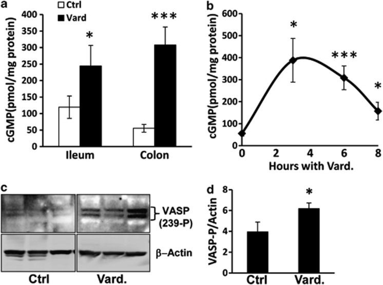
To test the hypothesis that PKG2 regulates homeostasis in the colon, it was necessary to identify an efficient method for increasing cGMP in the epithelial compartment. The possibility that inhibiting cGMP hydrolysis with PDE-5 inhibitors could accomplish this was tested by intraperitoneal injection of mice with Vard (Levitra). A single dose of Vard significantly increased mucosal cGMP levels in the ileum (2.1-fold, P<0.05) and colon (5.6-fold, P<0.001) when measured at 6 h after injection (Figure 1a). The effect of Vard on the colon was rapid, with maximal cGMP levels observed at 3–4 h after administration, and this slowly declined to near-basal levels after 8 h (Figure 1b). Vasodilator-stimulated phosphoprotein (VASP) has target phosphorylation sites for both PKA (Ser157) and PKG (Ser239), which are widely used to measure PKG activation. Treatment of mice with Vard led to significant increases in the level of VASP phosphorylated on Ser239 in the colon mucosa (Figures 1c and d). These data indicate that systemic Vard administration in mice was able to elevate cGMP levels high enough to activate PKG in the colon mucosa.
Figure 1.
Vard increases cGMP levels and PKG2 activation in the colon mucosa. (a) cGMP levels in the intestinal mucosa of CD-1 mice treated with Vard for 6 h or untreated (Ctrl). (b) cGMP levels in the proximal colon mucosa of mice over time following Vard treatment. (c) Western blot showing phospho-VASP (Ser239) in the colon mucosa of three mice with (Vard) or without (Ctrl) Vard treatment for 4 h. (d) Relative levels of phospho-VASP(Ser239) detected from western blot were quantitated and are expressed as a function of total β-actin as protein loading control. *P<0.05, ***P<0.001
PKG2 activation alters homeostasis in the colon mucosa
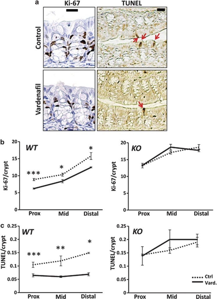
We previously reported that Prkg2−/− mice exhibit crypt hyperplasia, increased apoptosis and reduced differentiation in the colon epithelium.10 This suggested that PKG2 has a critical role in homeostatic regulation in adult colon mucosa, but the contribution of adaptation artifacts associated with PKG2 deficiency could not be ruled out. To explore this possibility further, WT and PKG2-deficient mice were treated with Vard daily, and proliferation and apoptosis were measured in the colon epithelium using immunohistochemistry (IHC; Figure 2a). Vard treatment significantly reduced proliferation by 20–30% throughout the colon epithelium of WT mice, but had no effect in Prkg2−/− mice (Figure 2b). Vard treatment also inhibited basal levels of apoptosis by 40–60% with increasing efficacy along the rostral-caudal axis, but was similarly ineffective in the colon of PKG2-deficient animals (Figure 2c).
Figure 2.
Vard inhibits cell proliferation and apoptosis in the colon mucosa. (a) IHC staining of Ki-67 (proliferative cells) and TUNEL (apoptotic cells, red arrows) in colon mucosa of WT mice treated with Vard for 7 days. Control mice were injected with vehicle. Scale bars, 20 μm. (b) Quantification of Ki-67-positive cells and (c) TUNEL-positive cells from the stained tissue sections from the proximal (prox), middle (mid) and distal colons from WT and PKG2 KO mice. Solid lines show Vard treated, and dashed lines show Controls. Numbers are presented as nuclei per crypt. *P<0.05, **P<0.01, ***P<0.001
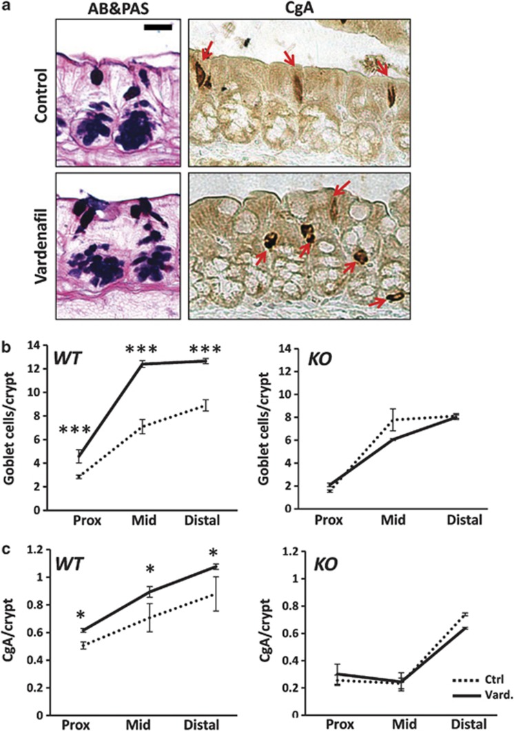
Central to homeostasis in the colon is the differentiation of cells as they move to the luminal surface and withdraw from the cell cycle. To determine whether Vard treatment affects differentiation, the colon sections were also analyzed for goblet and enteroendocrine cell content (Figure 3). The mature goblet cells were identified in the upper half of colonic crypts by staining for mucus with Alcian blue-periodic acid–Schiff, and enteroendocrine cells were identified by immunostaining for chromogranin A (CgA). Vard treatment markedly increased mature goblet cell numbers in the colons of WT but not Prkg2−/− mice (Figure 3b). Although goblet cell density was significantly higher in the proximal and distal regions of the colon from treated mice (50% increase, P<0.001), the effect was most pronounced in the mid-colon with a two fold increase. Despite the higher goblet cell density induced by Vard treatment, no significant difference in the thickness of the luminal mucus layer in the colon was observed (data not shown). Vard treatment also increased CgA-positive enteroendocrine cells in the colon mucosa of WT but not Prkg2−/− animals (Figure 3c). The effect of Vard on enteroendocrine cell density was less than on goblet cells, with increases more uniform throughout the colon and ranging from 20 to 29% (P<0.05). Despite the relatively potent effect of Vard on cGMP levels in the terminal ileum, this agent did not significantly affect homeostasis in that compartment (data not shown). Taken together, these data demonstrate that the PDE-5 inhibitor Vard is capable of increasing cGMP levels in the colon mucosa, and that this regulates homeostasis in a PKG2-dependent manner.
Figure 3.
Vard increases differentiation in the colon mucosa. (a) Staining of Alcian blue-periodic acid–Schiff (AB-PAS; mucus producing goblet cells) and CgA (enteroendocrine cells) in the colon mucosa of WT mice treated with Vard for 7 days. Control mice were injected with vehicle. Scale bar 10 μm. Quantification of (b) mature goblet cells in the upper region of crypts and (c) enteroendocrine cells (CgA) from the stained tissue sections from the colons of WT and PKG2 KO mice. Solid lines show Vard treated, and dashed lines show Controls. Numbers presented as cell counts per crypt. *P<0.05, ***P<0.001
PKG2 regulates colon homeostasis by attenuating JNK signaling
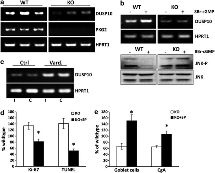
To better understand the mechanism(s) by which PKG2 controls homeostasis, cDNA microarray analysis was used to compare gene expression in the colon mucosa of WT and Prkg2−/− mice (Supplementary Data, Table 2). The expression analysis strongly supported the histological data in that most of the genes showing higher expression in knockout animals were associated with proliferation, whereas those with reduced expression were associated with a differentiated phenotype. The gene array study also identified DUSP10 as strongly reduced in expression in Prkg2−/− mice relative to WT siblings, and this was confirmed using reverse transcriptase polymerase chain reaction (RT-PCR; Figure 4a). The DUSP10 gene encodes a MAPK phosphatase (MKP5) that inactivates JNK by dephosphorylating serine/threonine and tyrosine residues in the active enzyme.16, 17 We tested the possibility that PKG2 activates DUSP10 expression by incubating colon explants from WT and Prkg2−/− mice with 8Br-cGMP for 2-h in vitro. These experiments showed that DUSP10 mRNA levels increased only in the colon mucosa of WT animals, and this corresponded with reduced levels of phospho-JNK (Figure 4b).
Figure 4.
DUSP10 is a PKG2 effector in the colon epithelium. (a) RT-PCR detecting mRNA levels of DUSP10 and PKG2 in colon mucosa of three WT/KO siblings. (b) WT/KO mice colon mucosa explants were treated with (+) or without (−) 8Br-cGMP. RT-PCR showing mRNA levels of DUSP10 (upper panel) and western blots showing phospho-JNK T183/Y185 (lower panel). HPRT1 and total JNK were used as loading controls respectively. (c) RT-PCR showing expression levels of DUSP10 in mucosa from ileum (I) and colon (C) of CD-1 mice treated with Vard or with vehicle (Ctrl). (d and e) PKG2 KO mice were treated with the JNK inhibitor SP600125 (KO+SP, dark bars) or vehicle (KO, open bars) and the colon was processed for quantification of (d) proliferative cells (Ki-67), apoptotic cells (TUNEL) and (e) mature goblet and enteroendocrine cells (CgA). Numbers are presented as a percentage of the respective mean in WT mice. *P<0.05
Treatment of mice with Vard for 48 h also led to increased DUSP10 mRNA levels in the terminal ileum and proximal colon (Figure 4c). This suggested that increased MKP5 levels could mediate the effects of PKG2 on homeostasis, and that sub-threshold levels of MKP5 could contribute to the aberrant homeostasis observed in the PKG2-deficient animals. To further explore this possibility, histological analysis was used to determine the effect of the JNK inhibitor SP600125 on homeostasis in the colons of Prkg2−/− mice. In agreement with our recent report, Prkg2−/− mice exhibited 1.5-fold higher proliferation (Ki-67-staining cells) and apoptosis (TUNEL-positive cells) relative to WT siblings. However, treatment with the JNK inhibitor reduced both proliferation and apoptosis to 80% and 60% of the WT (Figure 4d). The Prkg2−/− mice were also deficient in differentiated goblet and enteroendocrine cells in the colon relative to WT siblings (approximately half). Treatment of the Prkg2−/− mice with SP600125 significantly increased the density of differentiated goblet cells (150%, P<0.05) and enteroendocrine cells (110%, P<0.01) in the colon epithelium relative to WT siblings (Figure 4e). Taken together, these findings highlight the importance of JNK suppression in the regulation of colon homeostasis by PKG2.
Activation of MUC2 expression by PKG2 requires DUSP10
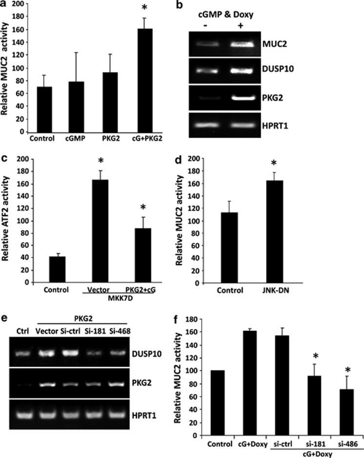
The significance of DUSP10 in the regulation of differentiation by PKG2 was further tested in vitro using LS174T cells. These cells are of a goblet lineage and can be made to differentiate as reflected by increased CDX2 and Muc2 expression.10, 18 Activation of ectopic PKG2 in LS174T cells increased transcription from a reporter construct driven by the MUC2 promoter, and this was associated with increased mRNA for both MUC2 and DUSP10 in these cells (Figures 5a and b). In addition, PKG2 significantly inhibited JNK activity (by 64%, P<0.05) as measured using an ATF2-reporter (Figure 5c). Co-transfection of the LS174T cells with a dominant-negative JNK construct increased the MUC2 reporter activity by 40% (Figure 5d). This indicates that MUC2 expression is inhibited by JNK in these cells. To test whether DUSP10 regulates MUC2 expression downstream of PKG2, siRNA was used to block the PKG2-dependent increase in DUSP10 mRNA (Figure 5e). In these experiments, DUSP10-specific siRNA significantly inhibited (P<0.05) the PKG2-dependent increase in MUC2 reporter activity (Figure 5f). These data demonstrate that DUSP10-mediated suppression of basal JNK activity can promote the expression of the differentiated goblet cell marker MUC2.
Figure 5.
Activation of MUC2 expression by PKG2 in LS174T cells requires DUSP10. (a) The effect of PKG2 activity on transcription from the MUC2 promoter was measured by luciferase reporter assay. (b) The ability of PKG2 to regulate MUC2 and DUSP10 mRNA levels was assessed by RT-PCR. PKG2 expression was induced by treating cells with doxycycline (doxy) and activated by 8Br-cGMP (cGMP). (c) The effect of PKG2 on JNK-dependent ATF2 transcription was measured by luciferase assay. (d) Effect of dominant-negative JNK (JNK-DN) on MUC2 promoter transcription was measured by luciferase assay. The effect of DUSP10-specific siRNA on (e) DUSP10 mRNA levels measured by RT-PCR, and on (f) MUC2 promoter transcription activity measured by luciferase assay. For RT-PCR studies HPRT1 was used as a loading control. Error bars indicate S.E.M., *P<0.05
Activation of PKG2 blocks DSS-induced apoptosis in the colon epithelium
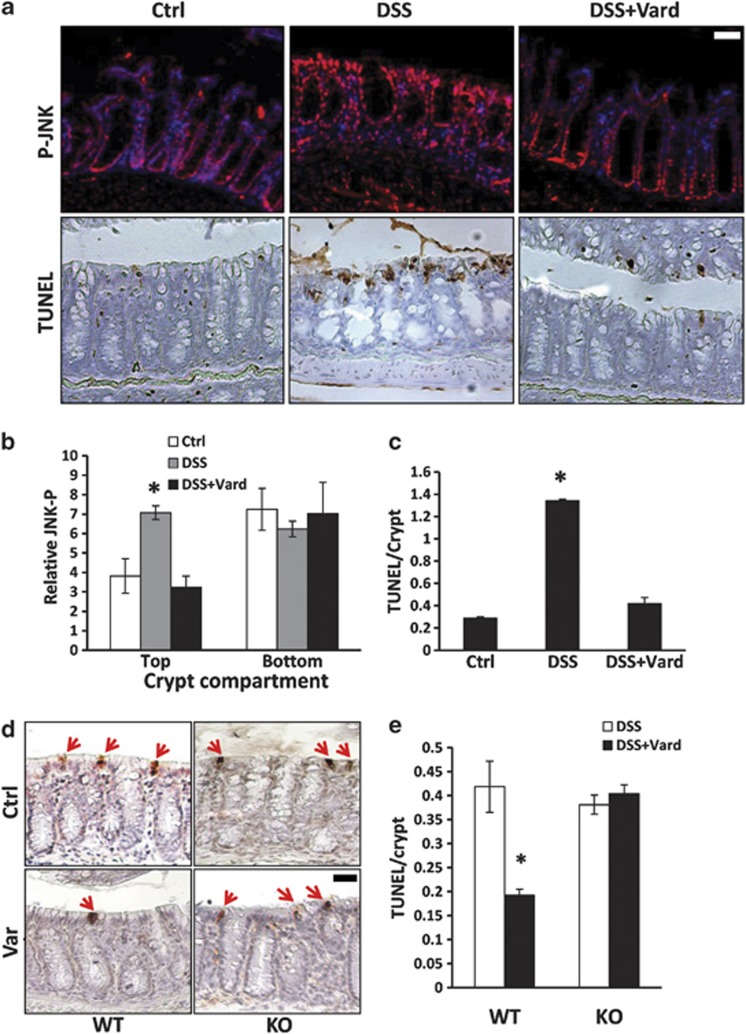
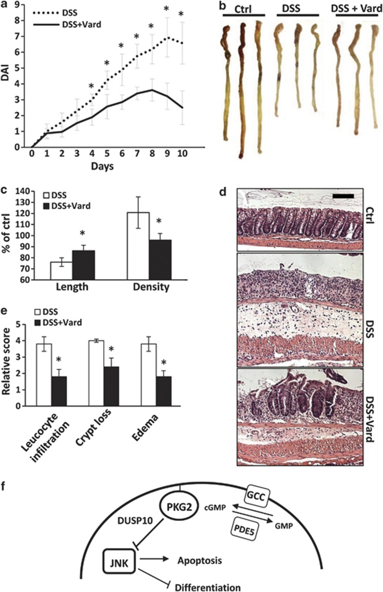
JNK signaling has many context-specific functions, but it is well established that this pathway is pro-apoptotic in most systems.19 It was shown here that Vard treatment significantly reduced apoptosis in the luminal epithelium of the colon in mice. Vard also increased DUSP10 expression, suggesting that the effect of this agent on apoptosis could involve suppression of phospho-JNK levels. This idea was explored further using immunofluorescence (IF) to measure the phosphorylation state of JNK in the colon mucosa (Figure 6). The majority of phospho-JNK staining was observed in the nuclei of cells predominantly in the lower half of the crypts with little staining at the luminal surface. This observation supports previous work suggesting that crypt-associated JNK activity may have a paradoxical proliferative role by potentiating β-catenin (β-cat)/TCF signaling.20 The absence of phospho-JNK staining at the luminal surface precluded our ability determine the effect of Vard in this region. To elicit a more robust activation of JNK in the luminal epithelium we used dextran sodium sulfate (DSS), which activates the stress response and causes apoptosis in the colonic epithelium.21 After 5 days of DSS treatment, a marked increase in nuclear phospho-JNK staining was observed in the luminal epithelium but no effect was observed deeper in the crypt (Figure 6a, upper panels). The intense phospho-JNK staining at the luminal surface colocalized with abundant apoptosis in the DSS-treated mouse colon (Figure 6a, lower panels). The increase in phospho-JNK levels and the associated apoptosis in response to DSS treatment were almost completely prevented by simultaneous Vard treatment (Figures 6b and c). Furthermore, the protective effect of Vard on DSS-mediated epithelial apoptosis in the colon was not observed in Prkg2−/− mice, indicating an important role for PKG2 in this process (Figures 6d and e). Damaging the colon epithelium of mice with DSS compromises the barrier leading to acute inflammation, and is a highly regarded model for experimental colitis.21 In this study, Vard treatment was able to significantly block gross disease symptoms such as loss of body weight, diarrhea and bleeding (Figure 7a). The protective effect of Vard was also evident in physical and histological changes to the colon in response to DSS, including edema, leukocyte infiltration and crypt loss (Figures 7b–e). Together these data indicate that activation of PKG2 signaling in the luminal epithelium of the colon reduces phospho-JNK levels by increasing DUSP10 expression, and this suppresses apoptosis in these cells (Figure 7f).
Figure 6.
Vard treatment inhibits JNK-associated apoptosis in the colon mucosa. (a) Control mice were treated with vehicle (Ctrl), experimental groups were treated with either DSS (DSS) or with both DSS and Vard (DSS+Vard) for 5 days. Upper panels show IF staining of phospho-JNK (red) in the colon mucosa and sections were counter stained with DAPI (blue). Lower panels show parallel sections stained for apoptotic cells using TUNEL. (b and c) Quantification of phospho-JNK and TUNEL staining, respectively. (d) TUNEL staining (red arrows) and (e) quantitation of apoptosis in the colon mucosa of WT and PKG2 knock out (KO) mice treated with DSS. Scale bars represent 20 μm. *P<0.05
Figure 7.
Vard treatment inhibits DSS-induced colitis in mice. CD-1 mice were either untreated (Ctrl), exposed to 5% DSS (DSS) or treated with Vard and exposed to 5% DSS (DSS+Vard). DSS was removed on day 6 and replaced by water. (a) Disease activity index (DAI); including body weight loss, rectal bleeding and stool consistency, was measured over time. (b and c) On day 10, the colons were harvested and the length and density were measured and presented as percentage of control mice. (d and e) Colons were then processed for histological staining with H&E and subjected to pathological scoring for leukocyte infiltration, crypt loss and edema. Scale bar represents 100 μm. *P<0.05. (f) Model depicting regulation of homeostasis by PKG2 signaling in the colon epithelium
Discussion
There is a significant body of genetic evidence demonstrating an important role for cGMP signaling in the control of gut homeostasis, but the underlying mechanisms remain poorly understood because few studies have examined the effects of direct cGMP modulation. In this study, systemic administration of the PDE-5 inhibitor Vard was used to increase cGMP in the colon mucosa of mice. We reasoned that the basal cGMP levels derived from endogenous guanylin/GCC activity in the colon epithelium might be potentiated using PDE-5 inhibitors. Our results showed that systemic delivery of Vard significantly increased cGMP levels and activated PKG in the colon mucosa. Furthermore, sustained Vard treatment suppressed proliferation and increased differentiation in the colon epithelium of WT mice. This clearly demonstrates the importance of cGMP signaling in colon homeostasis, and agrees with predictions arising from studies of knockout animals.8, 9, 10 It is possible that the effect of Vard on colon homeostasis did not involve signaling in epithelial cells directly because the drug was administered systemically. However, this is unlikely because homeostasis was not significantly affected by Vard treatment of Prkg2−/− mice. This demonstrates that epithelial PKG2 has a critical role in the regulation of colon homeostasis by cGMP.
PKG2 activation led to increased DUSP10 expression and reduced phospho-JNK levels in the colon epithelium of mice, indicating that this phosphatase is a novel downstream effector. An important role for DUSP10 in the regulation of gut homeostasis was suggested by the ability of the JNK inhibitor SP600125 to reverse the growth and differentiation defects in the colon epithelium of PKG2-deficient mice. These results strongly suggest that the defects in colon homeostasis in PKG2-deficient mice10 results from aberrant JNK activity arising from insufficient DUSP10 expression. Although this experiment did not definitively prove the specific involvement of DUSP10 in promoting differentiation in the mouse colon, DUSP10 expression was clearly shown to be essential for the cGMP/PKG2-dependent activation of the goblet cell marker MUC2 in LS174T cells. It has been reported that JNK can inhibit MUC2 expression via a repressive site in the promoter.10, 22, 23 It is therefore plausible that the increased MUC2 expression in LS174T cells is the result of derepression of the MUC2 gene rather than a true indicator of differentiation. Similarly, the increased numbers of MUC2-positive cells in the luminal epithelium after Vard treatment might reflect increased mucus production in existing goblet cells instead of an effect on differentiation. However, PKG2 was also able to induce enterocyte differentiation marker expression in Caco-2 cells, which are not related to goblet cell lineage (see Supplementary Figure 1). In addition, Vard treatment also increased CgA-positive enteroendocrine cells in the colon epithelium. Taken together, these data strongly support the idea that cGMP/PKG2 signaling more generally promotes differentiation in the colon mucosa.
Vard treatment reduced basal levels of both apoptosis and proliferation by activating PKG2 in the colon epithelium of mice. The JNK inhibitor had a similar effect on proliferation and apoptosis in Prkg2−/− mice as Vard did on the WT mice. This suggests that elevated JNK activity is a central driver of the phenotype resulting from PKG2 deficiency, and that inhibition of JNK by the PKG2-induced DUSP10 expression is likely to mediate these homeostatic effects of Vard treatment. Previous reports have shown that JNK enhances proliferation in the colonic crypt through interactions with the β-cat/TCF pathway.20, 24 Although it is possible that JNK inhibition by PKG2/DUSP10 could have directly affected proliferation in our studies, this is unlikely because PKG2 is predominantly expressed in the differentiated luminal epithelium rather than in the proliferative compartment.4, 25 In contrast to its role in the mitotic compartment, in the luminal epithelium JNK becomes activated in inflammatory bowel disease (IBD), where it promotes inflammatory cytokine production, apoptosis and barrier breakdown.26, 27, 28, 29 Proliferation and apoptosis in the colon epithelium are coordinated in order to maintain barrier integrity.30 It is therefore plausible that the suppression of proliferation by Vard treatment is indirectly because of a PKG2-mediated inhibition of spontaneous apoptosis at the luminal surface. Indeed, the increased numbers of apoptotic cells previously observed in the colons of PKG2 knockout mice were localized to the luminal surface.10 This idea is supported by a report that cGMP protects the epithelial barrier against radiation-induced damage,12 and another showing that GCC-deficient mice have a dysfunctional intestinal barrier.13 Although this model explains the effects of Vard in the mouse colon, we did not observe significantly increased numbers of phospho-JNK-staining cells in the colon mucosa of PKG2-deficient mice (unpublished results). A likely explanation is that JNK activity is tightly linked to apoptosis in the luminal epithelium, and the phospho-JNK-staining cells are rapidly shed into the luminal space. More conclusive evidence for the ability of Vard/PKG2 to inhibit phospho-JNK in vivo came from experiments using oral DSS, which is widely known to induce epithelial apoptosis in the distal colon. Our results showed that DSS caused massive JNK activation specifically in the luminal epithelium in association with equally profound apoptosis. The near complete blockade of both JNK-P activation and apoptosis by Vard treatment in WT but not PKG2-deficient animals strongly supports the idea that cGMP/PKG2 signaling inhibits colonocyte apoptosis by suppressing JNK.
IBD (Crohn's disease and ulcerative colitis) is a significant health issue that is characterized by bouts of inflammation affecting the mucosal lining of the gut. It is not fully understood whether a compromised epithelial barrier leads to mucosal inflammation, or whether aberrant inflammation precedes barrier breakdown. In either case, suppressing JNK signaling would be expected to increase the threshold for stress-induced barrier breakdown. This idea was demonstrated here using the PDE5 inhibitor Vard to suppress JNK signaling in the gut mucosa, which markedly reduced disease in the DSS model of experimental colitis. This exciting translational finding underscores the potential therapeutic value of PDE5 inhibitors in the gut, but further study with other disease models is indicated.
Taken together, our results show a novel function of cGMP in the colon epithelium and identify at least part of the mechanism. The cGMP/PKG2 signaling pathway is well established in the small intestine where it regulates solute balance, but its role in the colon is less clear.5, 31 We show here that PKG2 regulates homeostasis in the colon mucosa at least in part by inhibiting colonocyte apoptosis. Furthermore, suppression of JNK signaling by a PKG2-mediated increase in MKP5 expression was shown to be a critical part of the underlying mechanism. A growing body of evidence indicates that cGMP signaling could be barrier protective13, 14 and tumor suppressive11, 32 in the colon. The ability of the PDE-5 inhibitor Vard to increase cGMP levels and activate protective PKG2-mediated signaling in the colon epithelium therefore has significant translational potential. As several PDE-5 inhibitors are already used in the clinic to treat erectile dysfunction and pulmonary hypertension,33 the present results highlight additional therapeutic potential for these drugs in gastrointestinal diseases.
Materials and Methods
Animals
The type 2 PKG knockout (Prkg2−/−) mouse was provided by Dr. Franz Hofmann (Technische Universität München, Munich, Germany) with 129S1/Sv × 129 × 1/SvJ genetic background.5 Genotyping PCR was performed using genomic DNA isolated from tail-clip tissue using primer sets described previously.10 Age-matched CD-1 mice were purchased from Harlan (Prattville, AL, USA). All mouse procedures were approved by the GRU Institutional Animal Care and Use Committee.
Drug administration in vivo and in vitro
Pharmaceutical grade PDE5 inhibitors Vard (Levitra) was dissolved in DMSO and diluted 1/10 with PBS immediately before use. Animals were injected (IP) with vehicle or with a Vard dose of 0.6 mg/kg at 12-h intervals for up to 7 days. For time course studies, CD-1 mice were treated with a single dose of Vard and tissues were harvested at different time points. The JNK inhibitor SP600125 (Sigma, St. Louis, MO, USA) was similarly dissolved in DMSO and diluted 1/10 with PBS immediately before use, and injected IP (30 mg/kg body weight) twice a day for 3 days. For in vitro studies, the gut was removed, rinsed with PBS, divided into sections and the explants were incubated under tissue culture conditions. For all studies, the mucosa was harvested from underlying tissue by scraping.
Tissue culture and reagents
LS174T and Caco-2 cells were obtained from the American Type Culture Collection (ATCC, Manassas, VA, USA) and maintained in 5% CO2 in RPMI-1640 medium containing 10% FBS, and supplemented with 2 mM L-glutamine, 10 IU/ml penicillin and 10 mg/ml streptomycin. Cell lines made inducible for PKG2 expression were described previously.10 The doxycycline (doxy) and 8Br-cGMP were from Calbiochem (San Diego, CA, USA). NP-40, Tween-20 and puromycin were from Sigma. G418 was from Hyclone (Logan, UT, USA), and all other chemicals were from Fisher Scientific (Pittsburgh, PA, USA). The cGMP level in 0.1 M HCL extracts of gut mucosa were quantitated using a cyclic GMP EIA Kit according to the manufacturer instructions (Cayman, Ann Arbor, MI, USA) and standardized to protein measured by the Bradford assay (Bio-Rad, Hercules, CA, USA).
Western blotting and IHC
Cell and tissue lysates were prepared and separated on PAGE gels as described previously.10 Antibodies used for immunoblotting recognized PKG2 (1 : 200, Santa Cruz Biotechnology, Santa Cruz, CA, USA), β-actin (1 : 2000, Sigma) and all others were from Cell Signaling (1 : 1000, Danvers, MA, USA). Animal tissues were processed for histological analysis as described previously10 and probed using antibodies to Ki-67 (1 : 100 dilution; Dako Cytomation, Carpinteria, CA, USA), Phospho-JNK (1 : 100, Cell Signaling), and CgA (1 : 200, Immunostar, Hudson, WI, USA). Apoptotic cells were stained with a TUNEL kit (Millipore, Billerica, MA, USA). Histological analysis was done independently by two individuals who were blinded to treatments and mouse genotypes. To quantify immunoreactivity for western blotting, Licor IRDye Infrared Dye-conjugated secondary antibodies were used and images were captured and quantified by Odyssey Infrared imaging system version 3.0 (Lincoln, NB, USA). Quantitation of phospho-JNK staining in the colon was done from images captured from two animals per group. The intensity of red fluorescence was measured using ImageJ software (National Institutes of Health, Bethesda, MD, USA; http://imagej.nih.gov/ij/) relative to background, for at least 10-fields containing at least 10 crypts per each field for each animal.
Analysis of gene expression
Steady-state RNA levels were measured by semiquantitative RT-PCR using GeneAmp PCR kits (Applied Biosystems, Foster City, CA, USA) and primers designed using Primer Blast Software (National Center for Biotechnology Information, National Library of Medicine, Bethesda, MD, USA; see Supplementary Data Table 1). SiRNAs for DUSP10 were designed and purchased from BLOCK-iT RNAi Designer (Invitrogen, Carlsbad, CA, USA). The MUC2-luciferase reporter assay was described previously.18, 34 c-Jun- and ATF2-luciferase reporter assays were performed using PathDetect c-JUN trans-Reporting System (Agilent Technologies, Santa Clara, CA, USA) according to the manufacturer's protocols. Dominant-negative JNK was a generous gift from Dr. Shuang Huang (Georgia Regents University, Augusta, GA, USA). Active MEK7 (MKK7D) construct was obtained from Dr. Jiahui Han (Scripps Research Institute, La Jolla, CA, USA).
DSS-induced colonic injury
Six- to 8-week-old male mice were treated with DSS (m.w. 36 000–50 000; MP Biomedicals, Santa Ana, CA, USA) by dissolving in drinking water ad libitum. (3.5% w/v for strain 129, and 5% for CD-1). Vard or vehicle was administered IP starting 2 days before DSS treatment and continued for the duration of the experiment. For histological analysis of JNK and apoptosis, mice were treated with DSS for 5 days before harvesting and processing of colon tissues. For assessment of subsequent colitis symptoms, mice were treated with DSS for 6 days and switched to normal drinking water for an additional 4 days of recovery. Scoring of colitis endpoints was performed essentially as described previously.21, 35 Disease activity index included three components: weight loss (0=0%, 1=1–5%, 2=5–10%, 3=10–20%, 4=>20%), diarrhea (0=normal stool, 1=soft stool, 2=very soft stool, 3=diarrhea, 4=dysenteric diarrhea) and rectal bleeding (0=none, 1=occult bleeding, 2=blood on anus, 3=blood on fur/tail, 4=gross bleeding). After 10 days, the colons were removed and both weight and length were recorded. Colon tissue was then processed as Swiss Rolls for histopathological analysis of edema, leukocyte infiltration and crypt loss.
Statistical analysis
All quantitative experiments were reproduced in at least three independent experiments with multiple measures in each replicate. The resulting data were expressed as means with error bars indicating S.E.M. Two means were considered to be statistically significant when two-tailed Student's T-test had P-value <0.05.
Acknowledgments
This work was supported by a grants from the Georgia Cancer Coalition and from the NIH 1RO1CA172627-01A1 to DDB.
Glossary
- β-cat
β-catenin
- cGMP
3′,5′cyclic guanosine monophosphate
- DSS
dextran sodium sulfate
- DUSP
dual specificity protein phosphatase
- GCC
guanylyl cyclase C
- IF
immunofluorescence
- IHC
immunohistochemistry
- JNK
c-Jun N-terminal kinase
- PKG
protein kinase G
- RT-PCR
reverse transcriptase polymerase chain reaction
- Vard
Vardenafil
- WT
wild-type animals
The authors declare no conflict of interest.
Footnotes
Supplementary Information accompanies this paper on Cell Death and Differentiation website (http://www.nature.com/cdd)
Edited by JP Medema
Supplementary Material
References
- Forte LR., Jr Uroguanylin and guanylin peptides: pharmacology and experimental therapeutics. Pharmacol Ther. 2004;104:137–162. doi: 10.1016/j.pharmthera.2004.08.007. [DOI] [PubMed] [Google Scholar]
- Steinbrecher KA, Cohen MB. Transmembrane guanylate cyclase in intestinal pathophysiology. Curr Opin Gastroenterol. 2011;27:139–145. doi: 10.1097/MOG.0b013e328341ead5. [DOI] [PubMed] [Google Scholar]
- Lucas KA, Pitari GM, Kazerounian S, Ruiz-Stewart I, Park J, Schulz S, et al. Guanylyl cyclases and signaling by cyclic GMP. Pharmacol Rev. 2000;52:375–414. [PubMed] [Google Scholar]
- Markert T, Vaandrager AB, Gambaryan S, Pohler D, Hausler C, Walter U, et al. Endogenous expression of type II cGMP-dependent protein kinase mRNA and protein in rat intestine. Implications for cystic fibrosis transmembrane conductance regulator. J Clin Invest. 1995;96:822–830. doi: 10.1172/JCI118128. [DOI] [PMC free article] [PubMed] [Google Scholar]
- Pfeifer A, Aszodi A, Seidler U, Ruth P, Hofmann F, Fassler R. Intestinal secretory defects and dwarfism in mice lacking cGMP-dependent protein kinase II. Science. 1996;274:2082–2086. doi: 10.1126/science.274.5295.2082. [DOI] [PubMed] [Google Scholar]
- Simons BD, Clevers H. Stem cell self-renewal in intestinal crypt. Exp Cell Res. 2011;317:2719–2724. doi: 10.1016/j.yexcr.2011.07.010. [DOI] [PubMed] [Google Scholar]
- Medema JP, Vermeulen L. Microenvironmental regulation of stem cells in intestinal homeostasis and cancer. Nature. 2011;474:318–326. doi: 10.1038/nature10212. [DOI] [PubMed] [Google Scholar]
- Li P, Lin JE, Chervoneva I, Schulz S, Waldman SA, Pitari GM. Homeostatic control of the crypt-villus axis by the bacterial enterotoxin receptor guanylyl cyclase C restricts the proliferating compartment in intestine. Am J Pathol. 2007;171:1847–1858. doi: 10.2353/ajpath.2007.070198. [DOI] [PMC free article] [PubMed] [Google Scholar]
- Steinbrecher KA, Wowk SA, Rudolph JA, Witte DP, Cohen MB. Targeted inactivation of the mouse guanylin gene results in altered dynamics of colonic epithelial proliferation. Am J Pathol. 2002;161:2169–2178. doi: 10.1016/S0002-9440(10)64494-X. [DOI] [PMC free article] [PubMed] [Google Scholar]
- Wang R, Kwon IK, Thangaraju M, Singh N, Liu K, Jay P, et al. Type 2 cGMP-dependent protein kinase regulates proliferation and differentiation in the colonic mucosa. Am J Physiol Gastrointest Liver Physiol. 2012;303:G209–G219. doi: 10.1152/ajpgi.00500.2011. [DOI] [PubMed] [Google Scholar]
- Li P, Schulz S, Bombonati A, Palazzo JP, Hyslop TM, Xu Y, et al. Guanylyl cyclase C suppresses intestinal tumorigenesis by restricting proliferation and maintaining genomic integrity. Gastroenterology. 2007;133:599–607. doi: 10.1053/j.gastro.2007.05.052. [DOI] [PubMed] [Google Scholar]
- Garin-Laflam MP, Steinbrecher KA, Rudolph JA, Mao J, Cohen MB. Activation of guanylate cyclase C signaling pathway protects intestinal epithelial cells from acute radiation-induced apoptosis. Am J Physiol Gastrointest Liver Physiol. 2009;296:G740–G749. doi: 10.1152/ajpgi.90268.2008. [DOI] [PMC free article] [PubMed] [Google Scholar]
- Han X, Mann E, Gilbert S, Guan Y, Steinbrecher KA, Montrose MH, et al. Loss of guanylyl cyclase C (GCC) signaling leads to dysfunctional intestinal barrier. PLoS One. 2011;6:e16139. doi: 10.1371/journal.pone.0016139. [DOI] [PMC free article] [PubMed] [Google Scholar]
- Lin JE, Snook AE, Li P, Stoecker BA, Kim GW, Magee MS, et al. GUCY2C opposes systemic genotoxic tumorigenesis by regulating AKT-dependent intestinal barrier integrity. PLoS One. 2012;7:e31686. doi: 10.1371/journal.pone.0031686. [DOI] [PMC free article] [PubMed] [Google Scholar]
- Lin JE, Li P, Snook AE, Schulz S, Dasgupta A, Hyslop TM, et al. The hormone receptor GUCY2C suppresses intestinal tumor formation by inhibiting AKT signaling. Gastroenterology. 2010;138:241–254. doi: 10.1053/j.gastro.2009.08.064. [DOI] [PMC free article] [PubMed] [Google Scholar]
- Tanoue T, Moriguchi T, Nishida E. Molecular cloning and characterization of a novel dual specificity phosphatase, MKP-5. J Biol Chem. 1999;274:19949–19956. doi: 10.1074/jbc.274.28.19949. [DOI] [PubMed] [Google Scholar]
- Theodosiou A, Smith A, Gillieron C, Arkinstall S, Ashworth A. MKP5, a new member of the MAP kinase phosphatase family, which selectively dephosphorylates stress-activated kinases. Oncogene. 1999;18:6981–6988. doi: 10.1038/sj.onc.1203185. [DOI] [PubMed] [Google Scholar]
- Blache P, van de Wetering M, Duluc I, Domon C, Berta P, Freund JN, et al. SOX9 is an intestine crypt transcription factor, is regulated by the Wnt pathway, and represses the CDX2 and MUC2 genes. J Cell Biol. 2004;166:37–47. doi: 10.1083/jcb.200311021. [DOI] [PMC free article] [PubMed] [Google Scholar]
- Dhanasekaran DN, Reddy EP. JNK signaling in apoptosis. Oncogene. 2008;27:6245–6251. doi: 10.1038/onc.2008.301. [DOI] [PMC free article] [PubMed] [Google Scholar]
- Sancho R, Nateri AS, de Vinuesa AG, Aguilera C, Nye E, Spencer-Dene B, et al. JNK signalling modulates intestinal homeostasis and tumourigenesis in mice. EMBO J. 2009;28:1843–1854. doi: 10.1038/emboj.2009.153. [DOI] [PMC free article] [PubMed] [Google Scholar]
- Wirtz S, Neufert C, Weigmann B, Neurath MF. Chemically induced mouse models of intestinal inflammation. Nat Protoc. 2007;2:541–546. doi: 10.1038/nprot.2007.41. [DOI] [PubMed] [Google Scholar]
- Lee HY, Crawley S, Hokari R, Kwon S, Kim YS. Bile acid regulates MUC2 transcription in colon cancer cells via positive EGFR/PKC/Ras/ERK/CREB, PI3K/Akt/IkappaB/NF-kappaB and p38/MSK1/CREB pathways and negative JNK/c-Jun/AP-1 pathway. Int J Oncol. 2010;36:941–953. doi: 10.3892/ijo_00000573. [DOI] [PubMed] [Google Scholar]
- Ahn DH, Crawley SC, Hokari R, Kato S, Yang SC, Li JD, et al. TNF-alpha activates MUC2 transcription via NF-kappaB but inhibits via JNK activation. Cell Physiol Biochem. 2005;15:29–40. doi: 10.1159/000083636. [DOI] [PubMed] [Google Scholar]
- Li Y, Kundu P, Seow SW, de Matos CT, Aronsson L, Chin KC, et al. Gut microbiota accelerate tumor growth via c-jun and STAT3 phosphorylation in APCMin/+ mice. Carcinogenesis. 2012;33:1231–1238. doi: 10.1093/carcin/bgs137. [DOI] [PubMed] [Google Scholar]
- Golin-Bisello F, Bradbury N, Ameen N. STa and cGMP stimulate CFTR translocation to the surface of villus enterocytes in rat jejunum and is regulated by protein kinase G. Am J Physiol Cell Physiol. 2005;289:C708–C716. doi: 10.1152/ajpcell.00544.2004. [DOI] [PubMed] [Google Scholar]
- Apidianakis Y, Pitsouli C, Perrimon N, Rahme L. Synergy between bacterial infection and genetic predisposition in intestinal dysplasia. Proc Natl Acad Sci USA. 2009;106:20883–20888. doi: 10.1073/pnas.0911797106. [DOI] [PMC free article] [PubMed] [Google Scholar]
- Assi K, Pillai R, Gomez-Munoz A, Owen D, Salh B. The specific JNK inhibitor SP600125 targets tumour necrosis factor-alpha production and epithelial cell apoptosis in acute murine colitis. Immunology. 2006;118:112–121. doi: 10.1111/j.1365-2567.2006.02349.x. [DOI] [PMC free article] [PubMed] [Google Scholar]
- Karin M, Gallagher E. From JNK to pay dirt: Jun kinases, their biochemistry, physiology and clinical importance. IUBMB Life. 2005;57:283–295. doi: 10.1080/15216540500097111. [DOI] [PubMed] [Google Scholar]
- Waetzig GH, Seegert D, Rosenstiel P, Nikolaus S, Schreiber S. p38 mitogen-activated protein kinase is activated and linked to TNF-alpha signaling in inflammatory bowel disease. J Immunol. 2002;168:5342–5351. doi: 10.4049/jimmunol.168.10.5342. [DOI] [PubMed] [Google Scholar]
- Vereecke L, Beyaert R, van Loo G. Enterocyte death and intestinal barrier maintenance in homeostasis and disease. Trends Mol Med. 2011;17:584–593. doi: 10.1016/j.molmed.2011.05.011. [DOI] [PubMed] [Google Scholar]
- Vaandrager AB, Bot AG, Ruth P, Pfeifer A, Hofmann F, De Jonge HR. Differential role of cyclic GMP-dependent protein kinase II in ion transport in murine small intestine and colon. Gastroenterology. 2000;118:108–114. doi: 10.1016/s0016-5085(00)70419-7. [DOI] [PubMed] [Google Scholar]
- Shailubhai K, Yu HH, Karunanandaa K, Wang JY, Eber SL, Wang Y, et al. Uroguanylin treatment suppresses polyp formation in the Apc(Min/+) mouse and induces apoptosis in human colon adenocarcinoma cells via cyclic GMP. Cancer Res. 2000;60:5151–5157. [PubMed] [Google Scholar]
- Francis SH, Sekhar KR, Ke H, Corbin JD. Inhibition of cyclic nucleotide phosphodiesterases by methylxanthines and related compounds. Handb Exp Pharmacol. 2011;200:93–133. doi: 10.1007/978-3-642-13443-2_4. [DOI] [PubMed] [Google Scholar]
- Kwon IK, Wang R, Thangaraju M, Shuang H, Liu K, Dashwood R, et al. PKG inhibits TCF signaling in colon cancer cells by blocking beta-catenin expression and activating FOXO4. Oncogene. 2010;9:3423–3434. doi: 10.1038/onc.2010.91. [DOI] [PMC free article] [PubMed] [Google Scholar]
- Cooper HS, Murthy SN, Shah RS, Sedergran DJ. Clinicopathologic study of dextran sulfate sodium experimental murine colitis. Lab Invest. 1993;69:238–249. [PubMed] [Google Scholar]
Associated Data
This section collects any data citations, data availability statements, or supplementary materials included in this article.


