Abstract
Interleukin-6 (IL-6) is one of the most important cytokines which has been shown to play a critical role in the pathogenesis of cholesteatoma. In this study, we aimed to investigate the expression of interleukin-6 (IL-6) and phosphorylated signal transducer and activator of transcription 3 (p-STAT3) in middle ear cholesteatoma epithelium in an effort to determine the role of IL-6/JAK/STAT3 signaling pathway in the pathogenesis of cholesteatoma. Immunohistochemistry was used to examine the expression of IL-6 and p-STAT3 in 25 human middle ear cholesteatoma samples and 15 normal external auditory canal (EAC) epithelium specimens. We also analyzed the relation of IL-6 and p-STAT3 expression levels to the degree of bone destruction in cholesteatoma. We found that the expression of IL-6 and p-STAT3 were significantly higher in cholesteatoma epithelium than in normal EAC epithelium (p<0.05). In cholesteatoma epithelium, a significant positive association was observed between IL-6 and p-STAT3 expression (p<0.05). However, no significant relationships were observed between the degree of bone destruction and the levels of IL-6 and p-STAT3 expression (p>0.05). To conclude, our results support the concept that IL-6/JAK/STAT3 signaling pathway is active and may play an important role in the mechanisms of epithelial hyper-proliferation responsible for cholesteatoma.
Keywords: Cholesteatoma, IL-6, STAT3
Introduction
Cholesteatoma is a pathological condition associated with otitis media, accompanying hearing loss, facial paralysis, labyrinthine and brain abscess, and the recurrence after surgical treatment is very common. Cholesteatoma is morphologically characterized by epithelial cell proliferation and granulation tissue formation. Unfortunately, our understanding of the molecular mechanism underlying the pathogenesis of cholesteatoma is limited. Recently, many authors have demonstrated that cytokines and mediators secreted in the inflammatory responses may change the biochemical signaling pathways that mediate the pathogenesis of cholesteatoma [1-4].
Interleukin-6 (IL-6) is one of the most important cytokines which has been shown to play a critical role in the pathogenesis of cholesteatoma, such as epithelial hyper-proliferation and bone destruction. For example, increased IL-6 expression has been described in cholesteatoma tissues. Moreover, cholesteatoma keratinocytes showed higher production of IL-6 as compared with normal external auditory canal (EAC) skin. However, the exact mechanisms of IL-6 in the pathogenesis of cholesteatoma remain unknown. Interleukin-6 (IL-6) is a pleiotropic cytokine that plays an important role in a variety of cellular events including inflammatory reaction, immune response and cellular proliferation [4]. Signal transducer and activator of transcription 3 (STAT3) is a lipid kinase that controls cell growth, proliferation and survival, anabolic and autophagic activities and cytoskeletal organization [7], and is proved to have a major function in promoting cellular proliferation and inhibiting apoptosis in response to IL-6 [8]. Coupling with the IL-6 receptor and gp130, IL-6 activates a Janus kinase (JAK)-dependent signaling cascade, mediating tyrosine phosphorylated STAT3 (p-STAT3). STAT3 signaling pathway has been a hot spot in the pathogenesis exploration for proliferative diseases nowadays, but it is rare studied in the genesis and development of cholesteatoma.
In this study, we hypothesized that the IL-6/JAK/STAT3 signaling pathway may be activated and involved in the pathogenesis of cholesteatoma. To investigate this, we determined the expression of IL-6 and p-STAT3 in cholesteatoma and normal EAC skin by immunohistochemical analysis. We also analyzed the relation of IL-6 and p-STAT3 expression levels to the degree of bone destruction in cholesteatoma.
Materials and methods
Materials
A series of 25 acquired cholesteatoma tissues was obtained during middle ear surgery between January 2010 and June 2010 at the Department of Otolaryngology, Head and Neck Surgery. Of these, 16 were males and 9 were females with a mean age of 35 years. Meanwhile, 15 normal EAC skin samples were obtained to be used as controls. Specimens for hematoxylin-eosin (HE) staining and immunohistochemistry were immediately fixed in 10% buffered formalin and embedded in paraffin.
Immunohistochemistry
The avidin-biotin complex method was performed on tissue sections (4 μm thick). Sections were heated at 60°C for 2 h, deparaffinized with xylene for 15 min and rehydrated in a graded ethanol series (100%, 95%, 80%, 70%). The tissue sections were washed in phosphate-buffered saline (PBS) and endogenous peroxidase was inactivated by incubating 3% hydrogen peroxide at room temperature for 10 min, followed by being washed with PBS. Then tissue sections were treated in a microwave oven using 10 mmol/L citrate buffer (PH 6.0) for 30 min. Subsequently, the sections were pre-incubated with non-immune goat blood serum for 10 min at room temperature and then were incubated overnight at 4°C with primary rabbit anti-p-STAT3 polyclonal antibody or anti-IL-6 monoclonal antibody. After rinsing 3 times with PBS, sections were incubated with the secondary biotinylated goat-anti-rabbit antibody for 15 min at room temperature. Then sections were incubated with streptavidin conjugated with horse radish peroxidase after washing in PBS. Freshly prepared 3-amino-9-ethylcarbozole was used as a substrate for peroxidase. Finally, the sections were counterstained with hematoxylin, dehydrated and mounted. For the negative controls, PBS was used to substitute primary antibody [2].
Evaluation of immunohistochemistry
All tissue sections were analyzed independently by two experienced pathologists who were blind to the clinical data of patients. Cells with uniform red granules were regarded as positive cells. Both the percentage of positive cells and the intensity of staining were considered [9]. The percentage of positive cells was graded and scored as 0 for negative staining, 1 for <30%, 2 for 30-60%, and 3 for >60% positive cells. Staining intensity was graded and scored as 0 for negative staining, 1 for light red, 2 for red, and 3 for dark red. The overall score for each specimen was obtained by multiplying the percentage score and the staining intensity score. The final results were recorded as negative (-) with an overall score of 0, weak positive (+) with an overall score of 1-2, moderate positive (++) if the overall score was 3-5, and strong positive (+++) if the overall score was 6-9.
Analysis of the relation of IL-6 and p-STAT3 expression levels to the degree of bone destruction
The degree of bone destruction was evaluated as follows: grade I - cases without any bone destruction or with mild erosion of one ossicle; grade II - cases with moderate destruction of all ossicles; grade III - cases with severe and widespread destruction including all ossicles, tegmen, external ear canal, bony facial canal, horizontal semicircular canal and inner ear.
Statistical analysis
Statistical analysis was performed by SPSS 15.0 software. Protein expression patterns and relationships between the protein expression levels and the degree of bone destruction were both analyzed with the Chi-square test. Spearman’s rank correlation test was adopted to analyze correlations between the expression of p-STAT3 and IL-6. A value of P<0.05 was considered statistically significant.
Ethics
The study was approved by the Ethics Committee of Central South University and informed consent was obtained from all of the patients.
Results
Histopathological findings
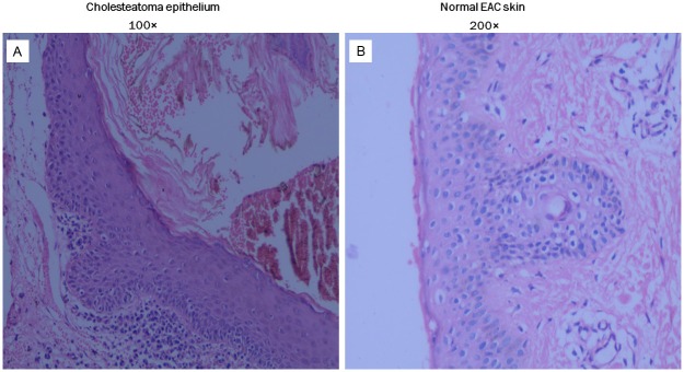
HE staining under light microscope showed that all 25 cholesteatoma specimens consisted of three parts: matrix, perimatrix and keratin debris (Figure 1A), confirming to the pathological criterion of diagnosis. Meanwhile, results of 15 normal EAC skin sections were also accorded with pathological diagnostic criterion (Figure 1B).
Figure 1.

HE staining of human cholesteatoma (A) and normal EAC skin (B). (A) The keratin debris, matrix epithelium and perimatrix subepithelial tissue of cholesteatoma are seen. Original magnification: 100×. (B) The epithelium and subepithelial tissue of normal EAC skin are seen. Original magnification: 200×.
Immunohistochemistry
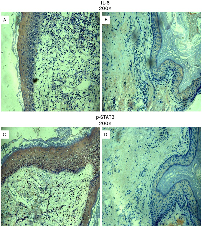
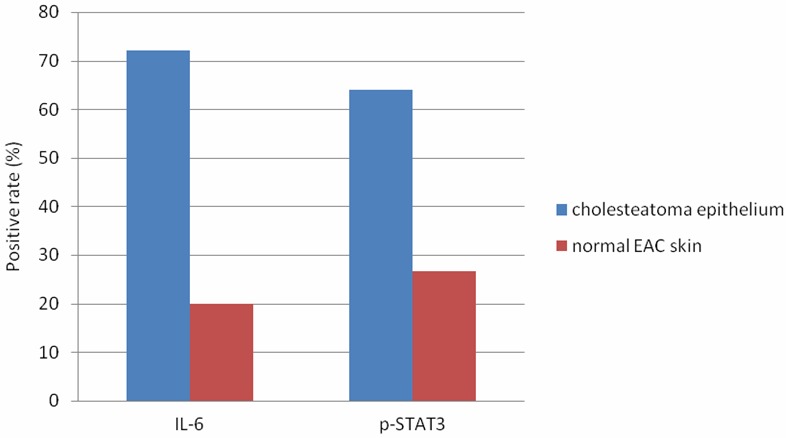
The expression of IL-6 in cholesteatoma was observed predominantly in the cytoplasm with light red or red staining and was localized in the basal and suprabasal layers of the epithelium (Figure 2A). Whereas in normal EAC skin epithelium, IL-6 expression was negative or weak positive (Figure 2B). The positive rate of IL-6 expression was 72% (18/25) in cholesteatoma epithelium compared to 20% (3/15) in normal EAC skin epithelium (Table 1, Figure 3). When compared with normal EAC skin epithelium, the positive rate of IL-6 expression in cholesteatoma epithelium was significantly increased (p=0.003).
Figure 2.

Immunohistochemical staining of IL-6 (A, B) and p-STAT3 (C, D) in human cholesteatoma epithelium and normal EAC skin. (A) IL-6 expression in cholesteatoma was observed predominantly in the cytoplasm with red staining and was localized in the basal and suprabasal layers of the epithelium. Original magnification: 200×. (B) IL-6 expression in normal EAC skin was negative. Original magnification: 200×. (C) p-STAT3 expression in cholesteatoma was observed in the cytoplasm and nucleus with red staining and was localized in the basal and suprabasal layers of the epithelium. Original magnification: 200×. (D) p-STAT3 expression in normal EAC skin was negative. Original magnification: 200×.
Table 1.
Protein expression of IL-6 and p-STAT3 in cholesteatoma epithelium and normal EAC skin
| Cases | IL-6 | p-STAT3 | |||||||
|---|---|---|---|---|---|---|---|---|---|
|
|
|
||||||||
| + | - | χ2 | p | + | - | χ2 | p | ||
| Chol | 25 | 18 | 7 | 10.165 | 0.003 | 16 | 9 | 5.227 | 0.022 |
| EAC | 15 | 3 | 12 | 4 | 11 | ||||
Chol = cholesteatoma; EAC = normal EAC skin.
Figure 3.

The positive rate of IL-6 and p-STAT3 in cholesteatoma epithelium and normal EAC skin: the positive rate of IL-6 expression was 72% (18/25) in cholesteatoma epithelium compared to 20% (3/15) in normal EAC skin epithelium.
In cholesteatoma epithelium, p-STAT3 expression was mainly observed in the cytoplasm and nucleus with red staining and was localized in the basal and suprabasal layers of the epithelium (Figure 2C). However, in normal EAC epithelium, p-STAT3 expression was barely detectable in the cytoplasm and nucleus (Figure 2D). The positive rate of p-STAT3 expression was 64% (16/25) in cholesteatoma epithelium and 26.67% (4/15) in normal EAC skin epithelium (Table 1). A significant difference was found in p-STAT3 expression between cholesteatoma epithelium and normal EAC skin epithelium (p=0.022).
Correlations between IL-6 and p-STAT3 in cholesteatoma epithelium
Among 16 cholesteatoma epithelium specimens with positive p-STATS3 expression, positive IL-6 expression was observed in 14 specimens; while among 18 cholesteatoma epithelium specimens with positive IL-6 expression, negative p-STAT3 expression was observed in 4 specimens. Using Spearman’s rank correlation test, a significant positive correlation was found between p-STAT3 and IL-6 in cholesteatoma epithelium (p=0.021) (Table 2).
Table 2.
Correlations between IL-6 and p-STAT3 in cholesteatoma epithelium
| IL-6 | p-STAT3 | r | p | |
|---|---|---|---|---|
|
|
||||
| + | - | |||
| + | 14 | 4 | 0.460 | 0.021 |
| - | 2 | 5 | ||
Relations of IL-6 and p-STAT3 expression levels to the degree of bone destruction
According to operative findings, 7 patients had grade I bone destruction, 14 had grade II bone destruction, and 4 had grade III bone destruction. We then analyzed the relationships between the protein expression levels and the degree of bone destruction. However, no relationship was found between the degree of bone destruction and the expression levels of IL-6 (p=0.313) and p-STAT3 (p=0.512) (Table 3).
Table 3.
Relation of IL-6 and p-STAT3 protein expression levels to the degree of bone destruction in cholesteatoma epithelium
| Degree | Cases | IL-6 | p-STAT3 | ||||||
|---|---|---|---|---|---|---|---|---|---|
|
|
|
||||||||
| + | - | χ2 | p | + | - | χ2 | p | ||
| I | 7 | 4 | 3 | 2.324 | 0.313 | 3 | 4 | 1.339 | 0.512 |
| II | 14 | 10 | 4 | 9 | 5 | ||||
| III | 4 | 4 | 0 | 3 | 1 | ||||
Discussion
IL-6 is a pro-inflammatory cytokine characterized as a potent activator of STAT3. They function cooperatively to promote cellular proliferation and inhibit apoptosis [10]. In this study, we focused on IL-6/JAK/STAT3 signaling pathway and studied its function in human cholesteatoma epithelium. As we expert, the significant presence of high IL-6 expression and p-STAT3 over-expression was observed in cholesteatoma epithelium. Consistent with our findings, previous studies also reported an increased expression of IL-6 and STAT3 in cholesteatoma epithelium [6,11]. Furthermore, a significantly positive association was observed between IL-6 and p-STAT3 expression, namely, obviously increased p-STAT3 expression existed in cholesteatoma epithelium with IL-6 high expression, indicating the persistent activation of IL-6/JAK/STAT3 signaling in the hyperplasia of cholesteatoma epithelium. Similar results were obtained by Jiang GX et al. [12], they reported that IL-6 promoted STAT3 activation significantly at a posttranslational level in vitro and indicated that IL-6/STAT3 signaling was involved in human biliary epithelial cell migration and wound healing.
Via the binding to gp130, IL-6 mainly activates JAK/STAT signal pathway to mediate a variety of cellular functions [10,12,13]. Combination of IL-6 and IL-6 receptor can phosphorylate the tyrosine of gp130, and further activate JAK family members including JAK1, JAK2, and tyrosine kinase2 (Tyk2). Then, STAT3 is phosphorylated by the activated JAK and translocates into the nucleus to control the expression of substrates [14]. As shown in Table 1, significant difference in the expression of IL-6 was observed between cholesteatoma epithelium and normal EAC epithelium (P=0.003). Marenda SA et al. [16] also found a positive rate of 100% and 25% for IL-6 expression in cholesteatoma epithelium and normal EAC skin, respectively, with a statistical significance. Moreover, they demonstrated a strong correlation between the expression level of IL-6 and ossicula auditus destruction, indicating a vital role of IL-6 in the bone destruction of cholesteatoma. All these results emphasize on the importance of IL-6 in the pathogenesis and development of cholesteatoma.
STAT3 mediates signal transduction and transcription via various cytokines as well as growth factor. p-STAT3 accelerates the cell cycle, promotes cellular differentiation, and inhibits apoptosis through regulating the expression of Cyclin D1, C-myc, Bcl-xl and vascular endothelial growth factor (VEGF) [15], which turns out to be an essential factor in cell over-proliferation. As shown in Table 1, significant difference in the expression of p-STAT3 was observed between cholesteatoma epithelium and normal EAC epithelium (P=0.022). Scanty research on the STAT3 expression in cholesteatoma has been carried out. David R et al. [17] detected the expression of STAT3 in cholesteatoma tissues and EAC skin, and discovered an obviously increased expression of p-STAT3 in cholesteatoma tissues as compared to normal EAC skin, indicating that the elevated expression of p-STAT3 promoted the genesis of cholesteatoma. The IL-6/JAK/STAT3 signaling pathway associates with various benign and malignant proliferative diseases. Recently, Liu ML et al. [18] demonstrated that IL-6/JAK/STAT3 signaling pathway prevented the genesis of breast cancer through inhibiting the Growth Regulation by Estrogen in Breast cancer (GREB1). da Silva CG et al. [19] also reported that liver regeneration was mediated by IL-6/JAK/STAT3 proliferative signaling. Our results are consistent with findings above and propose that IL-6/JAK/STAT3 signaling pathway is active in cholesteatoma epithelium and may represent a novel target for intratympanic drug therapies. In recent years, inhibitors of the IL-6/JAK/STAT3 signaling have emerged as a promising cancer treatment option. For instance, siRNA-STAT3 therapy was demonstrated effective in ovarian cancer [20], and suppressor of cytokine signaling treatment was also proved to be active in blocking the IL-6/JAK/STAT3 signaling in glioblastoma cells [21], such as JAK inhibitor (AG490) [22]. Meanwhile, the curcumin also has been proved to block small cell lung cancer cells migration, invasion, angiogenesis, and neoplasia through the IL-6/JAK/STAT3 pathway [23]. These strategies shed light on a new trend to cure tumors as well as benign proliferative diseases such as cholesteatoma.
Bone destruction is an essential characteristic in cholesteatoma. IL-6 is thought to be linked in the underlying pathology of bone destruction associated with cholesteatoma. Kuczkowski J et al [24] demonstrated the existence of a strong positive correlation between the increased expression of IL-6 in cholesteatoma tissues and the degree of bone destruction, and Nason R et al [25] revealed that the inhibition of IL-6 blocked the osteoclastogenesis which facilitating bone destruction in chronic otitis. However, in our study, no relationship was found between the degree of bone destruction and the expression levels of IL-6 and p-STAT3. This observation suggests that there may be pathways other than the IL-6/JAK/STAT3 signaling cascade that contribute to bone destruction in cholesteatoma.
In conclusion, increased protein expression of IL-6 and p-STAT3 was proved to exist in cholesteatoma epithelium. Furthermore, significantly positive correlation between IL-6 and p-STAT3 expression was confirmed. The increased protein expression of IL-6 and p-STAT3 and their association in cholesteatoma epithelium suggest that the activation of IL-6/JAK/STAT3 signaling pathway may be involved in cholesteatoma epithelial hyper-proliferation. We suggest that anti-IL-6/JAK/STAT3 pathway therapies will function as potent anti-proliferative agents in the cholesteatoma.
Disclosure of conflict of interest
None.
References
- 1.Olszewska E, Chodynicki S, Chyczewski L, Rogowski M. Some markers of proliferative activity in cholesteatoma epithelium in adults. Med Sci Monit. 2006 Aug;12:CR337–340. [PubMed] [Google Scholar]
- 2.Liu W, Yin T, Ren J, Li L, Xiao Z, Chen X, Xie D. Activation of the EGFR/Akt/NF-κB/cyclinD1 survival signaling pathway in human cholesteatoma epithelium. Eur Arch Otorhinolarygol. 2013 Mar 5; doi: 10.1007/s00405-013-2403-6. [Epub ahead of print] [DOI] [PubMed] [Google Scholar]
- 3.Caliman e Gurgel JD, Pereira SB, Alves AL, Riheiro FQ. Hyperproliferation markers in ear canal epidermis. Braz J Otorhinolaryngol. 2010 Sep-Oct;76:667–671. doi: 10.1590/S1808-86942010000500023. [DOI] [PMC free article] [PubMed] [Google Scholar]
- 4.Mihara M, Hashizume M, Yoshida H, Suzuki M, Shiina M. IL-6/IL-6 receptor system and its role in physiological and pathological conditions. Clin Sci (Lond) 2012 Feb;122:143–159. doi: 10.1042/CS20110340. [DOI] [PubMed] [Google Scholar]
- 5.Kuczkowski J, Sakowicz-Burkiewicz M, Izycka-Swieszewska E, Mikaszewski B, Pawełczyk T. Expression of tumor necrosis factor-α, interleukin-6 and interleukin-10 in chronic otitis media with bone osteolysis. ORL J Otorhinolaryngol Relat Spec. 2011;73:93–99. doi: 10.1159/000323831. [DOI] [PubMed] [Google Scholar]
- 6.Xu Y, Tao ZZ, Hua QQ, Wang XC, Xiao BK, Chen SM. Activation of nuclear factor-kappa B and aberrant gene expression of interleukin 6 in middle ear cholesteatoma. Zhonghua Er Bi Yan Hou Tou Jing Wai Ke Za Zhi. 2009 Mar;44:192–196. [PubMed] [Google Scholar]
- 7.Zoncu R, Efeyan A, Sabatini DM. mTOR: from growth signal integration to cancer, diabetes and aging. Nat Rev Mol Cell Biol. 2011 Jan;12:21–35. doi: 10.1038/nrm3025. [DOI] [PMC free article] [PubMed] [Google Scholar]
- 8.Tohyama M, Hanakawa Y, Shirakata Y, Dai X, Yang L, Hirakawa S, Tokumaru S, Okazaki H, Sayama K, Hashimoto K. IL-17 and IL-22 mediate IL-20 subfamily cytokine production in cultured keratinocytes via increased IL-22 receptor expression. Eur J Immunol. 2009 Oct;39:2779–2788. doi: 10.1002/eji.200939473. [DOI] [PubMed] [Google Scholar]
- 9.Hiraishi Y, Wada T, Nakatani K, Negoro K, Fujita S. Immunohistochemical expression of EGFR and p-EGFR in oral squamous cell carcinomas. Pathol Oncol Res. 2006;12:87–91. doi: 10.1007/BF02893450. [DOI] [PubMed] [Google Scholar]
- 10.Chang Q, Bournzou E, Sansone P, Berishaj M, Gao SP, Daly L, Wels J, Theilen T, Granitto S, Zhang X, Cotari J, Alpaugh ML, de Stanchina E, Manova K, Li M, Bonafe M, Ceccarelli C, Taffurelli M, Santini D, Altan-Bonnet G, Kaplan R, Norton L, Nishimoto N, Huszar D, Lyden D, Bromberg J. The IL-6/JAK/STAT3 feed-forward loop drives tumorigenesis and metastasis. Neoplasia. 2013 Jul;15:848–862. doi: 10.1593/neo.13706. [DOI] [PMC free article] [PubMed] [Google Scholar]
- 11.Ho KY, Huang HH, Hung KF, Chen JC, Chai CY, Chen WT, Tsai SM, Chien CY, Wang HM, Wu YJ. Cholesteatoma growth and proliferation: relevance with serpin B3. Laryngoscope. 2012 Dec;122:2818–1823. doi: 10.1002/lary.23547. [DOI] [PubMed] [Google Scholar]
- 12.Jiang GX, Zhong XY, Cui YF, Liu W, Tai S, Wang ZD, Shi YG, Zhao SY, Li CL. IL-6/STAT3/TFF3 signaling regulates human biliary epithelial cell migration and wound healing in vitro. Mol Biol Rep. 2010 Dec;37:3813–3818. doi: 10.1007/s11033-010-0036-z. [DOI] [PubMed] [Google Scholar]
- 13.Xiong H, Zhang ZG, Tian XQ, Sun DF, Liang QC, Zhang YJ, Lu R, Chen YX, Fang JY. Inhibition of JAK1, 2/STAT3 signaling induces apoptosis, cell cycle arrest, and reduces tumor cell invasion in colorectal cancer cells. Neoplasia. 2008 Mar;10:287–297. doi: 10.1593/neo.07971. [DOI] [PMC free article] [PubMed] [Google Scholar]
- 14.Januma N, Shima H, Nakamura K, Kikuchi K. Protein tyrosine phosphatase eC selectively inhibits interleukin-6- and IL-10- induced JAK-STAT signaling. Blood. 2001 Nov;98:3030–3034. doi: 10.1182/blood.v98.10.3030. [DOI] [PubMed] [Google Scholar]
- 15.Takeda K, Akim S. Stat family of transcription factors in cytokine - mediated biological responses. Cytokine Growth Factor Rev. 2000 Sep;11:199–207. doi: 10.1016/s1359-6101(00)00005-8. [DOI] [PubMed] [Google Scholar]
- 16.Marenda SA, Aufdemorte TB. Localization of cytokines in cholesteatoma tissue. Otolaryngol Head Neck Surg. 1995 Mar;112:359–368. doi: 10.1016/S0194-59989570268-7. [DOI] [PubMed] [Google Scholar]
- 17.Friedland DR, Eernisse R, Erbe C, Gupta N, Cioffi JA. Cholesteatoma growth and proliferation: posttranscriptional regulation by microRNA-21. Otol Neurotol. 2009 Oct;30:998–1005. doi: 10.1097/MAO.0b013e3181b4e91f. [DOI] [PMC free article] [PubMed] [Google Scholar]
- 18.Liu M, Wang G, Gomez-Fernandez CR, Guo S. GREB1 functions as a growth promoter and is modulated by IL6/STAT3 in breast cancer. PLoS One. 2012;7:e46410. doi: 10.1371/journal.pone.0046410. [DOI] [PMC free article] [PubMed] [Google Scholar] [Retracted]
- 19.da Silva CG, Studer P, Skroch M, Mahiou J, Minussi DC, Peterson CR, Wilson SW, Patel VI, Ma A, Csizmadia E, Ferran C. A20 promotes liver regeneration by decreasing SOCS3 expression to enhance IL-6/STAT3 proliferative signals. Hepatology. 2013 May;57:2014–2025. doi: 10.1002/hep.26197. [DOI] [PMC free article] [PubMed] [Google Scholar]
- 20.Huang F, Tong X, Fu L, Zhang R. Knockdown of STAT3 by shRNA inhibits the growth of CAOV3 ovarian cancer cell line in vitro and in vivo. Acta Biochim Biophys Sin (Shanghai) 2008 Jun;40:519–525. doi: 10.1111/j.1745-7270.2008.00424.x. [DOI] [PubMed] [Google Scholar]
- 21.Ehrmann J, Strakova N, Vrzalikova K, Hezova R, Kolar Z. Expression of STATs and their inhibitors SOCS and PIAS in brain tumors. In vitro and in vivo study. Neoplasma. 2008;55:482–487. [PubMed] [Google Scholar]
- 22.Tu B, Du L, Fan QM, Tang Z, Tang TT. STAT3 activation by IL-6 from mesenchymal stem cells promotes the proliferation and metastasis of osteosarcoma. Cancer Lett. 2012 Dec;325:80–88. doi: 10.1016/j.canlet.2012.06.006. [DOI] [PubMed] [Google Scholar]
- 23.Yang CL, Liu YY, Ma YG, Xue YX, Liu DG, Ren Y, Liu XB, Li Y, Li Z. Curcumin blocks small cell lung cancer cells migration, invasion, angiogenesis, cell cycle and neoplasia through Janus kinase-STAT3 signalling pathway. PLoS One. 2012;7:e37960. doi: 10.1371/journal.pone.0037960. [DOI] [PMC free article] [PubMed] [Google Scholar]
- 24.Kuczkowski J, Sakowicz-Burkiewica M, Izycka-Swieszewska E, Mikaszewski B, Pawełczyk T. Expression of tumor necrosis factor-α, interleukin-1α, interleukin-6 and interleukin-10 in chronic otitis media with bone osteolysis. ORL J Otorhinolaryngol Relat Spec. 2011;73:93–99. doi: 10.1159/000323831. [DOI] [PubMed] [Google Scholar]
- 25.Nason R, Jung JY, Chole RA. Lipopolysaccharide-induced osteoclastogenesis from mononuclear precursors: a mechanism for osteolysis in chronic otitis. J Assoc Res Otolaryngol. 2009 Jun;10:151–160. doi: 10.1007/s10162-008-0153-8. [DOI] [PMC free article] [PubMed] [Google Scholar]


