Abstract
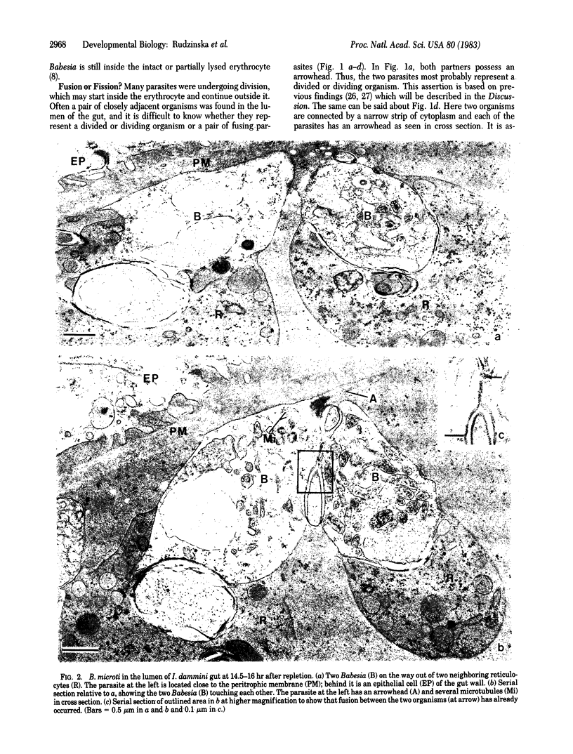
Protozoa of the closely related genera Babesia and Theileria are intraerythrocytic parasites of vertebrates. They have a complex life cycle that includes development in an intermediate vector host, a tick. Whether sexual stages occur in the tick has been a subject of great controversy. The small size of the organism and the complexity of developmental stages in the gut of the tick have prevented a definitive solution of this problem. By means of a simple and straightforward although time-consuming method, it became possible to demonstrate gametes and their sexual fusion in Babesia microti developing in the gut of larvae of the tick Ixodes dammini. Tick larvae fed on hamsters infected with a human strain of B. microti were fixed and processed for electron microscopy. It was found that some of the parasites formed a unique structure shaped like an arrowhead. Because it was suspected that these forms might represent gametes, a search was made for pairs of parasites that were fusing and with each member of the pair emerging from a different erythrocyte. Such a fusing pair could not possibly represent a parasite undergoing division. By study of serial sections such pairs were indeed found. In every case one member of the pair of gametes had an arrowhead structure. This proves sexuality of B. microti and makes highly likely its existence in all members of the genera Babesia and Theileria.
Full text
PDF


Images in this article
Selected References
These references are in PubMed. This may not be the complete list of references from this article.
- Fawcett D. W., Büscher G., Doxsey S. Salivary gland of the tick vector of East Coast fever. III. The ultrastructure of sporogony in Theileria parva. Tissue Cell. 1982;14(1):183–206. doi: 10.1016/0040-8166(82)90017-9. [DOI] [PubMed] [Google Scholar]
- Friedhoff K. T., Büscher G. Rediscovery of Koch's "strahlenörper" of Babesia bigemina. Z Parasitenkd. 1976 Oct 12;50(3):345–347. doi: 10.1007/BF02462979. [DOI] [PubMed] [Google Scholar]
- Garnham P. C., Bird R. G., Baker J. R. Electron microscope studies of motile stages of malaria parasites. V. Exflagellation in Plasmodium, Hepatocystis and Leucocytozoon. Trans R Soc Trop Med Hyg. 1967;61(1):58–68. doi: 10.1016/0035-9203(67)90054-5. [DOI] [PubMed] [Google Scholar]
- Mehlhorn H., Moltmann U., Schein E., Voigt W. P. Fine structure of supposed gametes and syngamy of Babesia canis (Piroplasmea) after in-vitro development. Zentralbl Bakteriol Mikrobiol Hyg A. 1981;250(1-2):248–255. [PubMed] [Google Scholar]
- Mehlhorn H., Schein E. Electron microscopic studies of the development of kinetes in Theileria annulata Dschunkowsky & Luhs, 1904 (Sporozoa, Piroplasmea). J Protozool. 1977 May;24(2):249–257. doi: 10.1111/j.1550-7408.1977.tb00974.x. [DOI] [PubMed] [Google Scholar]
- Mehlhorn H., Schein E. Elektronenmikroskopische untersuchungen an entwicklungsstadien von theileria parva (theiler, 1904) im darm der uberträgerzecke hyalomma anatolicum excavatum (koch, 1844) Tropenmed Parasitol. 1976 Jun;27(2):182–191. [PubMed] [Google Scholar]
- Mehlhorn H., Schein E., Voigt W. P. Light and electron microscopic study on developmental stages of Babesia canis within the gut of the tick Dermacentor reticulatus. J Parasitol. 1980 Apr;66(2):220–228. [PubMed] [Google Scholar]
- Mehlhorn H., Schein E., Warnecke M. Electron microscopic studies on the development of kinetes of Theileria parva Theiler, 1904 in the gut of the vector ticks Rhipicephalus appendiculatus Neumann, 1901. Acta Trop. 1978 Jun;35(2):123–136. [PubMed] [Google Scholar]
- Mehlhorn H., Schein E., Warnecke M. Electron-microscopic studies on Theileria ovis Rodhain, 1916: development of kinetics in the gut of the vector tick, Rhipicephalus evertsi evertsi Neumann, 1897, and their transformation within cells of the salivary glands. J Protozool. 1979 Aug;26(3):377–385. doi: 10.1111/j.1550-7408.1979.tb04640.x. [DOI] [PubMed] [Google Scholar]
- Mehlhorn H., Weber G., Schein E., Büscher G. Elektronenmikroskopische Untersuchung an Entwicklungsstadien von Theileria annulata (Dschunkowsky, Luhs, 1904) im Darm und in der Hämolymphe von Hyalomma anatolicum excavatum (Koch, 1844) Z Parasitenkd. 1975 Dec 23;48(2):137–150. doi: 10.1007/BF00389644. [DOI] [PubMed] [Google Scholar]
- Moltmann U. G., Mehlhorn H., Friedhoff K. T. Electron microscopic study on the development of Babesia ovis (Piroplasmia) in the salivary glands of the vector tick Rhipicephalus bursa. Acta Trop. 1982 Mar;39(1):29–40. [PubMed] [Google Scholar]
- Moltmann U. G., Mehlhorn H., Friedhoff K. T. Ultrastructural study of the development of Babesia ovis (Piroplasmia) in the ovary of the vector tick Rhipicephalus bursa. J Protozool. 1982 Feb;29(1):30–38. doi: 10.1111/j.1550-7408.1982.tb02877.x. [DOI] [PubMed] [Google Scholar]
- Piesman J., Spielman A., Etkind P., Ruebush T. K., 2nd, Juranek D. D. Role of deer in the epizootiology of Babesia microti in Massachusetts, USA. J Med Entomol. 1979 Sep 4;15(5-6):537–540. doi: 10.1093/jmedent/15.5-6.537. [DOI] [PubMed] [Google Scholar]
- Rudzinska M. A., Spielman A., Lewengrub S., Piesman J., Karakashian S. Penetration of the peritrophic membrane of the tick by Babesia microti. Cell Tissue Res. 1982;221(3):471–481. doi: 10.1007/BF00215696. [DOI] [PubMed] [Google Scholar]
- Rudzinska M. A., Spielman A., Riek R. F., Lewengrub S. J., Piesman J. Intraerythrocytic 'gametocytes' of Babesia microti and their maturation in ticks. Can J Zool. 1979 Feb;57(2):424–434. doi: 10.1139/z79-050. [DOI] [PubMed] [Google Scholar]
- Rudzinska M. A., Trager W. Formation of merozoites in intraerythrocytic Babesia microti: an ultrastructural study. Can J Zool. 1977 Jun;55(6):928–938. doi: 10.1139/z77-121. [DOI] [PubMed] [Google Scholar]
- Rudzinska M. A. Ultrastructure of intraerythrocytic Babesia microti with emphasis on the feeding mechanism. J Protozool. 1976 May;23(2):224–233. doi: 10.1111/j.1550-7408.1976.tb03759.x. [DOI] [PubMed] [Google Scholar]
- Ruebush T. K., 2nd, Juranek D. D., Spielman A., Piesman J., Healy G. R. Epidemiology of human babesiosis on Nantucket Island. Am J Trop Med Hyg. 1981 Sep;30(5):937–941. doi: 10.4269/ajtmh.1981.30.937. [DOI] [PubMed] [Google Scholar]
- Schein E., Büscher G., Friedhoff K. T. Lichtmikroskopische Untersuchungen über die Entwicklung von Theileria annulata (Dschunkowsky and Luhs, 1904) in Hyalomma anatolicum excavatum (Koch, 1844). I. Die Entwicklung im Darm vollgesogener Nymphen. Z Parasitenkd. 1975 Dec 23;48(2):123–136. doi: 10.1007/BF00389643. [DOI] [PubMed] [Google Scholar]
- Schein E. On the life cycle of Theileria annulata (Dschunkowsky and Luhs, 1904) in the midgut and hemolymph of Hyalomma anatolicum excavatum (Koch, 1844). Z Parasitenkd. 1975 Sep 12;47(2):165–167. doi: 10.1007/BF00382639. [DOI] [PubMed] [Google Scholar]
- Spielman A., Clifford C. M., Piesman J., Corwin M. D. Human babesiosis on Nantucket Island, USA: description of the vector, Ixodes (Ixodes) dammini, n. sp. (Acarina: Ixodidae). J Med Entomol. 1979 Mar 23;15(3):218–234. doi: 10.1093/jmedent/15.3.218. [DOI] [PubMed] [Google Scholar]
- Weber G., Friedhoff K. T. Preliminary observations on the ultrastructure of suppossed sexual stages of Babesia bigemina (Piroplasmea). Z Parasitenkd. 1977 Aug 25;53(1):83–92. doi: 10.1007/BF00383118. [DOI] [PubMed] [Google Scholar]
- Weber G., Walter G. Babesia microti (apicomplexa: piroplasmida): electron microscope detection in salivary glands of the tick vector Ixodes ricinus (Ixodoidea: Ixodidae). Z Parasitenkd. 1980;64(1):113–115. doi: 10.1007/BF00927061. [DOI] [PubMed] [Google Scholar]


