

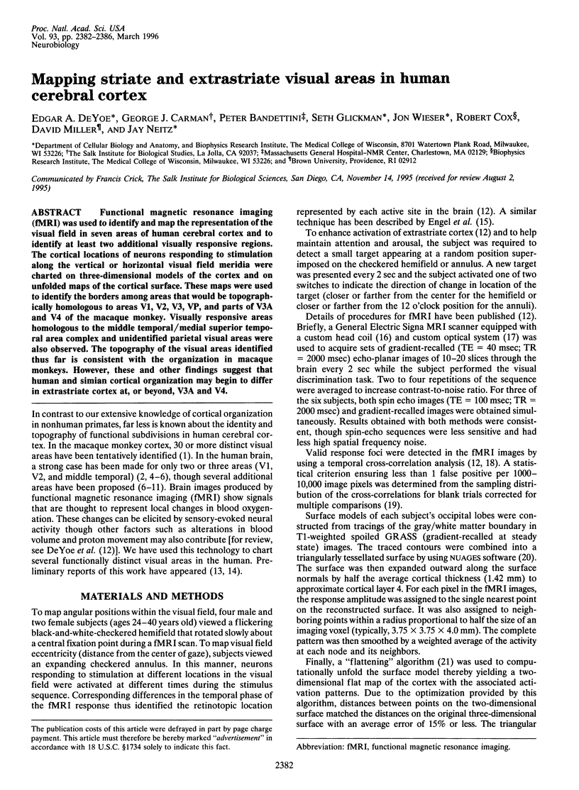
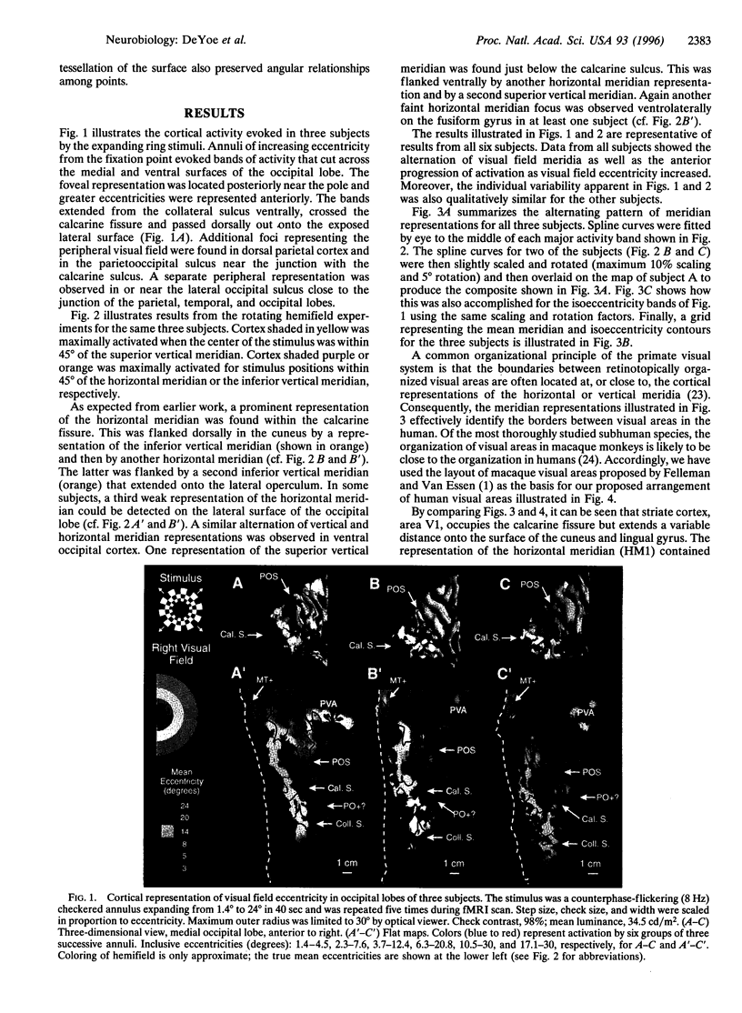
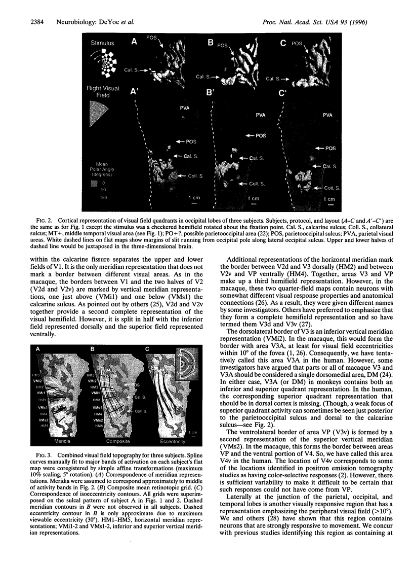
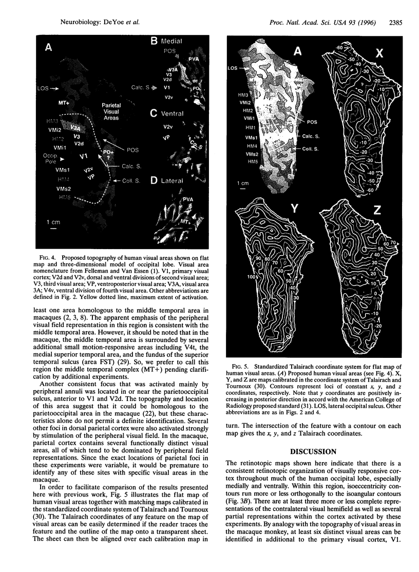
Abstract
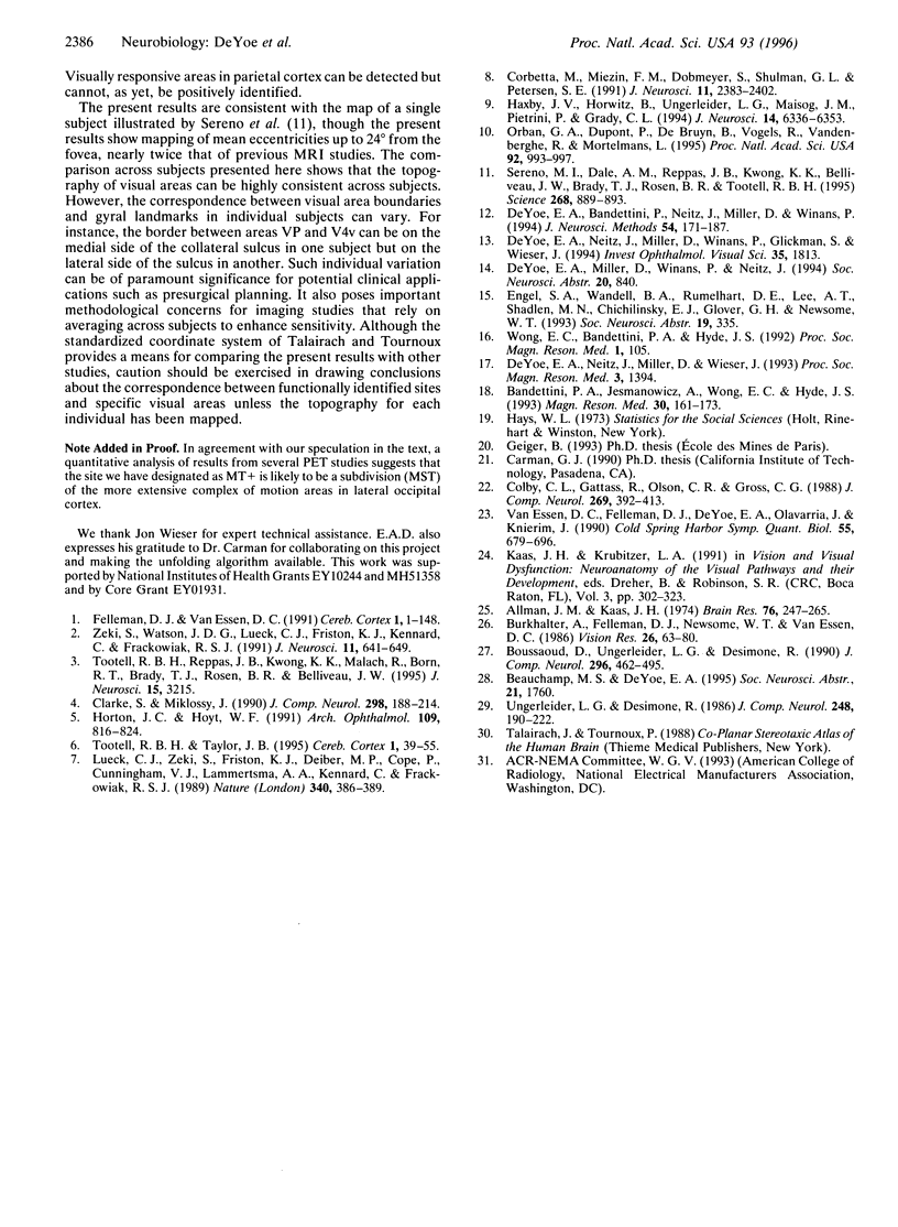
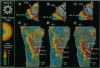
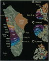
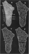
Functional magnetic resonance imaging (fMRI) was used to identify and map the representation of the visual field in seven areas of human cerebral cortex and to identify at least two additional visually responsive regions. The cortical locations of neurons responding to stimulation along the vertical or horizontal visual field meridia were charted on three-dimensional models of the cortex and on unfolded maps of the cortical surface. These maps were used to identify the borders among areas that would be topographically homologous to areas V1, V2, V3, VP, and parts of V3A and V4 of the macaque monkey. Visually responsive areas homologous to the middle temporal/medial superior temporal area complex and unidentified parietal visual areas were also observed. The topography of the visual areas identified thus far is consistent with the organization in macaque monkeys. However, these and other findings suggest that human and simian cortical organization may begin to differ in extrastriate cortex at, or beyond, V3A and V4.
Full text
PDF
Images in this article
Selected References
These references are in PubMed. This may not be the complete list of references from this article.
- Allman J. M., Kaas J. H. The organization of the second visual area (V II) in the owl monkey: a second order transformation of the visual hemifield. Brain Res. 1974 Aug 16;76(2):247–265. doi: 10.1016/0006-8993(74)90458-2. [DOI] [PubMed] [Google Scholar]
- Bandettini P. A., Jesmanowicz A., Wong E. C., Hyde J. S. Processing strategies for time-course data sets in functional MRI of the human brain. Magn Reson Med. 1993 Aug;30(2):161–173. doi: 10.1002/mrm.1910300204. [DOI] [PubMed] [Google Scholar]
- Boussaoud D., Ungerleider L. G., Desimone R. Pathways for motion analysis: cortical connections of the medial superior temporal and fundus of the superior temporal visual areas in the macaque. J Comp Neurol. 1990 Jun 15;296(3):462–495. doi: 10.1002/cne.902960311. [DOI] [PubMed] [Google Scholar]
- Burkhalter A., Felleman D. J., Newsome W. T., Van Essen D. C. Anatomical and physiological asymmetries related to visual areas V3 and VP in macaque extrastriate cortex. Vision Res. 1986;26(1):63–80. doi: 10.1016/0042-6989(86)90071-4. [DOI] [PubMed] [Google Scholar]
- Clarke S., Miklossy J. Occipital cortex in man: organization of callosal connections, related myelo- and cytoarchitecture, and putative boundaries of functional visual areas. J Comp Neurol. 1990 Aug 8;298(2):188–214. doi: 10.1002/cne.902980205. [DOI] [PubMed] [Google Scholar]
- Colby C. L., Gattass R., Olson C. R., Gross C. G. Topographical organization of cortical afferents to extrastriate visual area PO in the macaque: a dual tracer study. J Comp Neurol. 1988 Mar 15;269(3):392–413. doi: 10.1002/cne.902690307. [DOI] [PubMed] [Google Scholar]
- Corbetta M., Miezin F. M., Dobmeyer S., Shulman G. L., Petersen S. E. Selective and divided attention during visual discriminations of shape, color, and speed: functional anatomy by positron emission tomography. J Neurosci. 1991 Aug;11(8):2383–2402. doi: 10.1523/JNEUROSCI.11-08-02383.1991. [DOI] [PMC free article] [PubMed] [Google Scholar]
- DeYoe E. A., Bandettini P., Neitz J., Miller D., Winans P. Functional magnetic resonance imaging (FMRI) of the human brain. J Neurosci Methods. 1994 Oct;54(2):171–187. doi: 10.1016/0165-0270(94)90191-0. [DOI] [PubMed] [Google Scholar]
- Felleman D. J., Van Essen D. C. Distributed hierarchical processing in the primate cerebral cortex. Cereb Cortex. 1991 Jan-Feb;1(1):1–47. doi: 10.1093/cercor/1.1.1-a. [DOI] [PubMed] [Google Scholar]
- Haxby J. V., Horwitz B., Ungerleider L. G., Maisog J. M., Pietrini P., Grady C. L. The functional organization of human extrastriate cortex: a PET-rCBF study of selective attention to faces and locations. J Neurosci. 1994 Nov;14(11 Pt 1):6336–6353. doi: 10.1523/JNEUROSCI.14-11-06336.1994. [DOI] [PMC free article] [PubMed] [Google Scholar]
- Horton J. C., Hoyt W. F. The representation of the visual field in human striate cortex. A revision of the classic Holmes map. Arch Ophthalmol. 1991 Jun;109(6):816–824. doi: 10.1001/archopht.1991.01080060080030. [DOI] [PubMed] [Google Scholar]
- Lueck C. J., Zeki S., Friston K. J., Deiber M. P., Cope P., Cunningham V. J., Lammertsma A. A., Kennard C., Frackowiak R. S. The colour centre in the cerebral cortex of man. Nature. 1989 Aug 3;340(6232):386–389. doi: 10.1038/340386a0. [DOI] [PubMed] [Google Scholar]
- Orban G. A., Dupont P., De Bruyn B., Vogels R., Vandenberghe R., Mortelmans L. A motion area in human visual cortex. Proc Natl Acad Sci U S A. 1995 Feb 14;92(4):993–997. doi: 10.1073/pnas.92.4.993. [DOI] [PMC free article] [PubMed] [Google Scholar]
- Sereno M. I., Dale A. M., Reppas J. B., Kwong K. K., Belliveau J. W., Brady T. J., Rosen B. R., Tootell R. B. Borders of multiple visual areas in humans revealed by functional magnetic resonance imaging. Science. 1995 May 12;268(5212):889–893. doi: 10.1126/science.7754376. [DOI] [PubMed] [Google Scholar]
- Tootell R. B., Reppas J. B., Kwong K. K., Malach R., Born R. T., Brady T. J., Rosen B. R., Belliveau J. W. Functional analysis of human MT and related visual cortical areas using magnetic resonance imaging. J Neurosci. 1995 Apr;15(4):3215–3230. doi: 10.1523/JNEUROSCI.15-04-03215.1995. [DOI] [PMC free article] [PubMed] [Google Scholar]
- Tootell R. B., Taylor J. B. Anatomical evidence for MT and additional cortical visual areas in humans. Cereb Cortex. 1995 Jan-Feb;5(1):39–55. doi: 10.1093/cercor/5.1.39. [DOI] [PubMed] [Google Scholar]
- Ungerleider L. G., Desimone R. Cortical connections of visual area MT in the macaque. J Comp Neurol. 1986 Jun 8;248(2):190–222. doi: 10.1002/cne.902480204. [DOI] [PubMed] [Google Scholar]
- Van Essen D. C., Felleman D. J., DeYoe E. A., Olavarria J., Knierim J. Modular and hierarchical organization of extrastriate visual cortex in the macaque monkey. Cold Spring Harb Symp Quant Biol. 1990;55:679–696. doi: 10.1101/sqb.1990.055.01.064. [DOI] [PubMed] [Google Scholar]
- Zeki S., Watson J. D., Lueck C. J., Friston K. J., Kennard C., Frackowiak R. S. A direct demonstration of functional specialization in human visual cortex. J Neurosci. 1991 Mar;11(3):641–649. doi: 10.1523/JNEUROSCI.11-03-00641.1991. [DOI] [PMC free article] [PubMed] [Google Scholar]