Sir,
The sub-Tenon block (STB) is the most widely used anaesthetic technique for cataract surgery.1 This technique traditionally involves making an incision in the conjunctiva with blunt-tipped sprung Westcott scissors and blunt dissecting the sub-Tenon layer away from the sclera.2
We have previously developed a new minimally invasive technique of STB, which uses a blunt ‘pencil point' cannula to allow access to the sub-Tenon space without prior conjunctival incision3 (Supplementary video 1). We investigated the spread and distribution of local anaesthetic via the incisionless STB, and compared it with standard techniques, using B-scan ultrasonography.
Case report
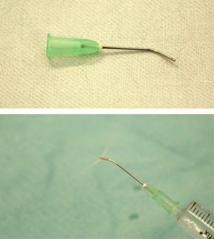
Patients having routine cataract extraction at the West of England Eye Unit were selected, and informed consent obtained. Dynamic B-scan ultrasonography was performed on the eye during administration of the STB by either the incisionless technique utilising the ‘pencil point' sub-Tenon cannula (21G × 25 mm angled Tri-Port sub-Tenon Anaesthetic Cannula; Eagle Laboratories, Cucamonga, CA, USA; Figure 1), or by standard methods.
Figure 1.
Eagle Tri-port sub-Tenon cannula used to perform minimally invasive sub-Tenon anaesthesia without prior conjunctival incision.
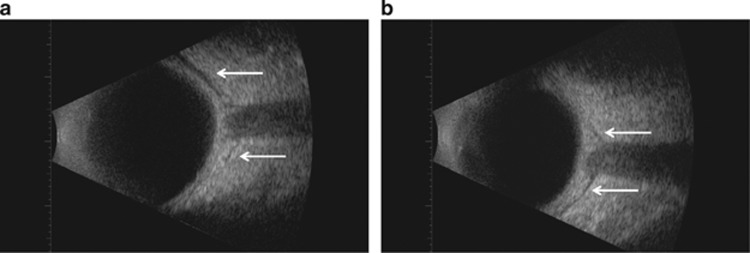
B-scan ultrasonography was chosen as a simple and non-invasive method for visualising the distribution of anaesthetic fluid in the posterior sub-Tenon space during an STB.4 We found that the anaesthetic fluid was easily visualised on ultrasonography as a dark outline tracking behind the globe in the retrobulbar space (Supplementary video 2).
Comment
The STB has gained popularity with both ophthalmic surgeons and anaesthetists as providing adequate anaesthesia and akinesia for intraocular procedures, while avoiding the risk of complications from sharp needles associated with other regional orbital block techniques.5 As the technique has further evolved and novel cannulae are introduced, it is important to objectively assess the safety and efficacy of these innovations.
We found that both standard and incisionless techniques of STB achieved similar ocular anaesthesia, and real-time visualisation of anaesthetic fluid localisation by B-scan ultrasonography demonstrated no significant differences in the spread and distribution of anaesthetic fluid between standard and incisionless techniques (Figure 2).
Figure 2.
B-scan ultrasound images with arrows showing expanded posterior sub-Tenon space and ‘T-sign' following (a) standard sub-Tenon technique and (b) incisionless sub-Tenon technique.
The incisionless STB has the advantage of reduced conjunctival trauma compared with traditional techniques, and reducing anterior refluxing of local anaesthetic improves the onset, quality, and reproducibility of the block.3 We feel that the incisionless STB is therefore recommended in view of its advantages over the traditional sub-Tenon anaesthetic techniques.
The authors declare no conflict of interest.
Footnotes
Supplementary Information accompanies this paper on Eye website (http://www.nature.com/eye)
This work was previously presented at the 44th RANZCO Annual Scientific Congress, November 2012, Melbourne, Australia.
Supplementary Material
References
- El-Hindy N, Johnston RL, Jaycock P, Eke T, Braga AJ, Tole DM, et al. The Cataract National Dataset Electronic Multi-centre Audit of 55,567 operations: anaesthetic techniques and complications. Eye (Lond) 2009;23:50–55. doi: 10.1038/sj.eye.6703031. [DOI] [PubMed] [Google Scholar]
- Kumar CM, Dodds C. Sub-Tenon's anesthesia. Ophthalmol Clin North Am. 2006;19:209–219. doi: 10.1016/j.ohc.2006.02.008. [DOI] [PubMed] [Google Scholar]
- Allman KG, Theron AD, Byles DB. A new technique of incisionless minimally invasive sub-Tenon's anaesthesia. Anaesthesia. 2008;63:782–783. doi: 10.1111/j.1365-2044.2008.05592.x. [DOI] [PubMed] [Google Scholar]
- Kumar CM, McNeela BJ. Ultrasonic localization of anaesthetic fluid using sub-Tenon's cannulae of three different lengths. Eye (Lond) 2003;17:1003–1007. doi: 10.1038/sj.eye.6700501. [DOI] [PubMed] [Google Scholar]
- Jeganathan VSE, Jeganathan VP. Sub-Tenon's anaesthesia: a well tolerated and effective procedure for ophthalmic surgery. Curr Opin Ophthalmol. 2009;20:205–209. doi: 10.1097/ICU.0b013e328329b6af. [DOI] [PubMed] [Google Scholar]
Associated Data
This section collects any data citations, data availability statements, or supplementary materials included in this article.


