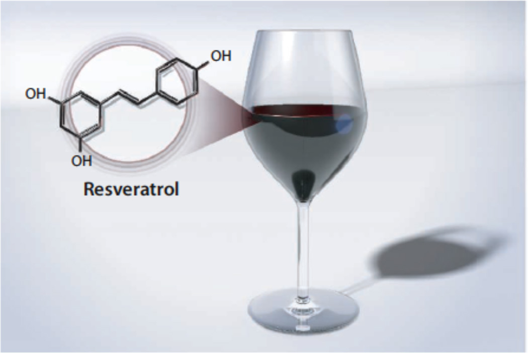
Once of interest only to yeast geneticists studying transcriptional regulation, Silent Information Regulator 2 (Sir2) received considerable attention from the broader scientific community when it was demonstrated that increased dosage of Sir2 increased yeast replicative life span (1). Sir2 homologues (sirtuins) are conserved from bacteria to man, and subsequent studies showed that overexpression of orthologous proteins in worms (2) and flies (3) also increased lifespan in these organisms. In each case, sirtuin-mediated lifespan extension was shown to mimic the effect of a diet of reduced calories (termed calorie restriction), the only previously known regimen known to increase lifespan in many organisms including mice (4). The fact that sirtuins are conserved in humans and the observation that calorie restriction in humans is correlated with physiological and behavioral changes that are linked to longer and healthier living , led to the notion that small molecule sirtuin activators might increase human healthspan and possibly also lifespan.
Sirtuins are NAD+ dependent protein deacetylases, although some members have recently been demonstrated to carry out other related enzymatic reactions (5). Humans have seven sirtuins, with SIRT1 being the most similar to yeast Sir2. SIRT1 targets a wide range of protein substrates and has been demonstrated to play a role in many age-related diseases including cancer, Alzheimer disease and type II diabetes. In 2003 Howitz et al. set out to identify sirtuin activating compounds (STACs) using recombinant SIRT1 in a biochemical assay with a fluorophore-tagged p53 substrate (6). This assay led to the identification of a family of polyphenols including resveratrol, a natural product found in red wine and previously known to exhibit positive health benefits. Subsequent studies using a related fluorophore identified an unrelated family of synthetic STACs that were more potent than resveratrol (7). These results on SIRT1 activators were called into question when several groups reported that resveratrol and the other STACs failed to activate SIRT1 in vitro when non-fluorophore-tagged substrates were used (8–11). Despite these contradictory in vitro results on the ability of STACs to directly activate SIRT1, other studies demonstrated that STACs caused pharmacological changes in cells consistent with SIRT1 activation (6,7,12,13). These findings lead to speculation that the cellular effects of STACs do not work through SIRT1 binding but instead work indirectly by binding other proteins.
In the current issue of Science on page XXX, Hubbard et. al. demonstrate that SIRT1 can indeed be activated by resveratrol and other STACs in vitro on substrates without a fluorophore tag, but only on certain natural peptide substrates. Hubbard et al. hypothesized that the fluorophore tags attached to the substrates employed for the SIRT1 activator screens might mimic hydrophobic amino acids of natural substrates at the same position as the fluorophore (+1 relative to the acetyl-lysine). With this in mind, the authors found that natural SIRT1 substrates that had large hydrophobic residues (Trp, Tyr or Phe) at positions +1 and +6 (PGC-1α-778) and +1 (FOXO3a-K290 ), as well as other peptides that conformed to this substrate signature were selectively activated by several STACs. Kinetic analysis of SIRT1 activation by STACs in the presence of these peptide substrates revealed that rate enhancement was mediated primarily through an improvement in peptide biding (lowering of peptide KM), consistent with an allosteric mechanism. This prompted the authors to screen for SIRT1 mutants that would be resistant to activation by STACs, leading to the identification of a single glutamate residue (E230) just N-terminal to the conserved sirtuin catalytic core that was critical for the activation of SIRT1 by over 100 STACs tested. Biophysical studies employing hydrogen/deuterium exchange confirmed that in addition to the conserved catalytic core domain and a C-terminal segment, a small rigid N-terminal region from 190 to 244 encompassing E230 was also protected from exchange, consistent with a structured role of this region for SIRT1 function and also consistent with previous studies demonstrating a role for this region in catalysis by SIRT1 (14). To demonstrate SIRT1-E230-dependent activity of STACs in cells, the authors used SIRT1 knockout cells to demonstrate that several STACs elicited pharmacological changes that were consistent with SIRT1 activation when cells carried wild-type mouse SIRT1 but that these changes were blocked when cells were reconstituted with mouse SIRT1 harboring the mouse equivalent of the human SIRT1-E230K mutant. Taken together, these studies demonstrated that STACs can increase the catalytic activity of SIRT1 towards certain substrates through an allosteric mechanism involving a SIRT1 region N-terminal to the catalytic core domain and through direct binding to SIRT1 both in vitro and in cells.
These studies have important implications for the further development of SIRT1 modulators. Allosteric activation of SIRT1 through a non-conserved N-terminal region suggests that SIRT1-selective activators can be developed. Although the current STACs only work against a subset of SIRT1 substrates that contain hydrophobic amino acids at position +1 to the acetyl-lysine, this is likely due to the bias of the initial screen that contained a fluorophore, hydrophobic residue mimic, at this position of the substrate peptide. Another screen that is not biased in this way may lead to the identification of STACs for SIRT1 substrates containing other sequence signatures. Moreover, if allosteric activators can be developed, then appropriate modifications of these molecules could lead to allosteric SIRT1 inhibitors with comparable protein selectivity. Needless to say, a structure of SIRT1 containing the catalytic domain and N-terminal segment bound to these STACs would facilitate a rational approach to the development of such molecules. We already know that some STACs, like resveratrol, modulate the activity of other molecules (9,15), further arguing for the need for structural information to facilitate the development of more potent and selective compounds.
These studies also have implications for the understanding some of the seemingly contradictory findings on SIRT1 biology. For example, SIRT1 has been reported to have a dual role in cell survival with properties of an oncoprotein while also playing a role in cell death with properties of a tumor suppressor (16). This might be related to the fact that particular cellular effectors modulate SIRT1 to deacetylate certain protein targets, just like STACs increase SIRT1’s ability to deacetylate proteins that contain hydrophobic residues at certain amino acid position. One could imagine that other stimuli might promote SIRT1 deacetylation of other substrates. Indeed, the N-terminal segment of SIRT1, the region critical to STACs activation, might be an important regulatory switch for SIRT1 function, consistent with recent studies demonstrating that the N-terminal segment of SIRT1 potentiates the proteins’ deacetylase activity (14). Along the same lines, a C-terminal segment of SIRT1 has also been shown to be important for optimal SIRT1 activity so might also play an important regulatory function (14,17). One thing is clear; SIRT1 activation by STACs is back in the news. Perhaps we should toast with a glass of red wine.
Figure 1.
References
- 1.Kaeberlein M, McVey M, Guarente L. Genes Dev. 1999;13:2570–2580. doi: 10.1101/gad.13.19.2570. [DOI] [PMC free article] [PubMed] [Google Scholar]
- 2.Tissenbaum HA, Guarente L. Nature. 2001;410:227–230. doi: 10.1038/35065638. [DOI] [PubMed] [Google Scholar]
- 3.Rogina B, Helfand SL. Proc Natl Acad Sci U S A. 2004;101:15998–16003. doi: 10.1073/pnas.0404184101. [DOI] [PMC free article] [PubMed] [Google Scholar]
- 4.Sinclair DA. Mechanisms of Ageing and Development. 2005;126:987–1002. doi: 10.1016/j.mad.2005.03.019. [DOI] [PubMed] [Google Scholar]
- 5.Yuan H, Marmorstein R. J Biol Chem. 2012;287:42428–42435. doi: 10.1074/jbc.R112.372300. [DOI] [PMC free article] [PubMed] [Google Scholar]
- 6.Howitz KT, Bitterman KJ, Cohen HY, Lamming DW, Lavu S, Wood JG, Zipkin RE, Chung P, Kisielewski A, Zhang LL, Scherer B, Sinclair DA. Nature. 2003;425:191–196. doi: 10.1038/nature01960. [DOI] [PubMed] [Google Scholar]
- 7.Milne JC, Lambert PD, Schenk S, Carney DP, Smith JJ, Gagne DJ, Jin L, Boss O, Perni RB, Vu CB, Bemis JE, Xie R, Disch JS, Ng PY, Nunes JJ, Lynch AV, Yang H, Galonek H, Israelian K, Choy W, Iffland A, Lavu S, Medvedik O, Sinclair DA, Olefsky JM, Jirousek MR, Elliott PJ, Westphal CH. Nature. 2007;450:712–716. doi: 10.1038/nature06261. [DOI] [PMC free article] [PubMed] [Google Scholar]
- 8.Beher D, Wu J, Cumine S, Kim KW, Lu SC, Atangan L, Wang M. Chem Biol Drug Des. 2009;74:619–624. doi: 10.1111/j.1747-0285.2009.00901.x. [DOI] [PubMed] [Google Scholar]
- 9.Pacholec M, Chrunyk BA, Cunningham D, Flynn D, Griffith DA, Griffor M, Loulakis P, Pabst B, Qiu X, Stockman B, Thanabal V, Varghese A, Ward J, Withka J, Ahn K. Journal of Biological Chemistry. 2010 doi: 10.1074/jbc.M109.088682. [DOI] [PMC free article] [PubMed] [Google Scholar]
- 10.Kaeberlein M, McDonagh T, Heltweg B, Hixon J, Westman EA, Caldwell SD, Napper A, Curtis R, DiStefano PS, Fields S, Bedalov A, Kennedy BK. J Biol Chem. 2005;280:17038–17045. doi: 10.1074/jbc.M500655200. [DOI] [PubMed] [Google Scholar]
- 11.Borra MT, Smith BC, Denu JM. J Biol Chem. 2005;280:17187–17195. doi: 10.1074/jbc.M501250200. [DOI] [PubMed] [Google Scholar]
- 12.Baur JA, Pearson KJ, Price NL, Jamieson HA, Lerin C, Kalra A, Prabhu VV, Allard JS, Lopez-Lluch G, Lewis K, Pistell PJ, Poosala S, Becker KG, Boss O, Gwinn D, Wang M, Ramaswamy S, Fishbein KW, Spencer RG, Lakatta EG, Le Couteur D, Shaw RJ, Navas P, Puigserver P, Ingram DK, de Cabo R, Sinclair DA. Nature. 2006;444:337–342. doi: 10.1038/nature05354. [DOI] [PMC free article] [PubMed] [Google Scholar]
- 13.Minor RK, Baur JA, Gomes AP, Ward TM, Csiszar A, Mercken EM, Abdelmohsen K, Shin YK, Canto C, Scheibye-Knudsen M, Krawczyk M, Irusta PM, Martin-Montalvo A, Hubbard BP, Zhang Y, Lehrmann E, White AA, Price NL, Swindell WR, Pearson KJ, Becker KG, Bohr VA, Gorospe M, Egan JM, Talan MI, Auwerx J, Westphal CH, Ellis JL, Ungvari Z, Vlasuk GP, Elliott PJ, Sinclair DA, de Cabo R. Scientific reports. 2011;1:70. doi: 10.1038/srep00070. [DOI] [PMC free article] [PubMed] [Google Scholar]
- 14.Pan M, Yuan H, Brent M, Ding EC, Marmorstein R. J Biol Chem. 2012;287:2468–2476. doi: 10.1074/jbc.M111.285031. [DOI] [PMC free article] [PubMed] [Google Scholar]
- 15.Park SJ, Ahmad F, Philp A, Baar K, Williams T, Luo H, Ke H, Rehmann H, Taussig R, Brown AL, Kim MK, Beaven MA, Burgin AB, Manganiello V, Chung JH. Cell. 2012;148:421–433. doi: 10.1016/j.cell.2012.01.017. [DOI] [PMC free article] [PubMed] [Google Scholar]
- 16.Deng CX. International journal of biological sciences. 2009;5:147–152. doi: 10.7150/ijbs.5.147. [DOI] [PMC free article] [PubMed] [Google Scholar]
- 17.Kang H, Suh J-Y, Jung Y-S, Jung J-W, Kim Myung K, Chung Jay H. Molecular Cell. 2011;44:203–213. doi: 10.1016/j.molcel.2011.07.038. [DOI] [PMC free article] [PubMed] [Google Scholar]


