ABSTRACT
Fertility is dependent on the hypothalamic-pituitary-gonadal axis. Each component of this axis is essential for normal reproductive function. Mice with a mutation in the forkhead transcription factor gene, Foxp3, exhibit autoimmunity and infertility. We have previously shown that Foxp3 mutant mice have significantly reduced expression of pituitary gonadotropins. To address the role of Foxp3 in gonadal function, we examined the gonadal phenotype of these mice. Foxp3 mutant mice have significantly reduced seminal vesicle and testis weights compared with Foxp3+/Y littermates. Spermatogenesis in Foxp3 mutant males is arrested prior to spermatid elongation. Activation of luteinizing hormone signaling in Foxp3 mutant mice by treatment with human chorionic gonadotropin significantly increases seminal vesicle and testis weights as well as testicular testosterone content and seminiferous tubule diameter. Interestingly, human chorionic gonadotropin treatments rescue spermatogenesis in Foxp3 mutant males, suggesting that their gonadal phenotype is due primarily to a loss of pituitary gonadotropin stimulation rather than an intrinsic gonadal defect.
Keywords: fertility, forkhead, FOXP3, gonadotropin, pituitary, spermatogenesis, transcription factor
The forkhead transcription factor FOXP3 is essential for normal pituitary gonadotropin expression and, consequently, spermatogenesis in male mice.
INTRODUCTION
Central to reproductive function is the hypothalamic-pituitary-gonadal axis, in which hypothalamic gonadotropin-releasing hormone (GnRH) binds to specific receptors on the surface of gonadotroph cells to stimulate synthesis of the gonadotropins: luteinizing hormone (LH) and follicle-stimulating hormone (FSH). Pituitary gonadotropins are heterodimers consisting of a common α subunit and unique β subunits, which confer their specialized functions. Luteinizing hormone binds to receptors on Leydig cells to stimulate testosterone production, which is essential for spermatogenesis to proceed [1, 2]. Follicle-stimulating hormone regulates Sertoli cell number and stimulates maintenance of spermatogenesis [3–5].
Balance between immune function and endocrine function is essential for normal homeostasis. When these systems become unbalanced—for example, in cases of increased immune function, such as autoimmunity, or decreased endocrine function, such as pituitary hormone deficiency—neither system functions properly. For this reason, many autoimmune diseases result in subfertility in males and females [6, 7].
The forkhead factor, FOXP3, plays important roles in the differentiation and function of regulatory T cells [8]. The gene encoding FOXP3 is located on the X chromosome in humans and mice. Mutations in the human FOXP3 gene result in an autoimmune syndrome referred to as immunodysregulation, polyendocrinopathy, and enteropathy, X-linked (IPEX). Symptoms include diarrhea, eczema, hemolytic anemia, diabetes mellitus, and thyroid autoimmunity leading to hypothyroidism [9]. Death often occurs during the first years of life [9].
A spontaneously occurring mutation, referred to as scurfy (sf), results in an IPEX-like syndrome in mice. This mutation has been mapped to the Foxp3 gene [10]. Interestingly, affected males (Foxp3sf/Y) have small testes, are sterile, and appear hypogonadal; however, no hormonal studies have been done [11, 12]. Recently, we found that pituitary expression of Lhb, Fshb, and Cga is significantly reduced in Foxp3sf/Y male mice [13]. In the following studies we characterize the gonadal phenotype in Foxp3sf/Y male mice to determine whether any intrinsic testicular defects are present.
MATERIALS AND METHODS
Mice
Foxp3 mutant mice were purchased from the Jackson Laboratory and maintained on a C57BL/6J background. Foxp3sf/Y females were mated to Foxp3+/Y males to obtain Foxp3sf/Y male offspring. Foxp3sf/Y male mice were left with dams to increase survival time. Mice were maintained on a 12L:12D cycle. To genotype mice, a Custom TaqMan SNP Genotyping Assay (Applied Biosystems) was used according to the manufacturer's instructions. Male mice were used for all studies.
All procedures using mice were approved by the Southern Illinois University Animal Care and Use Committee. All experiments were conducted in accordance with the principles and procedures outlined in the National Institutes of Health Guidelines for the Care and Use of Experimental Animals.
Histology and Immunohistochemistry
Each testis was incubated in Bouin fixative overnight at room temperature. Each testis was washed in 50% ethanol, then in 80% ethanol on ice before embedding. Serial sections were cut to 5 μm and stained with periodic acid-Schiff-hematoxylin (PASH).
Immunohistochemistry was performed by incubating tissue sections with specific antibodies that recognize 3 hydroxysteroid dehydrogenase (HSD3B1; provided by the late Dr. Anita Payne, Stanford University) at a dilution of 1:800 for 1 h at room temperature. Biotinylated secondary antibody was applied for 10 min, followed by Vectastain Elite ABC reagent (Vector Laboratories). Diaminobenzidine was added to visualize HSD3B1, and tissue sections were counterstained with hematoxylin.
Human Chorionic Gonadotropin Treatment
Foxp3sf/Y mice and Foxp3+/Y littermates were injected with 5 IU of human chorionic gonadotropin (hCG) starting at Postnatal Day 28 (P28) or P14. Animals were injected with hCG dissolved in NaCl (0.15 M) or with vehicle every 48 h for the duration of the treatment period. At P42, seminal vesicles and testes were collected and weighed. Testis and seminal vesicle weights were adjusted for total body weight. For each individual, one testis was snap frozen for testosterone measurement, and one was incubated in Bouin fixative and stained with PASH as described above. Each treatment group contained at least four animals.
To count elongated spermatids, 1 testis was analyzed per individual, 4 sections from each PASH-stained testis were analyzed, and 17 seminiferous tubules were counted per section. Counts for each testis were averaged together to obtain one value per individual. Individuals in each treatment group were averaged together to calculate the average number of elongated spermatids per treatment group and to determine the standard error around the mean. Four individuals were analyzed per treatment group.
Seminiferous tubule diameter was measured for the same tubules that were analyzed for elongated spermatid number. Seminiferous tubule diameter was measured using QCapture Pro software (version 6.0; QImaging). Measurements are expressed in micrometers. Measurements for each testis were averaged together to get one value per individual. Individuals in each treatment group were averaged together to calculate the average seminiferous tubule diameter per treatment group and to determine the standard error around the mean. Four individuals were analyzed per treatment group.
Testicular Testosterone Assays
Each testis was dissected, weighed, and snap frozen in liquid nitrogen. Testis tissue was homogenized, extracted with diethyl ether, and measured using the Parameter Testosterone Assay (R&D Systems) as per manufacturer instructions. At least four animals were included in each group.
RT-PCR
Pituitary, thymus, and testis tissues were dissected from mice and stored in RNAlater (Ambion Inc.) at −20°C. Total RNA was extracted and DNase treated using the RNAqueous-Micro Kit (AM1931; Ambion by Life Technologies,) per the manufacturer's instructions, and RNA concentrations were determined by spectrophotometry. The RT-PCR procedure employed the TaqMan RNA-to-CT 1-Step Kit (4392938; Applied Biosystems by Life Technologies Inc.) according to the manufacturer's directions and CFX96 Real Time System (Bio-Rad). Expression levels for Foxp3 (TaqMan probe Mm00475156_m1; Applied Biosystems by Life Technologies) Lhb (Mm00656868_g1), Fshb (Mm00433361_m1), and Foxo1 (Mm00490672_m1) were determined using β-actin (Actb; 4352933E) as the endogenous control (all TaqMan probes from Applied Biosystems Inc.). Fifty nanograms of cDNA was used in 20-μl reaction volumes in triplicate, and no-template and no-reverse transcriptase controls were used to ensure the absence of contamination and efficacy of the DNase treatment, respectively. At least four individuals were included in each group for all real-time experiments. Amplification was achieved by the following protocol: 48°C for 15 min, 95°C for 10 min, 40 cycles of 95°C for 15 sec, and 60°C for 1 min. Foxp3 and correlating Actb reactions were visualized on a 1% agarose gel. Relative quantification analysis was performed using the comparative CT method 2−ΔΔCT. The values for ΔΔCT were calculated by subtracting the average ΔCT of wild-type controls from the ΔCT for each sample.
Thyroid Treatment
Purina Test Diets provided thyroid hormone-enriched mouse chow consisting of special pelleted AIN-76A diet with thyroid gland powder added at a concentration of 25 mg/kg chow. Foxp3+/sf female mice were mated to C57BL6/J males. The date the copulatory plug was detected was considered to be Embryonic Day 0.5. Pregnant Foxp3+/sf female mice were fed either thyroid chow or control chow ad libitum starting at Embryonic Day 16.5. Foxp3sf/Y pups were housed with dams throughout the treatment period. At 6 wk of age pituitary tissue was collected. At least five animals were included in each treatment group.
Statistical Analysis
Data are expressed as a mean ± SEM. Data were analyzed by Student t-test to determine significant difference between Foxp3+/Y and Foxp3sf/Y mice or between different treatment groups (Microsoft Excel 2004 for Mac version 11.6.6). P values of less than 0.05 were considered statistically significant (*); P values less than 0.01 were considered very significant (**).
RESULTS
Gonadal Phenotype of Foxp3sf/Y Male Mice
Studies of Foxp3sf/Y male mice have suggested that they are hypogonadal and sterile [11, 12]. Consistent with this, Foxp3sf/Y mice have very low gonadotropin expression [13], suggesting that their hypogonadism may be hypogonadotropic in nature.
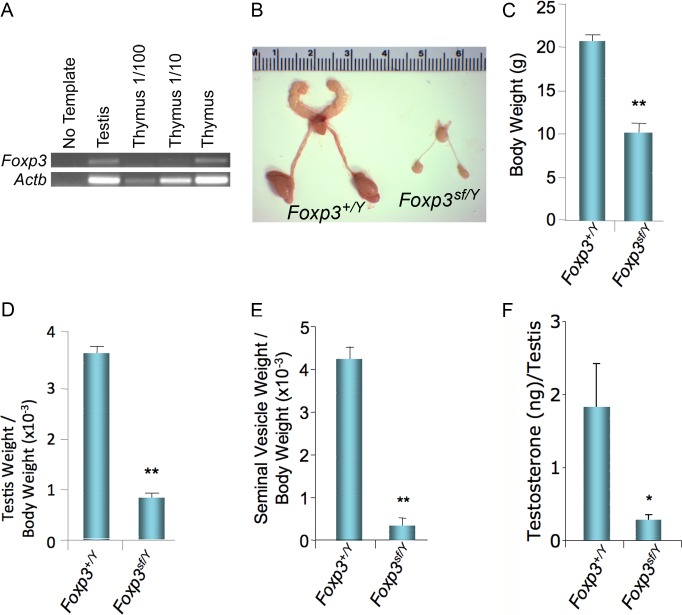
To determine whether Foxp3 is expressed in the testis, RT-PCR was performed using specific primers for Foxp3 and Actb. Foxp3 is expressed in thymus; thus, thymus was used as a positive control for the Foxp3 primers. Foxp3 primers amplified a band from thymus and from testis, suggesting that Foxp3 transcript is present in testis (Fig. 1A). We next performed immunohistochemistry to determine which cell types in the testis express FOXP3. Although FOXP3 protein was detected in spleen, none was detected in testis (Supplemental Fig. S1, available online at www.biolreprod.org). This suggests that although Foxp3 transcript is produced in the testis, FOXP3 protein is not.
FIG. 1.
A) Foxp3 mRNA is detected in the testis at 6 wk of age. B) Reproductive tracts from Foxp3+/Y and Foxp3sf/Y mice at 6 wk of age. Foxp3sf/Y mice exhibit a reduction in body weight (C; 20.55 ± 0.52 vs. 9.90 ± 1.06 g), testis weight/body weight (D; 3.70 × 10⁻³ ± 0.09 × 10⁻³ vs. 0.82 × 10⁻³ ± 0.09 × 10⁻³), and seminal vesicle weight/body weight (E; 4.26 × 10⁻³ ± 0.25 × 10⁻³ vs. 0.31 × 10⁻³ ± 0.17 × 10⁻³) at 6 wk of age. F) Testicular testosterone content is significantly reduced in Foxp3sf/Y mice compared with Foxp3+/Y littermates (1.97 ± 0.56 vs. 0.27 ± 0.06 ng per testis). Data are expressed as mean ± SEM of at least seven animals per genotype. The data were analyzed by Student t-test to determine significant difference between Foxp3sf/Y and Foxp3+/Y littermates (*P < 0.05, **P < 0.01).
To more carefully characterize the testicular phenotype and determine whether intrinsic testicular defects occur in the absence of Foxp3, the anatomy of Foxp3sf/Y reproductive tracts was observed (Fig. 1B). Seminal vesicles and testes from Foxp3sf/Y mice were much smaller than those of Foxp3+/Y littermates. The epididymis of Foxp3sf/Y mice was hypoplastic. Consistent with previous reports [14], overall body size of Foxp3sf/Y males was significantly reduced (Fig. 1C). Testicular size and seminal vesicle size were both significantly reduced, even when adjusted for body size (Fig. 1, D and E). Testicular testosterone content was significantly reduced in Foxp3sf/Y mice compared with Foxp3+/Y littermates (Fig. 1F).
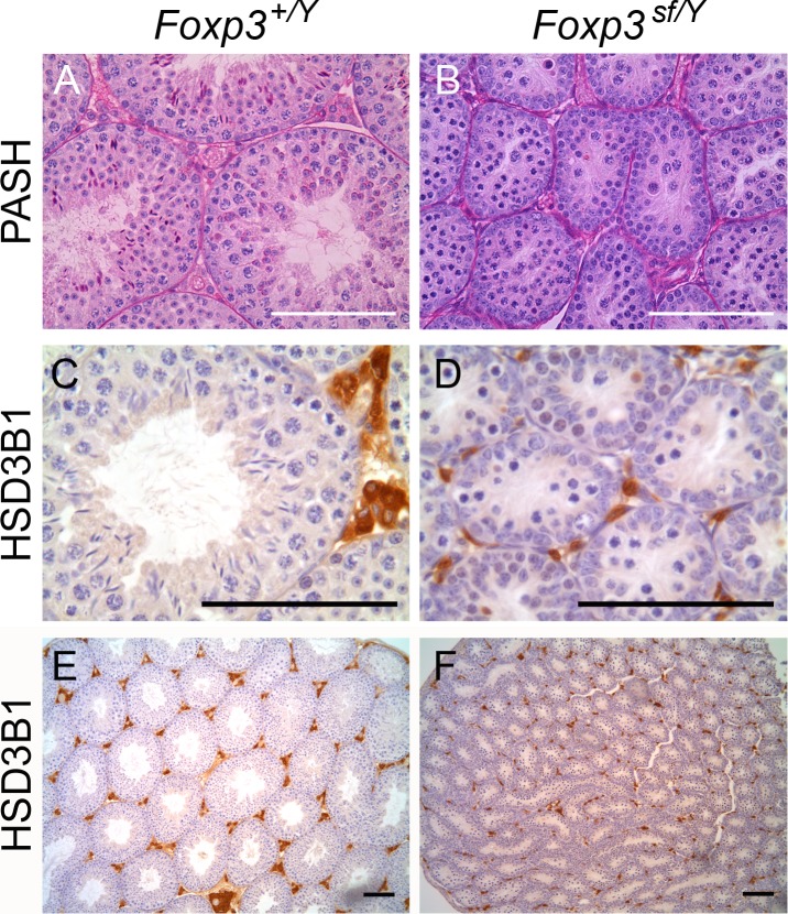
In light of their decreased testis and seminal vesicle weights, we hypothesized that Foxp3sf/Y mice would have abnormal testicular morphology. To assess testicular morphology, a PASH stain was performed on testis sections from Foxp3sf/Y mice and Foxp3+/Y littermates. These studies revealed that spermatogenesis is disrupted in Foxp3sf/Y mice (Fig. 2, A–C). Although spermatogonia and spermatocytes were present in Foxp3sf/Y testis, elongated spermatids were not observed. Because testicular testosterone levels were reduced in Foxp3sf/Y mice, we next sought to determine whether Leydig cells were present in normal numbers. Leydig cells were labeled using antibodies that specifically recognize HSD3B1. Although Leydig cells were present in Foxp3sf/Y testis, the size of the clusters was apparently reduced (Fig. 2, D–F).
FIG. 2.
Testis sections from Foxp3+/Y (A) and Foxp3sf/Y (B) mice were stained with PASH, revealing that seminiferous tubule diameter is reduced in Foxp3sf/Y mice and spermatogenesis is arrested. Immunohistochemistry for HSD3B1 was used to label Leydig cells in testis from Foxp3+/Y (C and E) and Foxp3sf/Y (D and F) mice. Foxp3sf/Y mice exhibit smaller clusters of Leydig cells than their wild-type littermates. Four animals were analyzed per genotype. Original magnifications ×400 (A and B), ×630 (C and D), and ×100 (E and F); bar = 100 μm.
Activation of LH Receptor Signaling in Foxp3sf/Y Mice
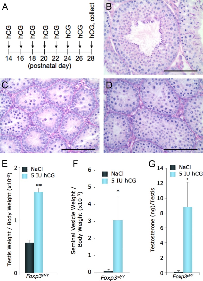
Previously, we found that pituitary gonadotropin production is significantly reduced in Foxp3sf/Y mice [13]. To determine whether the arrest in spermatogenesis is due entirely to loss of gonadotropin stimulation, these mice were treated with hCG to stimulate the LH signaling pathway. Foxp3sf/Y mice and Foxp3+/Y littermates were injected with 5 IU of hCG every other day starting at P28 and continuing until P42, when tissues were collected (Fig. 3A). Treatment with hCG rescued the testicular phenotype by increasing seminiferous tubule size, but it did not rescue spermatogenesis (Fig. 3, B–G). Treatment with hCG increased testis weight, seminal vesicle weight, and testicular testosterone content in Foxp3sf/Y mice (Fig. 3, E–G). These data suggest that treating Foxp3sfY mice with hCG for 14 days begins to rescue their testicular phenotype.
FIG. 3.
A) Foxp3sf/Y mice were treated with 5 IU of hCG (n = 5) or vehicle (n = 5) every other day for 14 days. Testis sections from Foxp3+/Y mouse treated with vehicle (B), Foxp3sf/Y mouse treated with vehicle (C), and Foxp3sf/Y mouse treated with 5 IU of hCG (D) are shown. Original magnification ×400; bar = 100 μm. E) Average testis weight/body weight of Foxp3+/Y mice is 3.63 × 10⁻³ ± 0.08 × 10⁻³ (data not shown). Testis weights are increased significantly in Foxp3sf/Y mice treated with 5 IU of hCG (1.55 × 10⁻³ ± 0.05 × 10⁻³) compared with Foxp3sf/Y mice treated with vehicle (0.56 × 10⁻³ ± 0.06 × 10⁻³). F) Average seminal vesicle weight/body weight of Foxp3+/Y mice is 2.98 × 10⁻³ ± 0.34 × 10⁻³ (data not shown). Seminal vesicle weights are significantly increased with 5 IU of hCG (3.06 × 10⁻³ ± 0.14 × 10⁻³) compared with Foxp3sf/Y mice treated with vehicle (0.10 × 10⁻³ ± 0.02 × 10⁻³). G) Average testicular testosterone levels for Foxp3+/Y mice treated with vehicle are 7.13 ± 4.12 ng per testis (data not shown). Testicular testosterone levels increased significantly in Foxp3sf/Y mice treated with 5 IU of hCG for 2 wk (8.81 ± 3.27 ng per testis) compared with Foxp3sf/Y mice treated with vehicle (0.21 ± 0.03 ng per testis). Data are expressed as mean ± SEM of four animals per group. The data were analyzed by Student t-test to determine significant difference between Foxp3sf/Y mice treated with hCG or vehicle (*P < 0.05, **P < 0.01).
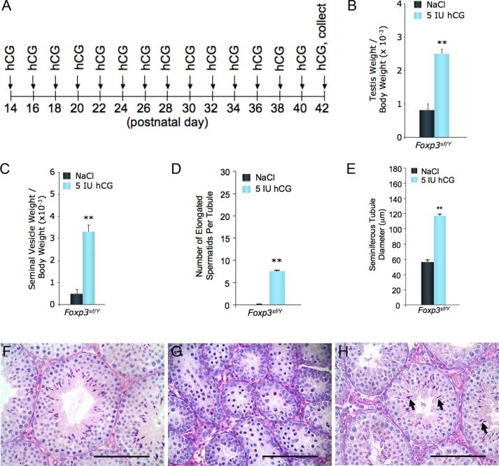
To determine whether longer treatment with hCG could rescue the testicular phenotype of Foxp3sfY mice more completely, mice were treated with 5 IU of hCG starting at P14 and continuing until P42 (Fig. 4A). The longer treatment regimen significantly increased testis weight, seminal vesicle weight, the number of elongated spermatids, and seminiferous tubule diameter (Fig. 4, B–E). Elongated spermatids were present in testis from all Foxp3sfY mice treated with 5 IU of hCG for 4 wk. Histological analysis of testis sections revealed that testis from Foxp3sfY mice is very similar to that of their Foxp3+/Y littermates (Fig. 4, F–H). Thus, spermatogenesis in Foxp3sfY mice is rescued by 28 days of hCG treatment. Considering the entire process of spermatogenesis is approximately 35 days in mice [15], it is likely that an even longer treatment would cause a more complete rescue. These data indicate that the testicular phenotype is largely due to loss of gonadotropin stimulation.
FIG. 4.
A) Foxp3sf/Y mice were treated with 5 IU of hCG (n = 5) or vehicle (n = 4) every other day starting at P14 and continuing to P42. B) Average testis weight/body weight for Foxp3+/Y mice treated with vehicle is 3.24 × 10⁻³ ± 0.50 × 10⁻³ (data not shown). Testis weights increased very significantly in Foxp3sf/Y mice treated with 5 IU of hCG for 4 wk (2.49 × 10⁻³ ± 0.15 × 10⁻³) compared with Foxp3sf/Y mice treated with vehicle (0.82 × 10⁻³ ± 0.18 × 10⁻³). C) Average seminal vesicle weight/body weight for Foxp3+/Y mice treated with vehicle is 4.30 × 10⁻³ ± 0.34 × 10⁻³ (data not shown). Seminal vesicle weights increased very significantly in Foxp3sf/Y mice treated with 5 IU of hCG for 28 days (3.31 × 10⁻³ ± 0.63 × 10⁻³) compared with Foxp3sf/Y mice treated with vehicle (0.50 × 10⁻³ ± 0.19 × 10⁻³). D) The average number of elongated spermatids per cross section of seminiferous tubule is 23 ± 5 for Foxp3+/Y mice treated with vehicle, whereas Foxp3sf/Y mice treated with vehicle lack elongated spermatids. However, spermatogenesis is rescued in five of five Foxp3sf/Y mice treated with 5 IU of hCG for 28 days, with an average of 7 ± 2 elongated spermatids per cross section of seminiferous tubule. E) Average seminiferous tubule diameter of Foxp3+/Y mice treated with vehicle is 164 ± 2 μm. Seminiferous tubule diameter was significantly increased in Foxp3sf/Y mice treated with 5 IU of hCG (117 ± 2 μm) compared with Foxp3sf/Y mice treated with vehicle (57 ± 3 μm). Data are expressed as mean ± SEM and were analyzed by Student t-test to determine significant difference between Foxp3sf/Y mice treated with 5 IU of hCG or vehicle (**P < 0.01). Testis sections from Foxp3+/Y mouse treated with vehicle (F), Foxp3sf/Y mouse treated with vehicle (G), and Foxp3sf/Y mouse treated with 5 IU of hCG (H) for 28 days are shown. Testis from Foxp3sf/Y mice treated with hCG for 28 days contains elongated spermatids (arrows). Original magnification ×400; bar = 100 μm.
Hypothyroidism and Infertility
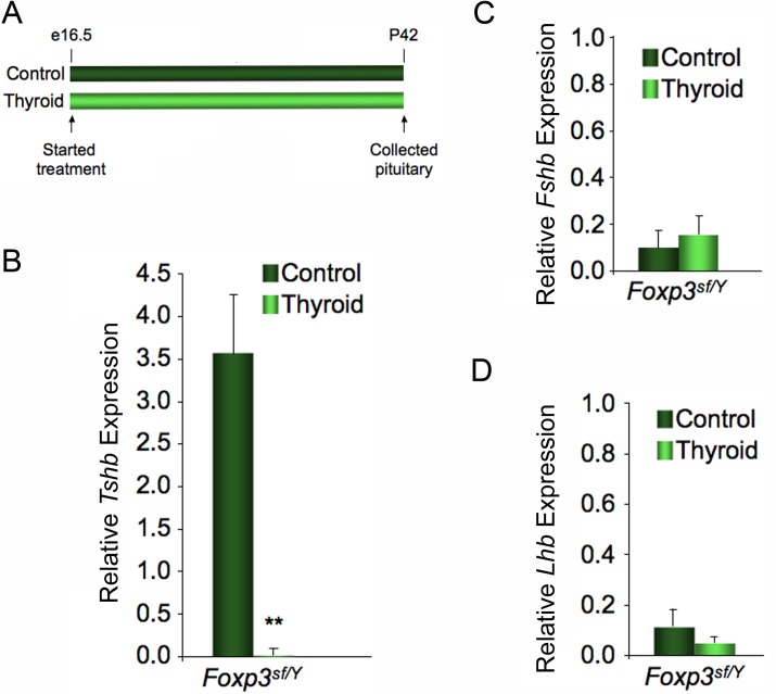
Humans with FOXP3 mutation are hypothyroid because of immune destruction of the thyroid gland [16]. Thyroid-stimulating hormone levels are a very sensitive indicator of hypothyroidism [17]. Previously, we found that Tshb expression is elevated in Foxp3sf/Y mice, suggesting that like many humans with IPEX, Foxp3sf/Y mice also exhibit hypothyroidism [13]. Evidence suggests hypothyroidism in males can cause abnormal gonadotropin levels [18, 19]. To determine whether treatment with thyroid hormone could rescue gonadotropin levels in Foxp3sf/Y mice, pregnant Foxp3sf/+ dams were fed chow containing thyroid powder or a control chow beginning at Embryonic Day 16.5 (Fig. 5A). Foxp3sf/Y pups remained with their dams and continued to receive their respective diets until 6 wk of age, when tissues were collected. Foxp3sf/Y mice that were fed thyroid chow exhibited a very significant reduction in Tshb expression, consistent with the reversal of hypothyroidism (Fig. 5B). No significant change in Fshb or Lhb expression levels occurred when Foxp3sf/Y mice were fed thyroid chow, indicating that hypothyroidism is not the cause of the infertility seen in Foxp3sf/Y mice (Fig. 5, C and D). Thyroid treatment did not affect body weight, seminal vesicle weight, or testis weight, nor did it rescue spermatogenesis (data not shown).
FIG. 5.
A) Pregnant dams were fed control chow or chow supplemented with thyroid powder beginning at Embryonic Day 16.5. Dams and pups were fed their respective diets until the pups reached 6 wk of age. Real-time RT-PCR was used to determine the relative expression levels of Tshb (B), Fshb (C), and Lhb (D) in pituitary gland tissue of Foxp3sf/Y mice on thyroid versus control chow diets. Thyroid hormone replacement significantly reduced expression of Tshb in Foxp3sf/Y mice (0.02) compared with Foxp3sf/Y mice fed control chow (3.57 ± 0.70), but it had no effect on expression of Fshb (0.15 ± 0.08) or Lhb (0.05 ± 0.03) compared with Foxp3sf/Y mice fed control diet (0.10 ± 0.08 and 0.11 ± 0.07, respectively). Expression level was calculated by the 2−ΔΔCT method and represents expression relative to the average ΔCT of samples from Foxp3+/Y mice fed control diet. Data are expressed as mean ± SEM of four animals per group. The data were analyzed by Student t-test to determine significant difference between control and thyroid chow diets (**P < 0.01).
DISCUSSION
Foxp3sf/Y Male Mice Are Hypogonadal and Infertile
Foxp3 mutant males are infertile and exhibit reduced gonadotropin expression [12, 13]. Although Foxp3 transcript is present in the testis, FOXP3 protein is not detected. The testicular phenotype of Foxp3sf/Y mice is very similar to that of mice lacking gonadotropins. Hpg mice are gonadotropin deficient because of a spontaneous mutation in the Gnrh gene [20, 21]. Treatment of hpg mice with hCG for 6 wk rescues spermatogenesis and testis size, but these parameters do not reach normal levels, suggesting that other factors, such as FSH, are important for quantitative normalization of spermatogenesis [22]. Mice with deletions of the genes encoding for LHB (Lhb) or the receptor for LH (Lhcgr) are hypogonadal and infertile, with reduced testosterone levels, resulting in spermatogenesis being arrested at the round spermatid stage [23–25]. The similarity between Foxp3sf/Y mice and Lhb and Lhcgr null mice, combined with the rescue of spermatogenesis in Foxp3sf/Y mice by activating LH signaling, suggests that the gonadal phenotype in Foxp3sf/Y mice is due primarily to a lack of pituitary LH stimulation. This does not rule out the possibility that the immune system is directly inhibiting gonadal function and that treatment with hCG causes hyperstimulation of the testis, overcoming immune suppression of testis.
When bred onto a nude mouse background, which eliminates their autoimmunity, Foxp3 mutant males become fertile, suggesting that their infertility is secondary to autoimmunity [12]. Nude mice have an autosomal recessive mutation of Foxn1, causing them to be athymic and immunosuppressed [26]. Godfrey et al. [14] bred scurfy mice onto a nude mouse background to create Foxp3sf/Y,Foxn1nu/nu mice. These mice no longer exhibited autoimmunity. They did exhibit immunosuppression consistent with that observed in Foxn1nu/nu mice. Interestingly, Foxp3sf/Y,Foxn1nu/nu mice were capable of siring progeny. This allowed for generation of Foxp3sf/sf,Foxn1+/nu female mice, which exhibited scurfylike lesions and life spans of <30 days. The microscopic anatomy in the female reproductive tracts was normal [12]. We find that Foxp3 is not expressed in the adult pituitary gland [13]. This, together with the fact that eliminating autoimmunity in Foxp3sf/Y mice by breeding them onto a nude mouse background rescues their fertility, suggests that the reproductive phenotype in Foxp3sf/Y mice is secondary due to loss of Foxp3 in regulatory T cells.
Foxp3sf/Y mice have elevated levels of many cytokines, including interleukin 2 (IL2), IL4, IL5, IL7, IL10, interferon γ (IFNγ), and tumor necrosis factor α (TNFα) [27–29]. There are many examples of cytokine regulation of reproductive function; for example, IL2 has been shown to stimulate Pomc expression and inhibit LH, FSH, and growth hormone release [30]. TNFα has been shown to inhibit release of growth hormone, LH, prolactin, and GnRH [31, 32]. Considering the lack of Foxp3 expression in hypothalamus and pituitary, it is unlikely that FOXP3 directly effects gonadotropin or Gnrh expression [13]. It is possible that the gonadal phenotype observed in Foxp3sf/Y mice is due to cytokine inhibition of gonadotropin expression or GnRH release.
Hypothyroidism with Loss of FOXP3
Humans with FOXP3 mutations often exhibit hypothyroidism due to autoimmune destruction of the thyroid gland [17]. Few studies address thyroid function in Foxp3sf/Y mice. Sharma et al. [33] found no inflammation in pancreas or thyroid. However, Lahl et al. [34] observed immune infiltrate and destruction of the islets in the acini of pancreas in Foxp3sf/Y mice. Unfortunately, they did not examine thyroid tissue from Foxp3sf/Y mice [34]. We find that Foxp3sf/Y mice have elevated Tshb expression, which is reversed by thyroid hormone replacement, suggesting that Foxp3 mutant mice are hypothyroid, like many human patients with IPEX. The thyroid and pancreatic phenotypes of these mice, like human patients with IPEX, may be variable. Thyroid hormone replacement did not change gonadotropin expression in Foxp3sf/Y mice, leading us to conclude that their infertility is not due to hypothyroidism.
Hypothyroidism is normally accompanied by increased PRL levels. This is because the lack of negative feedback from thyroid hormone causes an increase in hypothalamic thyroid-releasing hormone, which is a secretagogue for PRL [35]. In contrast, we observed reduced expression of Prl in Foxp3sf/Y mice [13]. This could mean that a prolactin inhibitory factor is being produced at high levels in Foxp3sf/Y mice.
Taken together, these data suggest that reduced gonadotropin levels are responsible for the arrest in spermatogenesis observed in Foxp3sf/Y mice. In the absence of Foxp3, pituitary gonadotropin expression decreases, resulting in hypogonadotropic hypogonadism and infertility. This hypogonadotropic hypogonadism is a secondary effect, most likely due to loss of Foxp3 in immune cells. Thus, loss of Foxp3, likely in regulatory T cells, results in hypogonadotropic hypogonadism and infertility.
Supplementary Material
ACKNOWLEDGMENT
We thank Maureen Doran and Dawn Grisley for their help with our histological studies.
Footnotes
Supported by startup funds from Southern Illinois University to B.S.E., and National Institutes of Health grant HD044119 to P.N. Presented in part at the 44th Annual Meeting of the Society for the Study of Reproduction, July 31-August 4, 2011, Portland, Oregon.
REFERENCES
- Kerr JB, Loveland KL, O'Bryan MK, de Krester DM. Cytology of the testis and intrinsic control mechanisms. : Knobil E, Neill JD. (eds.), Physiology of Reproduction, 3rd ed. Houston, TX: Gulf Professional Publishing; 2006: 827–920. [Google Scholar]
- Saez JM. Leydig cells: endocrine, paracrine, and autocrine regulation. Endocr Rev 1994; 15: 574–626. [DOI] [PubMed] [Google Scholar]
- Genuth SM. Physiology. St. Louis, MO: Mosby Year Book; 1993. [Google Scholar]
- Sairam MR, Krishnamurthy H. The role of follicle-stimulating hormone in spermatogenesis: lessons from knockout animal models. Arch Med Res 2001; 32: 601–608. [DOI] [PubMed] [Google Scholar]
- Simoni M, Gromoll J, Nieschlag E. The follicle-stimulating hormone receptor: biochemistry, molecular biology, physiology, and pathophysiology. Endocr Rev 1997; 18: 739–773. [DOI] [PubMed] [Google Scholar]
- Hubert FX, Kinkel SA, Crewther PE, Cannon PZ, Webster KE, Link M, Uibo R, O'Bryan MK, Meager A, Forehan SP, Smyth GK, Mittaz L, et al. Aire-deficient C57BL/6 mice mimicking the common human 13-base pair deletion mutation present with only a mild autoimmune phenotype. J Immunol 2009; 182: 3902–3918. [DOI] [PubMed] [Google Scholar]
- Pelletier RM, Yoon SR, Akpovi CD, Silvas E, Vitale ML. Defects in the regulatory clearance mechanisms favor the breakdown of self-tolerance during spontaneous autoimmune orchitis. Am J Physiol Regul Integr Comp Physiol 2009; 296: R743–R762. [DOI] [PubMed] [Google Scholar]
- Li B, Greene MI. FOXP3 actively represses transcription by recruiting the HAT/HDAC complex. Cell Cycle 2007; 6: 1432–1436. [PubMed] [Google Scholar]
- Powell BR, Buist NR, Stenzel P. An X-linked syndrome of diarrhea, polyendocrinopathy, and fatal infection in infancy. J Pediatr 1982; 100: 731–737. [DOI] [PubMed] [Google Scholar]
- Brunkow ME, Jeffery EW, Hjerrild KA, Paeper B, Clark LB, Yasayko SA, Wilkinson JE, Galas D, Ziegler SF, Ramsdell F. Disruption of a new forkhead/winged-helix protein, scurfin, results in the fatal lymphoproliferative disorder of the scurfy mouse. Nat Genet 2001; 27: 68–73. [DOI] [PubMed] [Google Scholar]
- Lyon MF. Hypogonadism in scurfy (sf) males. Mouse News Letter 1986; 74: 93. [Google Scholar]
- Godfrey VL, Wilkinson JE, Rinchik EM, Russell LB. Fatal lymphoreticular disease in the scurfy (sf) mouse requires T cells that mature in a sf thymic environment: potential model for thymic education. Proc Natl Acad Sci U S A 1991; 88: 5528–5532. [DOI] [PMC free article] [PubMed] [Google Scholar]
- Jung DO, Jasurda JS, Egashira N, Ellsworth BS. The forkhead transcription factor, FOXP3, is required for normal pituitary gonadotropin expression in mice. Biol Reprod 2012; 86: 144. [DOI] [PMC free article] [PubMed] [Google Scholar]
- Godfrey VL, Wilkinson JE, Russell LB. X-linked lymphoreticular disease in the scurfy (sf) mutant mouse. Am J Pathol 1991; 138: 1379–1387. [PMC free article] [PubMed] [Google Scholar]
- Oakberg EF. Duration of spermatogenesis in the mouse. Nature 1957; 180: 1137–1138. [DOI] [PubMed] [Google Scholar]
- Ferguson PJ, Blanton SH, Saulsbury FT, McDuffie MJ, Lemahieu V, Gastier JM, Francke U, Borowitz SM, Sutphen JL, Kelly TE. Manifestations and linkage analysis in X-linked autoimmunity-immunodeficiency syndrome. Am J Med Genet 2000; 90: 390–397. [DOI] [PubMed] [Google Scholar]
- Torgerson TR, Ochs HD. Immune dysregulation, polyendocrinopathy, enteropathy, X-linked: Forkhead box protein 3 mutations and lack of regulatory T cells. J Allergy Clin Immunol 2007; 120: 744–750. [DOI] [PubMed] [Google Scholar]
- Brent GA, Davies TF. Hyperthyroidism and thyroiditis. : Melmed S, Polonsky KS, Larsen PR, Kronenberg HM. (eds.), William's Textbook of Endocrinology, 12th ed. Philadelphia, PA: Elsevier Inc; 2013: 406–435. [Google Scholar]
- Wortsman J, Rosner W, Dufau ML. Abnormal testicular function in men with primary hypothyroidism. Am J Med 1987; 82: 207–212. [DOI] [PubMed] [Google Scholar]
- Cattanach BM, Iddon CA, Charlton HM, Chiappa SA, Fink G. Gonadotrophin-releasing hormone deficiency in a mutant mouse with hypogonadism. Nature 1977; 269: 338–340. [DOI] [PubMed] [Google Scholar]
- Mason AJ, Hayflick JS, Zoeller RT, Young WS, III, , Phillips HS, Nikolics K, Seeburg PH. A deletion truncating the gonadotropin-releasing hormone gene is responsible for hypogonadism in the hpg mouse. Science 1986; 234: 1366–1371. [DOI] [PubMed] [Google Scholar]
- Spaliviero JA, Jimenez M, Allan CM, Handelsman DJ. Luteinizing hormone receptor-mediated effects on initiation of spermatogenesis in gonadotropin-deficient (hpg) mice are replicated by testosterone. Biol Reprod 2004; 70: 32–38. [DOI] [PubMed] [Google Scholar]
- Lei ZM, Mishra S, Zou W, Xu B, Foltz M, Li X, Rao CV. Targeted disruption of luteinizing hormone/human chorionic gonadotropin receptor gene. Mol Endocrinol 2001; 15: 184–200. [DOI] [PubMed] [Google Scholar]
- Ma X, Dong Y, Matzuk MM, Kumar TR. Targeted disruption of luteinizing hormone beta-subunit leads to hypogonadism, defects in gonadal steroidogenesis, and infertility. Proc Natl Acad Sci U S A 2004; 101: 17294–17299. [DOI] [PMC free article] [PubMed] [Google Scholar]
- Zhang FP, Poutanen M, Wilbertz J, Huhtaniemi I. Normal prenatal but arrested postnatal sexual development of luteinizing hormone receptor knockout (LuRKO) mice. Mol Endocrinol 2001; 15: 172–183. [DOI] [PubMed] [Google Scholar]
- Nehls M, Pfeifer D, Schorpp M, Hedrich H, Boehm T. New member of the winged-helix protein family disrupted in mouse and rat nude mutations. Nature 1994; 372: 103–107. [DOI] [PubMed] [Google Scholar]
- Lin W, Truong N, Grossman WJ, Haribhai D, Williams CB, Wang J, Martin MG, Chatila TA. Allergic dysregulation and hyperimmunoglobulinemia E in Foxp3 mutant mice. J Allergy Clin Immunol 2005; 116: 1106–1115. [DOI] [PubMed] [Google Scholar]
- Kanangat S, Blair P, Reddy R, Daheshia M, Godfrey V, Rouse BT, Wilkinson E. Disease in the scurfy (sf) mouse is associated with overexpression of cytokine genes. Eur J Immunol 1996; 26: 161–165. [DOI] [PubMed] [Google Scholar]
- Blair PJ, Bultman SJ, Haas JC, Rouse BT, Wilkinson JE, Godfrey VL. CD4+CD8-T cells are the effector cells in disease pathogenesis in the scurfy (sf) mouse. J Immunol 1994; 153: 3764–3774. [PubMed] [Google Scholar]
- Savino W, Arzt E, Dardenne M. Immunoneuroendocrine connectivity: the paradigm of the thymus-hypothalamus/pituitary axis. Neuroimmunomodulation 1999; 6: 126–136. [DOI] [PubMed] [Google Scholar]
- Gaillard RC, Turnill D, Sappino P, Muller AF. Tumor necrosis factor alpha inhibits the hormonal response of the pituitary gland to hypothalamic releasing factors. Endocrinology 1990; 127: 101–106. [DOI] [PubMed] [Google Scholar]
- Watanobe H, Hayakawa Y. Hypothalamic interleukin-1 beta and tumor necrosis factor-alpha, but not interleukin-6, mediate the endotoxin-induced suppression of the reproductive axis in rats. Endocrinology 2003; 144: 4868–4875. [DOI] [PubMed] [Google Scholar]
- Sharma R, Jarjour WN, Zheng L, Gaskin F, Fu SM, Ju ST. Large functional repertoire of regulatory T-cell suppressible autoimmune T cells in scurfy mice. J Autoimmun 2007; 29: 10–19. [DOI] [PMC free article] [PubMed] [Google Scholar]
- Lahl K, Loddenkemper C, Drouin C, Freyer J, Arnason J, Eberl G, Hamann A, Wagner H, Huehn J, Sparwasser T. Selective depletion of Foxp3+ regulatory T cells induces a scurfy-like disease. J Exp Med 2007; 204: 57–63. [DOI] [PMC free article] [PubMed] [Google Scholar]
- Poppe K, Velkeniers B, Glinoer D. The role of thyroid autoimmunity in fertility and pregnancy. Nat Clin Pract Endocrinol Metab 2008; 4: 394–405. [DOI] [PubMed] [Google Scholar]
Associated Data
This section collects any data citations, data availability statements, or supplementary materials included in this article.


