Abstract
In the current investigation we examined the number and proportion of neuronal and non-neuronal cells in the primary sensory areas of the neocortex of a South American marsupial, the short-tailed opossum (Monodelphis domestica). The primary somatosensory (S1), auditory (A1), and visual (V1) areas were dissected from the cortical sheet and compared with each other and the remaining neocortex using the isotropic fractionator technique. We found that although the overall sizes of V1, S1, A1, and the remaining cortical regions differed from each other, these divisions of the neocortex contained the same number of neurons, but the remaining cortex contained significantly more non-neurons than the primary sensory regions. In addition, the percent of neurons was higher in A1 than in the remaining cortex and the cortex as a whole. These results are similar to those seen in non-human primates. Furthermore, these results indicate that in some respects, such as number of neurons, the neocortex is homogenous across its extent, whereas in other aspects of organization, such as non-neuronal number and percentage of neurons, there is non-uniformity. Whereas the overall pattern of neuronal distribution is similar between short-tailed opossums and eutherian mammals, short-tailed opossum have a much lower cellular and neuronal density than other eutherian mammals. This suggests that the high neuronal density cortices of mammals such as rodents and primates may be a more recently evolved characteristic that is restricted to eutherians, and likely contributes to the complex behaviors we see in modern mammals.
INDEXING TERMS: evolution, Monodelphis, isotropic fractionation
The six-layered neocortex is a general feature of the mammalian brain in most (but not all) mammals, and has been observed and studied in species as diverse as platypus, capybaras, and humans (DeFelipe et al., 2002; Hutsler et al., 2005; Krubitzer et al., 1995). One of the major functions of the neocortex is to process and integrate sensory signals coming from sensory receptor arrays in the skin, muscles, joints, ears, and eyes to effect context-appropriate behavior. In all mammals examined, these specific sensory modalities have at least one neocortical area that contains a map of the sensory receptor array, and is defined by dense myelination, a koniocellular layer 4, and a homologous pattern of thalamocortical connections (Kaas, 1983; Krubitzer, 2009). However, these primary sensory areas can also vary in a number of parameters including relative size, connectivity, and their number of sublaminae, to name a few. Although these traditional views of the cortical field have become textbook knowledge, important questions regarding the cellular composition of the neocortex and the evolution of basic processing circuitry persist.
The first question centers on the extent to which the neocortex has a basic composition of neurons and glial cells that is ubiquitous across its extent and across species. This question was first addressed by Rockel and colleagues in 1980. They posit (with the exception of area 17) that independent of cortical thickness, laminar specialization, or function, the absolute number of cells is the same across the cortical sheet regardless of the cortical field in question. Furthermore, they argue that this number is constant regardless of brain size or species. Although subsequent studies seem to refute this appealing hypothesis (Haug, 1987; Herculano-Houzel et al., 2008; Prothero, 1997), a recent study suggests that this early supposition is correct and that other studies that contest this idea suffered from methodological or quantification problems (Carlo and Stevens, 2013).
The second question addresses the evolution of the basic circuitry of the neocortex and seeks to uncover both the rules of construction as well as the constraints imposed on evolving brains. Although fossil records provide information on the size of the brain and gross morphological characteristics of the neocortex, the only way to appreciate the cortical networks that were present in early mammals and the types of cellular and systems level changes that have been made to these networks is to perform a comparative analysis. These types of studies reveal both fundamental features of processing as well as derivations that generate the remarkable diversity in behavior observed in extant species.
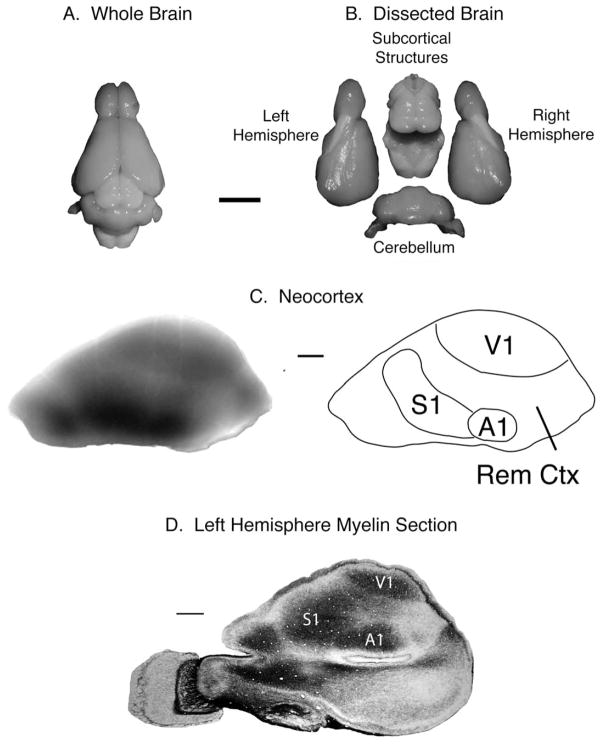
Unfortunately, most studies that address these questions of both basic cortical composition and evolution only consider a few species of eutherian mammals such as monkeys, cats, and rats (or mice). As a result, there are gaping holes in our knowledge of two major branches of mammals, monotremes and marsupials, the latter of which provides a better reflection of the ancestral state more than the commonly used eutherian models (Frost et al., 2000; Kaas, 2011; Karlen and Krubitzer, 2007). In the present investigation we utilized the isotropic fractionator method to examine the cellular composition of the neocortex of a marsupial, the South American short-tailed opossum, Monodelphis domestica. Like other mammals, the opossum has a six-layered cortex and primary sensory areas that can be clearly differentiated by their architectonic appearance, functional organization, and neuroanatomical connections (Catania et al., 2000; Kahn et al., 2000; Karlen et al., 2006; Saunders et al., 1989). Furthermore, because of their dense myelination, primary fields can be visualized in raw cortical tissue that is properly illuminated, and these areas can then be carefully dissected from the rest of the cortex (Fig. 1). In this experiment we dissected the neocortex into the three primary sensory areas, auditory (A1), somatosensory (S1), and visual (V1) cortices, as well as the remaining cortical areas and determined the number and density of neurons and non-neurons, as well as the percentage of neurons within both the entire neocortical hemisphere and a given structure.
Figure 1.
Dissection of the opossum brain for isotropic fractionation. A,B: The whole brain (A) is separated into the left and right cerebral hemispheres, the cerebellum, and subcortical structures (including the midbrain, thalamus, hypothalamus, and brainstem; B). C: The cerebral hemispheres are further dissected, removing the hippocampus and basal ganglia, until only the neocortex remains (left image). The darker, more densely myelinated regions correspond to the primary sensory areas (right image). D: The primary sensory areas can be clearly seen on section of flattened cortical tissue that has been stained for myelin. A1, primary auditory cortex; S1, somatosensory cortex; V1, visual cortex; Rem Ctx, remaining cortex. Scale bar = 5 mm in A (applies to A,B); 1 mm in C,D.
MATERIALS AND METHODS
As is the case with most methodologies, the isotropic fractionator technique has both costs and benefits associated with it, and we have addressed these points previously (Seelke et al., 2013).
The isotropic fractionation procedure consists of multiple stages. First, tissue is dissected into major structures. Next, tissue is processed, which includes homogenization, 4′,6-diamidino-2-phenylindole (DAPI) staining and NeuN immunohistochemistry. The third stage involves quantifying the number of DAPI-labeled nuclei within a sample, and the fourth stage involves determining the proportion of NeuN-labeled nuclei within that same sample. Finally, in the last stage we use these values to calculate the total number of cells, total number of neurons, total number of non-neurons, cell density, neuronal density, and non-neuronal density. A number of cell types fall under the grouping of “non-neuronal cells,” including endothelial, mesothelial, ependymal, and glial cells. Endothelial cells form the thin lining of blood vessels and compose the blood–brain barrier, mesothelial cells comprise the pia mater, and ependymal cells line the ventricles, and of these non-neuronal cell types, the glia are most prevalent (Morest and Silver, 2003; Temple, 2001). Thus, for the purposes of this study we will consider the non-neuronal cells to be predominantly glial in nature.
Subjects
Five adult South American short-tailed opossums (Monodelphis domestica) were used in these experiments (see Table 1 for ages, weights, and sexes). Subjects were born and raised in the Psychology Department Vivarium at the University of California, Davis. Animals were housed in standard laboratory cages with ad libitum access to food and water. All animals were maintained on a 14-hour light/dark cycle with the lights on at 7 AM. All experiments were performed under National Institutes of Health guidelines for the care of animals in research and were approved by the Institutional Animal Care and Use Committee of the University of California, Davis.
TABLE 1.
Characteristics of the Study Subjects
| Case no. | Age (days) | Weight (g) | Sex |
|---|---|---|---|
| 11–151 | 696 | 148 | M |
| 11–152 | 610 | 121 | F |
| 11–153 | 476 | 107 | F |
| 11–155 | 511 | 134 | M |
| 11–243 | 429 | 109 | M |
Tissue dissection
Animals were euthanized with an overdose of sodium pentobarbital (Beuthanasia; 250 mg/kg) and transcardially perfused with phosphate-buffered saline (PBS) followed by 4% paraformaldehyde. The brain was extracted, photographed, weighed, and dissected; the two cortical hemispheres, the subcortical structures, and the cerebellum were separated (Fig. 1). The neocortex was isolated by removing the hippocampus, basal ganglia, pyriform cortex (defined as all tissue lateral to the rhinal sulcus, including the amygdala), and olfactory bulbs from both cerebral hemispheres. In each case, one neocortical hemisphere was kept intact and the other neocortical hemisphere was dissected into primary sensory fields. The densely myelinated primary sensory fields were visualized by placing the tissue on a light box. This back-illuminated tissue was then dissected into the primary sensory fields, V1, A1, S1, and the remaining cortex. These regions were photographed and weighed, and then placed in 5% paraformaldehyde for storage.
Tissue processing
Samples were prepared as described previously (Campi et al., 2011; Seelke et al., 2013). Briefly, tissue was homogenized in a 15-ml glass KONTES* Tenbroek tissue grinder (Kimble Chase, Vineland, NJ) with Triton X-100 and sodium citrate in distilled water. This process broke down cell membranes to produce a suspension of isolated nuclei. The samples were centrifuged and resuspended in a solution of PBS and DAPI. In some cases dissected tissue was stored in fixative for more than 4 weeks. This tissue was homogenized and suspended in a boric acid solution then placed in an oven at 70°C for 1 hour for epitope retrieval. In all cases a subsample of the main suspension was stained for neuronal nuclei using immunocytochemical techniques with the anti-NeuN antibody (Millipore, Bedford, MA). Alexa Fluor 647 or Alexa Fluor 700 goat antimouse IgG secondary antibody (Invitrogen, Carlsbad, CA) was used to fluorescently visualize NeuN-labeled nuclei.
Antibody characterization
The NeuN antibody (NEUronal Nuclei; clone A60) specifically recognizes the DNA-binding, neuron-specific protein NeuN, which is present in most central and peripheral nervous system neuronal cell types of all vertebrates tested. In a western blot analysis, this antibody recognized two to three bands in the 46–48-kDa range and possibly another band at approximately 66 kDa (adapted from product information). Because NeuN specifically stains neuronal nuclei, staining of nonneuronal tissue was used as a negative control (see Table 2 for details).
TABLE 2.
Antibody Used in This Study
| Antigen | Immunogen | Maunfacturer, species, mono- vs. polyclonal, cat. no. | Dilution |
|---|---|---|---|
| NeuN | Purified cell nuclei from mouse brain | Millipore (Billerica, MA), mouse monoclonal, #MAB377 | 1:300 |
Nuclei quantification
For cell counting, samples were vortexed, and 10-μL aliquots were immediately loaded into a Neubauer cell-counting chamber (Labor Optik, Bad Homburg, Germany) and placed on a fluorescence microscope for visualization and counting of nuclei. Standard stereological protocols were used (Mouton, 2002). DAPI-labeled nuclei within 10 distinct 8 square by 8 square sections of the Neubauer chamber were counted (for details, see Campi et al., 2011; Seelke et al., 2013). The mean number of DAPI+ nuclei in each 8 × 8 section was then calculated, and was later used to calculate the total number of cells within a given sample.
Determining NeuN+ percent
To determine the ratio of neuronal to non-neuronal nuclei within a sample, a flow cytometer was used (UC Davis Flow Cytometry Shared Resource Center). This allowed us to automate the detection and counting of neuronal and non-neuronal nuclei in a faster and more reliable manner than visual inspection under a fluorescent microscope (Collins et al., 2010b). The proportion of neuronal nuclei was quantified by using a Becton Dickinson (San Jose, CA) 5-Laser LSRII flow cytometer. Specific lasers were used to excite DAPI-positive nuclei (50 mW, 405 nm) and Alexa Fluor 647- and Alexa Fluor 700–labeled nuclei (50 mW, 635 nm). For all samples, 1,000–10,000 DAPI-positive nuclei were evaluated for Alexa Fluor 647 and Alexa Fluor 700 label. Samples that were run on the flow cytometer were forced through a 35-μm mesh cell filter, and then vortexed and immediately taken into the LSRII. Selection gates were determined by a flow cytometry expert who was blind to the cortical areas of the samples, and the ratio of neuronal to non-neuronal nuclei was estimated from this gated population.
Equations
Estimates of cellular composition for a given cortical region were derived from the following equations.
Analysis
In each case, one neocortical hemisphere was left intact and the other was dissected into the primary sensory fields and remaining cortical areas. The intact neocortical hemisphere was compared against the summed values obtained from the dissected neocortical hemisphere to ensure consistency across samples. The weights of these regions were compared by using analysis of variance (ANOVA; JMP; SAS, Cary, NC), as were differences in the % neurons, total number of cells, total number of neurons, total number of non-neurons, cell density, neuronal density, and non-neuronal density. Differences between specific regions were determined by using Student’s t-tests. For all tests, alpha = 0.05.
RESULTS
In the following results we dissected different portions of the cortical sheet (Fig. 1) and quantified the cellular composition of the whole neocortical sheet, the primary sensory regions of the neocortex and the remaining cortex. We compared numbers, density and percentage of all cells, neurons and non-neurons, within a cortical field as well as across cortical fields. To ensure the reliability of these results, we compared the values obtained from intact neocortical hemispheres with the summed values for the primary sensory regions and remaining cortical regions, which we identify as the summed neocortex. Animal information can be found in Table 1.
Cortical field weights
We first compared the weights of the intact neocortical hemisphere (neocortex), the summed weight of the dissected neocortical hemisphere (ΣCtx), and the individual cortical fields, including the A1, S1, and V1 cortices, and the Rem Ctx (Fig. 2). The intact neocortical hemisphere was small and weighed 0.073 g, with little variability across animals ( ± 0.003 g; Table 3), and the summed neocortex displayed nearly identical values (ΣCtx = 0.072 ± 0.003 g). Together, the primary sensory fields comprised approximately half of the weight of the neocortex (V1 = 0.015 ± 0.002 g; A1 = 0.007 ± 0.001 g; S1 = 0.011 ± 0.001 g) with the balance consisting of the remaining cortex (0.039 ± 0.003 g; Fig. 2A). In terms of size of the different divisions of the neocortex, the remaining cortex was the largest and weighed significantly more than V1 and S1, and V1 weighed significantly more than A1 (which was the smallest field examined). The weight of S1 did not differ from V1 and A1 (F5,29 = 173.49, P < 0.0001; Fig. 2A). Similarly, the largest proportion (percentage) of the neocortex was comprised of the Rem Ctx, which was significantly larger than V1, which was in turn significantly larger than A1. The proportion of neocortex devoted to S1 significantly differed from Rem Ctx, but not V1 and A1 (F3,19 = 110.86, P < 0.0001; Fig. 2B).
Figure 2.
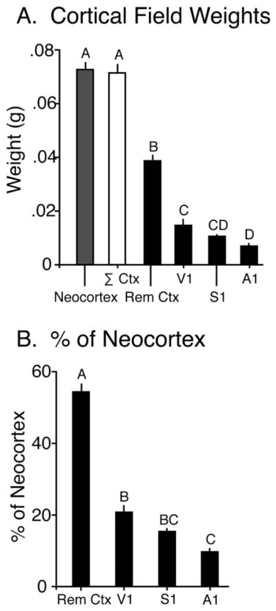
A: The weight (in grams) of the whole neocortex, the primary sensory areas, and the remaining cortical areas. The weight of the intact neocortical sheet is equal to the sum of the weight of V1, S1, A1, and Rem Ctx. B: The percentage of neocortex comprised by the primary sensory areas and the remaining cortical areas. Mean ± SEM. Values with different letters are significantly different. A1, primary auditory cortex; S1, somatosensory cortex; V1, visual cortex; Rem Ctx, remaining cortex.
TABLE 3.
Cortical Field Weights
| Area | Weight (g) | % of Σneocortex |
|---|---|---|
| Whole neocortex | 0.073 ± 0.003 | |
| Σ neocortex | 0.072 ± 0.003 | |
| Rem Ctx | 0.039 ± 0.003 | 54.1 ± 2.5 |
| V1 | 0.015 ± 0.002 | 20.7 ± 2.3 |
| S1 | 0.011 ± 0.001 | 15.3 ± 1.1 |
| A1 | 0.007 ± 0.001 | 9.8 ± 1.2 |
For abbreviations, see list.
Cellular composition
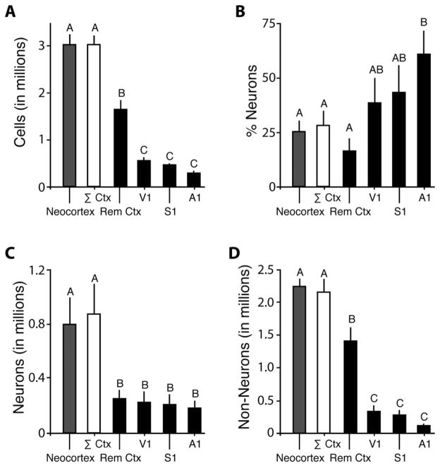
One intact neocortical hemisphere contained 3.03 ± 0.21 million cells, and the ΣCtx contained 3.02 ± 0.21 million cells (Table 3). V1, S1, and A1 contained 0.57 ± 0.09 million cells, 0.50 ± 0.03 million cells, and 0.30 ± 0.03 million cells, respectively, and Rem Ctx contained 1.66 ± 0.18 million cells. The total number of cells significantly differed across cortical regions (F5,29 = 73.53, P < 0.0001; Fig. 3A). The number of cells in the intact neocortex and ΣCtx did not differ from each other. The Rem Ctx contained more cells than V1, S1, and A1. However, there was no significant difference between the numbers of cells in V1, S1, and A1.
Figure 3.
The cellular composition of the intact neocortical hemisphere (neocortex; gray), summed neocortical hemisphere (ΣCtx; white), and the primary sensory and remaining cortical regions (Rem Ctx, V1, S1, A1; black). A: The total number of cells in millions. The number of cells varies with the size of each cortical region, with larger regions having more cells and smaller regions having fewer cells. The number of cells in the intact neocortical sheet is equal to the sum of the number of cells in V1, S1, A1, and Rem Ctx. B: The percentage of neurons in the entire neocortex and each cortical region. In general, the primary sensory regions contain a higher proportion of neurons than the remaining cortical areas. The intact neocortical sheet contains the same proportion of neurons as the weighted sum of V1, S1, A1, and Rem Ctx. C: The total number of neurons in millions. Despite their smaller size, the primary sensory areas contain the same number of neurons as the remaining cortical regions. The number of neurons in the intact neocortical sheet is equal to the sum of the number of neurons in V1, S1, A1, and Rem Ctx. D: The total number of non-neuronal cells in millions. The primary sensory regions contain significantly fewer non-neuronal cells than the remaining neocortical regions. The number of non-neuronal cells in the intact neocortical sheet is equal to the sum of the number of non-neuronal cells in V1, S1, A1, and Rem Ctx. Mean ± SEM. Values with different letters are significantly different. A1, primary auditory cortex; S1, somatosensory cortex; V1, visual cortex; Rem Ctx, remaining cortex.
We next determined the proportion of neurons contained within each cortical region. The intact neocortex contained 25.6 ± 4.9% neurons, and the ΣCtx contained 28.4 ± 6.4% neurons (Table 4). The Rem Ctx consisted of 16.6 ± 6.2% neurons, and V1, S1, and A1 contained 38.7 ± 11.2%, 43.2 ± 12.4%, and 60.9 ± 11.5%, respectively. There was a significant difference between the proportion of neurons in different cortical regions (F5,29 = 2.86, P < 0.05; Fig. 3B). Whereas the percent neurons did not differ between V1, S1 and the remaining cortex, the percent neurons in A1 was significantly higher than in the Rem Ctx as well as ΣCtx and the intact neocortical hemisphere.
TABLE 4.
Proportion of Neurons Contained Within Each Cortical Region
| Area | No. of cells (in millions) | % Neurons | No. of neurons (in millions) | No. of non-neurons (in millions) |
|---|---|---|---|---|
| Whole neocortex | 3.03 ± 0.21 | 25.6 ± 4.9 | 0.80 ± 0.20 | 2.22 ± 0.12 |
| Σ neocortex | 3.02 ± 0.21 | 28.4 ± 6.4 | 0.88 ± 0.22 | 2.14 ± 0.22 |
| Rem Ctx | 1.66 ± 0.18 | 16.6 ± 6.2 | 0.25 ± 0.07 | 1.41 ± 0.21 |
| V1 | 0.57 ± 0.09 | 38.7 ± 11.2 | 0.23 ± 0.08 | 0.34 ± 0.08 |
| S1 | 0.50 ± 0.03 | 43.2 ± 12.4 | 0.21 ± 0.07 | 0.28 ± 0.07 |
| A1 | 0.30 ± 0.03 | 60.9 ± 11.5 | 0.18 ± 0.04 | 0.11 ± 0.03 |
For abbreviations, see list.
By multiplying the total number of cells by the percentage of neurons within a structure, we determined the total number of neurons within a structure The intact neocortex contained 0.80 ± 0.20 million neurons and ΣCtx contained 0.88 ± 0.22 million neurons (Table 4). The remaining cortex contained 0.25 ± 0.07 million neurons, and V1, S1, and A1 contained 0.25 ± 0.08 million neurons, 0.21 ± 0.07 million neurons, and 0.18 ± 0.04 million neurons, respectively. The total numbers of neurons in V1, A1, S1, and the Rem Ctx did not significantly differ from each other (Fig. 3C).
The total number of non-neurons within a cortical region was determined by subtracting the total number of neurons from the total number of cells. The intact neocortex contained 2.22 ± 0.12 million non-neuronal cells and ΣCtx contained 2.14 ± 0.22 million non-neuronal cells (Table 4). The remaining cortex contained 1.41 ± 0.21 million non-neuronal cells. V1, S1, and A1 contained 0.34 ± 0.08 million, 0.28 ± 0.07 million, and 0.11 ± 0.03 million non-neuronal cells, respectively. The total number of non-neuronal cells significantly differed between cortical regions (F5,29 = 46.64, P < 0.0001; Fig. 3D). The Rem Ctx contained more non-neuronal cells than V1, S1, and A1. However, there was no significant difference between the number of non-neuronal cells in V1, S1, and A1.
Cellular density
Cellular density was determined by dividing the total number of cells in a given cortical structure by the weight of the given cortical structure. The cellular density of the intact neocortical hemisphere was 41.70 ± 2.07 million cells/g of tissue, and the cellular density of the ΣCtx was the same (42.39 ± 3.01 million cells/g of tissue; Table 5). The cellular densities of the Rem Ctx, V1, S1, and A1 did not significantly differ from each other (F5,29 = 0.17, P = NS; Fig. 4A).
TABLE 5.
Cellular Density
| Area | Cells/g (in millions) | Neurons/g (in millions) | Non-neurons/g (in millions) |
|---|---|---|---|
| Whole neocortex | 41.70 ± 2.07 | 10.92 ± 2.54 | 30.78 ± 1.68 |
| Σ neocortex | 42.39 ± 3.01 | 12.28 ± 2.89 | 30.11 ± 3.36 |
| Rem Ctx | 42.52 ± 2.42 | 6.73 ± 2.21 | 35.79 ± 3.97 |
| V1 | 40.98 ± 8.13 | 17.31 ± 6.32 | 23.68 ± 5.84 |
| S1 | 46.34 ± 4.77 | 20.29 ± 7.66 | 26.05 ± 7.28 |
| A1 | 44.04 ± 4.44 | 28.05 ± 5.76 | 15.99 ± 3.15 |
For abbreviations, see list.
Figure 4.

The cellular density of the intact neocortical hemisphere (neocortex; gray), summed neocortical hemisphere (ΣCtx; white), and the primary sensory and remaining cortical regions (Rem Ctx, V1, S1, A1; black). A: The cellular density of each cortical region, in millions of cells per gram of tissue. The total cellular density does not vary across different cortical regions. The cellular density of the intact neocortical sheet is equal to the cellular density of the summed neocortical regions. B: The neuronal density of each cortical region, in millions of cells per gram of tissue. Although there is not a significant difference in neuronal density across cortical regions, the data suggest that the primary sensory regions contain a higher density of neurons than remaining cortical regions. The neuronal density of the intact neocortical sheet is equal to the neuronal density of the summed neocortical regions. C: The non-neuronal density of each cortical region, in millions of cells per gram of tissue. Although there is not a significant difference in non-neuronal density across cortical regions, the data suggest that the primary sensory regions contain a lower density of non-neuronal cells than remaining cortical regions. The non-neuronal density of the intact neocortical sheet is equal to the non-neuronal density of the summed neocortical regions. Mean ± SEM. Values with different letters are significantly different. A1, primary auditory cortex; S1, somatosensory cortex; V1, visual cortex; Rem Ctx, remaining cortex.
Neuronal density was calculated by dividing the total number of neurons in a given cortical structure by the weight of that structure. As with the total number of cells, the neuronal density of the intact neocortical hemisphere was not significantly different from ΣCtx (Table 5). In addition, there were no significant differences in the neuronal density of the Rem Ctx, or the primary sensory regions, V1, S1, and A1 (Table 5). Whereas there was no significant difference between cortical regions (F5,29 = 2.30, P = 0.076; Fig. 4B), a preplanned comparison indicated that A1 had a significantly higher neuronal density than the Rem Ctx as well as ΣCtx and the intact neocortical hemisphere.
The density of non-neuronal cells was determined by dividing the number of non-neuronal cells in a given cortical region by the weight of a given cortical region. The non-neuronal density of the intact neocortical hemisphere was 30.78 ± 1.68 million cells/g, as was the density of the ΣCtx (Table 5). There was no significant difference in the non-neuronal density of the Rem Ctx, V1, S1, and A1. Whereas there was no significant difference between cortical regions (F5,29 = 2.21, P = 0.086; Fig. 4C), a preplanned comparison indicated that A1 had a significantly lower non-neuronal density than the Rem Ctx as well as ΣCtx and the intact neocortical hemisphere.
DISCUSSION
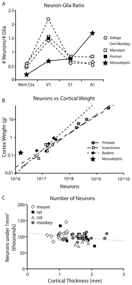
In the following discussion we provide comparative evidence indicating that there is a non-uniformity in the cellular composition of the neocortex within and across species. This is not surprising, because our own and other laboratories have used multiple criteria to subdivide the neocortex in a variety of species and have found similar features of organization as well as clear heterogeneity in structure, function, and connectivity. What is extraordinary is that despite years of debate, there is still no consensus on this fundamental issue in neuroscience. In 1980, Rockel, Hiorns, and Powell used stereological techniques to count the absolute number of cells in a specific volume of tissue in the brains of rats, cats, humans, and several non-human primates. These investigators sampled six cortical regions, including the frontal, parietal, temporal, motor, and somatosensory areas, as well as area 17, which is coextensive with the primary visual area. They found that, with the exception of area 17 in primates, the number of cells did not differ between cortical areas or mammalian species. The thickness of cortical layers did vary between species, which led to the conclusion that cells were more densely packed in smaller brains than in larger brains. These results (excluding the area 17 analysis) were recently replicated by Carlo and Stevens (2013), and are shown in Figure 5C.
Figure 5.
Comparative studies of cortical cellular composition. A: Changes in the neuron–glia ratio across the cortical sheet. The number of neurons in a specific cortical region was divided by the number of glia (or non-neurons) in that same region. In primates and Monodelphis the neuron–glia ratio was higher in the primary sensory areas than in remaining cortical areas, indicating that the primary sensory areas have a larger number of neurons that are more densely packed than the remaining cortical areas. Within the primary sensory areas, however, the neuron–glia ratio varies between primates and Monodelphis. In all of the primates examined, the ratio is highest in V1 and lower in S1 and A1, but in Monodelphis primary sensory regions the glia–neuron ratio is highest in A1 and lowest in V1. This indicates that the distribution of neurons, and possibly the overall organization and function of the neocortex, fundamentally differs between primates and Monodelphis. B: The number of neurons in both cortical hemispheres versus the weight of both cortical hemispheres in different primates (gray circles), insectivores (white squares), rodents (black diamonds), and Monodelphis (black star). Strikingly, Monodelphis does not appear to conform to the patterns of the other species, instead showing a lower than expected number of cortical neurons for its cortical weight. C: Data adapted from Carlo and Stevens (2012) showing the number of neurons under 1 mm2 of cortical surface as a function of cortical thickness. Neuron counts were taken from mice (diamonds), rats (black squares), cats (triangles), and monkeys (gray circles), and the mean for all data points, represented by the dashed line, is 97,780 neurons per mm2. Carlo and Stevens claim that these data show that the neuronal composition of the cortex does not vary between species, although the exact anatomical location from which each sample was taken was not provided. Data adapted from Carlo and Stevens, (2013), Collins et al. (2010b), Herculano-Houzel et al. (2006, 2011), and Sarko et al. (2009).
Although the data from the original study of Rockel et al. (1980) are intriguing, they did not provide a thorough description of their counting methods. A larger issue, and one that is inherent in many comparative studies, is the accurate and consistent identification of cortical field boundaries within a species, and making valid comparisons of homologous cortical fields across species. Unfortunately, this crucial piece of data was lacking in Rockel et al. (1980), so it is difficult to make valid comparisons with our own, or other modern studies that examine the cellular composition of the neocortex. As described above, the only cortical region they explicitly identified was area 17, which is readily identified with a variety of techniques in most, if not all mammals examined. Furthermore, area 17 contained 2.5 times the number of neurons as any other cortical region. These results comport with recent findings in primates, mice, and now opossums, which show that non-primary and association regions contain a smaller number of neurons than primary sensory areas (Collins et al., 2010a; Herculano-Houzel et al., 2008, 2013).
This important issue of accurately defining cortical field boundaries within and across species is raised in the more recent study by Carlo and Stevens (2013). However their resolution of this issue is a post hoc statistical analysis rather than a more direct anatomical method of cortical field segregation. Their analysis assumes that the composition of a given cortical area will be homogenous, and any variation between counting columns will be normally distributed. They took multiple samples from a given cortical region and calculated the variance of those samples. They then compared the observed variance with a predicted variance distribution. If counting columns with non-homogenous properties were grouped together, the observed variance would be greater than the predicted variance. Because, in all cases, the distribution of the predicted and observed variance matched, Carlo and Stevens concluded that their identification of cortical areas was correct. Ultimately, regardless of the method by which areal boundaries were confirmed, the important point is that cortical areas be accurately and consistently defined both within and across species.
There have been other studies that used similar stereological methods to examine differences in neuronal density across the cortical sheet; however, counter to the studies described above, these studies report that the neocortex does not exhibit uniform neuronal density (Beaulieu and Colonnier, 1989; Charvet et al., 2013; Collins, 2011; Collins et al., 2010a; Herculano-Houzel et al., 2013; Leuba and Garey, 1989; Ribeiro et al., 2013; Schuz and Palm, 1989). Although they used different methodologies, ranging from traditional stereology (Beaulieu and Colonnier, 1989; Charvet et al., 2013; Leuba and Garey, 1989; Schuz and Palm, 1989) to isotropic fractionation (Collins et al., 2010a; Herculano-Houzel et al., 2013; Ribeiro et al., 2013), and examined different species, including mice (Herculano-Houzel et al., 2013; Schuz and Palm, 1989), cats (Beaulieu and Colonnier, 1989), non-human primates (Charvet et al., 2013; Collins et al., 2010a), and humans (Leuba and Garey, 1989; Ribeiro et al., 2013), each of these studies reached the same general conclusion: cellular composition varies between different cortical regions. Significantly, each of these studies also found that the primary sensory areas (especially V1) had a greater neuronal density than nonprimary sensory areas.
Recently, the isotropic fractionator technique has been used to investigate the cellular composition of large heterogeneous brain regions, but to date only two other studies have examined differences in neuronal density in different portions of the cortical sheet. Collins and colleagues (2010a) examined the number of total cells, neurons, and neuronal density in the neocortex of four primate species including galagos, owl monkeys, macaques, and baboons. In all cases they found a great deal of variation in the number and density of neurons across the cortex, and in all cases the neuronal density was greatest in visual areas, followed by S1 and A1, and lowest in the other cortical areas. Similarly, Herculano-Houzel and colleagues (2013) examined the distribution of neurons across the cerebral cortex of the mouse. They found that, much like in primates, visual areas exhibited the highest neuronal density, followed by sensory areas and remaining cortical regions.
Interestingly, the data presented in the present study indicate that the conclusion of homogeneity versus heterogeneity depends on the metric examined. For example, we saw no difference in cellular density or the total number of neurons in the primary versus nonprimary areas of cortex. However, there were fewer non-neurons in primary areas, and a greater neuron/glia cell ratio in primary areas (particularly A1) in opossums compared with the remaining cortex (Fig. 5A). Furthermore, A1 had a larger percentage of neurons as well as a greater neuronal density, and a similar trend was observed in V1 and S1.
Comparative analysis of neuron number and density across species
In addition to similarities and differences across the cortical sheet within a species, different species also show tremendous variability in neocortical cellular composition (Figs. 5B, 6). For example, the combined weight of the cerebral hemispheres in the short-tailed opossum (0.15 g) is similar to that of the mouse (0.17 g), naked mole rat (0.18 g), and short-tailed shrew (0.18 g; Fig 6B) (Herculano-Houzel et al., 2011; Sarko et al., 2009). However, the total number of cells in both hemispheres is much lower in the short-tailed opossum (~6 million) than in the mouse (25.8 million), short-tailed shrew (25 million), and naked mole rat (14.5 million; Fig. 6E). Thus, the absolute number of cells (both neurons and non-neurons) is different in animals with comparably sized neocortices.
Figure 6.
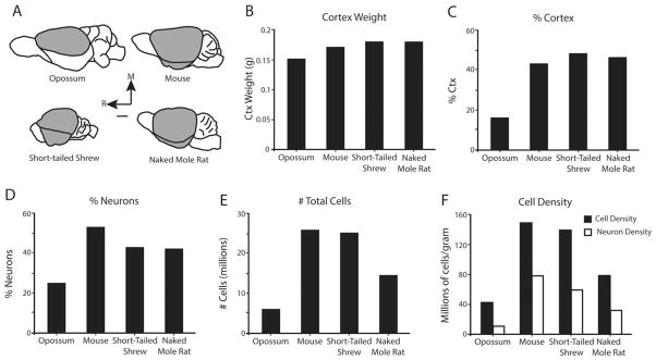
The cellular composition in small mammals with similarly sized cortices. A: Side views of the brains of four small mammals: the short-tailed opossum, mouse, naked mole rat, and short-tailed shrew. In each brain the region studied is shaded in gray. B,C: Although the cortices are all of a similar size (B), the short-tailed opossum neocortex comprises less of the brain than the cortices of the eutherian species (C). D–F: The short-tailed opossum has a lower proportion of cortical neurons (D) and fewer total cells (E) than the mouse, naked mole rat, and short-tailed shrew. The short-tailed opossum accordingly has a lower cellular density (black) and neuronal density (white) than the mouse, naked mole rat, and short-tailed shrew (F). Mouse and naked mole rat data were adapted from Herculano-Houzel et al. (2011), and short-tailed shrew data were adapted from Sarko et al. (2009). Scale bar = 2 mm in A.
In terms of cellular density, the short-tailed opossum neocortex has an overall cellular density of 42 million cells/g, and 11 million neurons/g (Table 5, Fig. 4A,B). In contrast, the mouse neocortex has 149 million cells/g, and 79 million neurons/g, the short-tailed shrew neocortex has 139 million cells/g, and 59 million neurons/g, and the naked mole rat neocortex has 79 million cells/g, and 33 million neurons/g (Fig. 6F). Thus, by all measures the short-tailed opossum has a lower number of cells, lower cellular density, lower proportion of neurons, and higher glia-to-neuron ratio than would be expected for an animal with a comparably sized neocortex (Fig. 5B).
In addition to differences in cellular composition of the neocortex, there are radical differences in the relative size of the neocortex across species. Although the combined weight of both neocortical hemispheres is similar in short-tailed opossums, mice, naked mole rats, and short-tailed shrews, the proportion of the brain comprised by the neocortex is different in different species. In short-tailed opossums, the combined neocortical hemispheres assume 16% of the entire brain (Seelke et al., 2013), 43% in mice, 46% in naked mole rates, and 48% in short-tailed shrew (Fig. 6C) (Herculano-Houzel et al., 2011; Sarko et al., 2009). Thus, despite a similar overall brain size, both the proportion of the brain assumed by the neocortex and the number and composition of cells are different for different species. It has been argued that there are within-order consistencies in cellular composition for rodents, insectivores, and primates (Herculano-Houzel et al., 2006, 2007, 2011; Sarko et al., 2009); however, dramatic differences in cortical sheet size, relative size of cortical fields, and cellular composition are observed between orders.
It should be noted that each study described here isolated the cortex in a slightly different manner. We removed the olfactory bulb, pyriform cortex, and hippocampus to isolate the neocortex. Sarko and her collaborators (2009) excluded the hippocampus and olfactory bulb but included the pyriform cortex in their neocortical analysis, and Herculano-Houzel and colleagues (2011) included everything caudal to the olfactory tract, including the pyriform cortex and hippocampus. However, these methodological differences do not substantially change the overall conclusion that short-tailed opossums’ neocortex contains fewer total cells and fewer neurons, and has a lower cellular and neuronal density and a lower proportion of neurons than other similarly sized animals.
The present study is the most quantitative measure of the number of cortical neurons in a marsupial species to date, and is in agreement with previous observations suggesting that marsupials (including short-tailed opossums) exhibit a low density of cortical neurons compared with other mammals, as well as a lower proportion of neurons, despite a prolonged period of cell division (Cheung et al., 2010; Haug, 1987; Nudo et al., 1995; Saunders et al., 1989). Although this would suggest that short-tailed opossums have a prolonged period of gliogenesis compared with the eutherian mammals studied, short-tailed opossums also have a low number and density of non-neuronal cells compared with other mammals (Collins et al., 2010a; Herculano-Houzel et al., 2011; Sarko et al., 2009). This prolonged period of cell division could be due to a number of factors such as a slower rate of cell division, fewer numbers of cells that re-enter the cell cycle, reduced numbers of intermediate progenitor cells, or some other feature of cell cycle kinetics that may be different in marsupials than in eutherian mammals.
The lower number of neurons and glia, as well as the smaller proportion of neurons observed in opossums indicates that the metabolic requirements of the marsupial cortex are significantly lower than those of their eutherian counterparts (Attwell and Laughlin, 2001; Belanger et al., 2011; Magistretti, 2006). For example, a single rat cortical neuron consumes 3.42 × 10⁸ molecules of ATP each second to maintain its resting potential, whereas a glial cell consumes 1.02 × 10⁸ molecules of ATP each second (Attwell and Laughlin, 2001). When we apply these numbers to a mouse (Herculano-Houzel et al., 2011), which is a eutherian mammal that has a neocortex of a similar size to that of short-tailed opossums, we find that the neurons and glial cells within the mouse cortex require a total of 59.1 × 10¹⁴ molecules of ATP per second (46.8 × 10¹⁴ for neurons and 12.3 × 10¹⁴ for glial cells). In contrast, the neurons and glial cells within the opossum cortex only consume a total of 5.0 × 10¹⁴ molecules of ATP per second (2.74 × 10¹⁴ for neurons and 2.26 × 10¹⁴ for glial cells), less than 10% of the metabolic requirement of the mouse cortex. This massive difference is due to both the lower number of total cells and the lower proportion of neurons found within the cortex, and clearly has important implications for neural processing and synaptic transmission (Seelke et al., 2013).
If the short-tailed opossum is an accurate reflection of the ancestral state, these data suggest that the neocortex of early mammals was relatively smaller and contained many fewer neurons and relatively more glial cells than modern eutherian mammals. Furthermore, these data imply that both temporal and mechanical aspects of cortical development have been altered in eutherians.
The notion that early mammals had brains that required significantly less energy is intriguing. Early monotremes radiated from the ancestral mammalian branch between 166 and 240 million years ago, and marsupials radiated somewhat later (between 148 and 180 million years ago; (Bininda-Emonds et al., 2007; Cifelli and Davis, 2003; Grutzner et al., 2003; Murphy et al., 2004). At these times the atmospheric oxygen levels were significantly lower and carbon dioxide levels were significantly higher than those found today (Berner, 2006; Rothman, 2002). Under these conditions animals that consumed less oxygen due to a lower metabolic rate would have a competitive advantage. Similarly, animals with a lower metabolic rate would require less food to maintain the body’s basic functions. In fact, monotremes have a lower metabolic rate than marsupials, which have a lower metabolic rate than eutherian mammals (Bennett, 1988). This trait would be particularly adaptive in areas that contain a low availability of food resources. Over the course of time atmospheric oxygen levels increased and resources became more plentiful. During this period the early eutherians underwent a massive evolutionary radiation (Archibald, 2003), and some of these new species evolved higher metabolic rates, which allowed for the expansion of a metabolically expensive structure such as the neocortex. This in turn allowed for a larger repertoire of complex and likely highly adaptive behaviors.
Acknowledgments
Grant sponsor: National Institutes of Health; Grant number: F32 NS064792 (to A.M.H.S.), R21 NS071225 and R01 EY022987 (to L.A.K.), and T32 EY015387 (to J.C.D.).
Thanks to Carol Oxford for her assistance at the UC Davis Flow Cytometry Shared Resource. Thanks also to Cindy Clayton, DVM, and the rest of the animal care staff at the UC Davis Psychology Department Vivarium.
Abbreviations
- A1
primary auditory cortex
- AF 647/700
AlexaFluor 647/700
- DAPI
4′,6-diamidino-2-phenylindole
- NeuN
neuronal nuclear protein
- PBS
phosphate-buffered saline
- Rem Ctx
remaining cortical areas
- S1
primary somatosensory cortex
- V1
primary visual cortex
- ΣCtx
sum of V1, A1, S1, and Rem Ctx
Footnotes
CONFLICT OF INTEREST STATEMENT
The authors have no conflicts of interest.
ROLE OF AUTHORS
All authors had full access to all of the data in the study and take responsibility for the integrity of the data and the accuracy of the data analysis. Study concept and design: AMHS, JCD, LAK. Acquisition of data: AMHS. Analysis and interpretation of data: AMHS. Drafting of the manuscript: AMHS, LAK. Critical revision of the manuscript for important intellectual content: AMHS, JCD, LAK. Statistical analysis: AMHS. Obtained funding: LAK. Administrative, technical, and material support: JCD. Study supervision: LAK.
LITERATURE CITED
- Archibald JD. Timing and biogeography of the eutherian radiation: fossils and molecules compared. Mol Phylogenet Evol. 2003;28:350–359. doi: 10.1016/s1055-7903(03)00034-4. [DOI] [PubMed] [Google Scholar]
- Attwell D, Laughlin SB. An energy budget for signaling in the grey matter of the brain. J Cereb Blood Flow Metab. 2001;21:1133–1145. doi: 10.1097/00004647-200110000-00001. [DOI] [PubMed] [Google Scholar]
- Beaulieu C, Colonnier M. Number of neurons in individual laminae of areas 3B, 4 gamma, and 6a alpha of the cat cerebral cortex: a comparison with major visual areas. J Comp Neurol. 1989;279:228–234. doi: 10.1002/cne.902790206. [DOI] [PubMed] [Google Scholar]
- Belanger M, Allaman I, Magistretti PJ. Brain energy metabolism: focus on astrocyte-neuron metabolic cooperation. Cell Metab. 2011;14:724–738. doi: 10.1016/j.cmet.2011.08.016. [DOI] [PubMed] [Google Scholar]
- Bennett AF. Structural and functional determinates of metabolic rate. Am Zool. 1988;28:699–708. [Google Scholar]
- Berner RA. GEOCARBSULF: a combined model for Phanerozoic atmospheric O2 and CO2. Geochim Cosmochim Acta. 2006;70:5653–5664. [Google Scholar]
- Bininda-Emonds OR, Cardillo M, Jones KE, MacPhee RD, Beck RM, Grenyer R, Price SA, Vos RA, Gittleman JL, Purvis A. The delayed rise of present-day mammals. Nature. 2007;446:507–512. doi: 10.1038/nature05634. [DOI] [PubMed] [Google Scholar]
- Campi KL, Collins CE, Todd WD, Kaas J, Krubitzer L. Comparison of area 17 cellular composition in laboratory and wild-caught rats including diurnal and nocturnal species. Brain Behav Evol. 2011;77:116–130. doi: 10.1159/000324862. [DOI] [PMC free article] [PubMed] [Google Scholar]
- Carlo CN, Stevens CF. Structural uniformity of neocortex, revisited. Proc Natl Acad Sci U S A. 2013;110:1488–1493. doi: 10.1073/pnas.1221398110. [DOI] [PMC free article] [PubMed] [Google Scholar]
- Catania KC, Jain N, Franca JG, Volchan E, Kaas JH. The organization of somatosensory cortex in the short-tailed opossum (Monodelphis domestica) Somatosens Motor Res. 2000;17:39–51. doi: 10.1080/08990220070283. [DOI] [PubMed] [Google Scholar]
- Charvet CJ, Cahalane DJ, Finlay BL. Systematic, cross-cortex variation in neuron numbers in rodents and primates. Cereb Cortex. 2013 doi: 10.1093/cercor/bht214. [DOI] [PMC free article] [PubMed] [Google Scholar]
- Cheung AF, Kondo S, Abdel-Mannan O, Chodroff RA, Sirey TM, Bluy LE, Webber N, DeProto J, Karlen SJ, Krubitzer L, Stolp HB, Saunders NR, Molnar Z. The subventricular zone is the developmental milestone of a 6-layered neocortex: comparisons in metatherian and eutherian mammals. Cereb Cortex. 2010;20:1071–1081. doi: 10.1093/cercor/bhp168. [DOI] [PubMed] [Google Scholar]
- Cifelli RL, Davis BM. Paleontology. Marsupial origins. Science. 2003;302:1899–1900. doi: 10.1126/science.1092272. [DOI] [PubMed] [Google Scholar]
- Collins CE. Variability in neuron densities across the cortical sheet in primates. Brain Behav Evol. 2011;78:37–50. doi: 10.1159/000327319. [DOI] [PubMed] [Google Scholar]
- Collins CE, Airey DC, Young NA, Leitch DB, Kaas JH. Neuron densities vary across and within cortical areas in primates. Proc Natl Acad Sci U S A. 2010a;107:15927–15932. doi: 10.1073/pnas.1010356107. [DOI] [PMC free article] [PubMed] [Google Scholar]
- Collins CE, Young NA, Flaherty DK, Airey DC, Kaas JH. A rapid and reliable method of counting neurons and other cells in brain tissue: a comparison of flow cytometry and manual counting methods. Front Neuroanat. 2010b;4:5. doi: 10.3389/neuro.05.005.2010. [DOI] [PMC free article] [PubMed] [Google Scholar]
- DeFelipe J, Alonso-Nanclares L, Arellano JI. Microstructure of the neocortex: comparative aspects. J Neurocytol. 2002;31:299–316. doi: 10.1023/a:1024130211265. [DOI] [PubMed] [Google Scholar]
- Frost SB, Milliken GW, Plautz EJ, Masterton RB, Nudo RJ. Somatosensory and motor representations in cerebral cortex of a primitive mammal (Monodelphis domestica): a window into the early evolution of sensorimotor cortex. J Comp Neurol. 2000;421:29–51. doi: 10.1002/(sici)1096-9861(20000522)421:1<29::aid-cne3>3.0.co;2-9. [DOI] [PubMed] [Google Scholar]
- Grutzner F, Deakin J, Rens W, El-Mogharbel N, Marshall Graves JA. The monotreme genome: a patchwork of reptile, mammal and unique features? Comp Biochem Physiology A. 2003;136:867–881. doi: 10.1016/j.cbpb.2003.09.014. [DOI] [PubMed] [Google Scholar]
- Haug H. Brain sizes, surfaces, and neuronal sizes of the cortex cerebri: a stereological investigation of man and his variability and a comparison with some mammals (primates, whales, marsupials, insectivores, and one elephant) Am J Anat. 1987;180:126–142. doi: 10.1002/aja.1001800203. [DOI] [PubMed] [Google Scholar]
- Herculano-Houzel S, Mota B, Lent R. Cellular scaling rules for rodent brains. Proc Natl Acad Sci U S A. 2006;103:12138–12143. doi: 10.1073/pnas.0604911103. [DOI] [PMC free article] [PubMed] [Google Scholar]
- Herculano-Houzel S, Collins CE, Wong P, Kaas JH. Cellular scaling rules for primate brains. Proc Natl Acad Sci U S A. 2007;104:3562–3567. doi: 10.1073/pnas.0611396104. [DOI] [PMC free article] [PubMed] [Google Scholar]
- Herculano-Houzel S, Collins CE, Wong P, Kaas JH, Lent R. The basic nonuniformity of the cerebral cortex. Proc Natl Acad Sci U S A. 2008;105:12593–12598. doi: 10.1073/pnas.0805417105. [DOI] [PMC free article] [PubMed] [Google Scholar]
- Herculano-Houzel S, Ribeiro P, Campos L, Valotta da Silva A, Torres LB, Catania KC, Kaas JH. Updated neuronal scaling rules for the brains of Glires (rodents/lagomorphs) Brain Behav Evol. 2011;78:302–314. doi: 10.1159/000330825. [DOI] [PMC free article] [PubMed] [Google Scholar]
- Herculano-Houzel S, Watson C, Paxinos G. Distribution of neurons in functional areas of the mouse cerebral cortex reveals quantitatively different cortical zones. Front Neuroanat. 2013;7:35. doi: 10.3389/fnana.2013.00035. [DOI] [PMC free article] [PubMed] [Google Scholar]
- Hutsler JJ, Lee DG, Porter KK. Comparative analysis of cortical layering and supragranular layer enlargement in rodent carnivore and primate species. Brain Res. 2005;1052:71–81. doi: 10.1016/j.brainres.2005.06.015. [DOI] [PubMed] [Google Scholar]
- Kaas JH. What, if anything, is S1? Organization of first somatosensory area of the cortex. Physiol Rev. 1983;63:206–231. doi: 10.1152/physrev.1983.63.1.206. [DOI] [PubMed] [Google Scholar]
- Kaas JH. Reconstructing the areal organization of the neocortex of the first mammals. Brain Behav Evol. 2011;78:7–21. doi: 10.1159/000327316. [DOI] [PMC free article] [PubMed] [Google Scholar]
- Kahn DM, Huffman KJ, Krubitzer L. Organization and connections of V1 in Monodelphis domestica. J Comp Neurol. 2000;428:337–354. [PubMed] [Google Scholar]
- Karlen SJ, Krubitzer L. The functional and anatomical organization of marsupial neocortex: evidence for parallel evolution across mammals. Prog Neurobiol. 2007;82:122–141. doi: 10.1016/j.pneurobio.2007.03.003. [DOI] [PMC free article] [PubMed] [Google Scholar]
- Karlen SJ, Kahn DM, Krubitzer L. Early blindness results in abnormal corticocortical and thalamocortical connections. Neuroscience. 2006;142:843–858. doi: 10.1016/j.neuroscience.2006.06.055. [DOI] [PubMed] [Google Scholar]
- Krubitzer L. In search of a unifying theory of complex brain evolution. Ann N Y Acad Sci. 2009;1156:44–67. doi: 10.1111/j.1749-6632.2009.04421.x. [DOI] [PMC free article] [PubMed] [Google Scholar]
- Krubitzer L, Manger P, Pettigrew J, Calford M. Organization of somatosensory cortex in monotremes: in search of the prototypical plan. J Comp Neurol. 1995;351:261–306. doi: 10.1002/cne.903510206. [DOI] [PubMed] [Google Scholar]
- Leuba G, Garey LJ. Comparison of neuronal and glial numerical density in primary and secondary visual cortex of man. Exp Brain Res. 1989;7:31–38. doi: 10.1007/BF00250564. [DOI] [PubMed] [Google Scholar]
- Magistretti PJ. Neuron-glia metabolic coupling and plasticity. J Exp Biol. 2006;209:2304–2311. doi: 10.1242/jeb.02208. [DOI] [PubMed] [Google Scholar]
- Morest DK, Silver J. Precursors of neurons, neuroglia, and ependymal cells in the CNS: what are they? Where are they from? How do they get where they are going? Glia. 2003;43:6–18. doi: 10.1002/glia.10238. [DOI] [PubMed] [Google Scholar]
- Mouton PR. Principles and practices of unbiased stereology: an introduction for bioscientists. Baltimore, MD: Johns Hopkins University Press; 2002. [Google Scholar]
- Murphy WJ, Pevzner PA, O’Brien SJ. Mammalian phylogenomics comes of age. Trends Genet. 2004;20:631–639. doi: 10.1016/j.tig.2004.09.005. [DOI] [PubMed] [Google Scholar]
- Nudo RJ, Sutherland DP, Masterton RB. Variation and evolution of mammalian corticospinal somata with special reference to primates. J Comp Neurol. 1995;358:181–205. doi: 10.1002/cne.903580203. [DOI] [PubMed] [Google Scholar]
- Prothero J. Scaling of cortical neuron density and white matter volume in mammals. J Hirnforsch. 1997;38:513–524. [PubMed] [Google Scholar]
- Ribeiro PF, Ventura-Antunes L, Gabi M, Mota B, Grinberg LT, Farfel JM, Ferretti-Rebustini RE, Leite RE, Filho WJ, Herculano-Houzel S. The human cerebral cortex is neither one nor many: neuronal distribution reveals two quantitatively different zones in the gray matter, three in the white matter, and explains local variations in cortical folding. Front Neuroanat. 2013;7:28. doi: 10.3389/fnana.2013.00028. [DOI] [PMC free article] [PubMed] [Google Scholar]
- Rockel AJ, Hiorns RW, Powell TP. The basic uniformity in structure of the neocortex. Brain. 1980;103:221–244. doi: 10.1093/brain/103.2.221. [DOI] [PubMed] [Google Scholar]
- Rothman DH. Atmospheric carbon dioxide levels for the last 500 million years. Proc Natl Acad Sci U S A. 2002;99:4167–4171. doi: 10.1073/pnas.022055499. [DOI] [PMC free article] [PubMed] [Google Scholar]
- Sarko DK, Catania KC, Leitch DB, Kaas JH, Herculano-Houzel S. Cellular scaling rules of insectivore brains. Front Neuroanat. 2009;3:8. doi: 10.3389/neuro.05.008.2009. [DOI] [PMC free article] [PubMed] [Google Scholar]
- Saunders NR, Adam E, Reader M, Mollgard K. Monodelphis domestica (grey short-tailed opossum): an accessible model for studies of early neocortical development. Anat Embryol. 1989;180:227–236. doi: 10.1007/BF00315881. [DOI] [PubMed] [Google Scholar]
- Schuz A, Palm G. Density of neurons and synapses in the cerebral cortex of the mouse. J Comp Neurol. 1989;286:442–455. doi: 10.1002/cne.902860404. [DOI] [PubMed] [Google Scholar]
- Seelke AMH, Dooley JC, Krubitzer L. Differential changes in the cellular composition of the developing marsupial brain. J Comp Neurol. 2013;52:2602–2620. doi: 10.1002/cne.23301. [DOI] [PMC free article] [PubMed] [Google Scholar]
- Temple S. The development of neural stem cells. Nature. 2001;414:112–117. doi: 10.1038/35102174. [DOI] [PubMed] [Google Scholar]


