

Summary
Lipid metabolism in Trypanosoma brucei, the causative agent of African sleeping sickness, differs from its human host in several fundamental ways. This has lead to the validation of a plethora of novel drug targets, giving hope of novel chemical intervention against this neglected disease. Cytidine diphosphate diacylglycerol (CDP‐DAG) is a central lipid intermediate for several pathways in both prokaryotes and eukaryotes, being produced by CDP‐DAG synthase (CDS). However, nothing is known about the single T. brucei CDS gene (Tb927.7.220/EC 2.7.7.41) or its activity. In this study we show TbCDS is functional by complementation of a non‐viable yeast CDS null strain and that it is essential in the bloodstream form of the parasite via a conditional knockout. The TbCDS conditional knockout showed morphological changes including a cell‐cycle arrest due in part to kinetoplast segregation defects. Biochemical phenotyping of TbCDS conditional knockout showed drastically altered lipid metabolism where reducing levels of phosphatidylinositol detrimentally impacted on glycoylphosphatidylinositol biosynthesis. These studies also suggest that phosphatidylglycerol synthesized via the phosphatidylglycerol‐phosphate synthase is not synthesized from CDP‐DAG, as was previously thought. TbCDS was shown to localized the ER and Golgi, probably to provide CDP‐DAG for the phosphatidylinositol synthases.
Introduction
Trypanosoma brucei, the causative agent of African sleeping sickness, expresses a variant surface glycoprotein (VSG) coat, which protects the parasite from the alternative complement pathway and from a specific immune response by antigenic variation (Cross, 1996). The 5 × 10⁶ VSG dimers are held in the outer leaflet plasma membrane of the parasite by glycosylphosphatidylinositol (GPI) anchors. The biosynthesis of this conserved GPI anchor has been previously genetically (Nagamune et al., 2000; Chang et al., 2002) and chemically validated (Smith et al., 2004) as a drug target in bloodstream form T. brucei. The importance of GPI anchors to T. brucei has lead our group to investigate various aspects of lipid metabolism, some enzymes of which have already been shown to be essential (reviewed in Smith and Buetikofer, 2010). The nucleolipid cytidine diphosphate diacylglycerol (CDP‐DAG) is a key metabolite, acting as a high energy donor in the synthesis of numerous phospholipids in both prokaryotes and eukaryotes. In T. brucei these include synthesis of phosphatidylinositol (PI), which is essential for the production of GPI anchors (Martin and Smith, 2006b), and potentially phosphatidyglycerol (PG), cardolipin (CL) and phosphatidylserine (PS). CDP‐DAG is synthesized by the enzyme CDP‐DAG synthase (CDS) (EC 2.7.7.41) from cytidine triphosphate (CTP) and phosphatidic acid (PA) (Fig. 1A).
Figure 1.
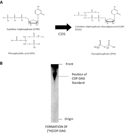
CDP‐DAG synthase activity.
A. Schematic of the enzymatic reaction catalysed by CDP‐DAG synthase.
B. TbCDS assay of T. brucei lysate, autoradiography of a representative HPTLC of lipid fraction shows [3H]CDP‐DAG formation from [3H]CTP and PA. The position of unlabelled CDP‐DAG standard run in the same solvent system and visualized by iodine is indicated on the right hand side.
A gene encoding CDS activity was first discovered in E. coli mutants deficient in CDP‐DAG synthesis (Ganong et al., 1980; Ganong and Raetz, 1982). In Drosophila a photoreceptor CDS is involved in phosphoinositide signalling and vision (Wu et al., 1995), which increased interest in the gene and led to its cloning and study in yeast (Shen et al., 1996) and a variety of mammals (Heacock et al., 1996; Saito et al., 1997; Halford et al., 1998; Volta et al., 1999; Inglis‐Broadgate et al., 2005). CDS homologues have now been identified in all eukaryotic genomes sequenced to date and some organisms have multiple CDS genes (Lykidis, 2007). The genes show high sequence similarity, suggesting a degree of conservation during evolution. All identified CDS enzymes are integral membrane proteins. In prokaryotes, CDS activity is associated with the inner membrane of the cell envelope (Bell et al., 1971; White et al., 1971) and in eukaryotes the majority of activity has been found in the ER and mitochondria (Bishop and Strickland, 1976; Kuchler et al., 1986; Kelley and Carman, 1987; Mok et al., 1992; Heacock and Agranoff, 1997).
The fact that the pool of CDP‐DAG is much smaller than that of CDS's lipid substrate PA (Raetz and Kennedy, 1973; Thompson and MacDonald, 1975; Nickels et al., 1994) along with the position of CDS at a branch point in phospholipid synthesis suggests CDP‐DAG synthesis may be a rate‐limiting step. In yeast, CDS is under tight transcriptional regulation via the inositol‐responsive cis‐acting element and trans‐acting factors (reviewed in Carman and Han, 2011), but in other organisms regulation varies greatly (Carter and Kennedy, 1966; Sribney et al., 1977; Langley and Kennedy, 1978; Ganong and Raetz, 1982; Baelee and Carman, 1984; Icho et al., 1985; Kelley and Carman, 1987; Carman and Baelee, 1992; Mok et al., 1992; Shen and Dowhan, 1996; Lykidis et al., 1997; Saito et al., 1997; Sato et al., 2000; Haselier et al., 2010).
CDS null mutants in yeast (Shen and Dowhan, 1996; Shen et al., 1996), E. coli (Ganong and Raetz, 1982) and Synechocystis (Sato et al., 2000) are inviable due to massive direct and indirect disruption of phospholipid synthesis. Due to the essential requirement for PI (Martin and Smith, 2006a,b), phosphorylated PIs (PIPs) (Rodgers et al., 2007) and GPI anchors (Nagamune et al., 2000; Chang et al., 2002; Smith et al., 2004) in T. brucei, it seemed likely that CDS would be essential for the parasite, therefore presenting a possible novel drug target. In order to validate CDS as a drug target we created a conditional knockout in the bloodstream form parasite, which has highlighted several novel aspects of phospholipid metabolism in this early divergent eukaryotic parasite.
Results
Cloning and sequencing T. brucei CDS (TbCDS)
A putative CDS was identified in the T. brucei genome database (Sanger Centre) using Blast (Tb927.2.220). This putative ORF was PCR amplified, cloned and sequenced. The sequence and its translation are shown in Fig. S1. An alignment of the predicted translated sequence with CDSs from other organisms is shown in Fig. S2A, with a corresponding phylogenetic tree in Fig. S2B. Unsurprisingly, TbCDS is closely related to other kinetoplastids, showing 99% identity with the T. b. brucei strain 927 and T. b. gambiense; 64% with T. cruzi, 60% with T. vivax and 49–50% with various Leishmania species, while significantly less to the two human CDS homologues. The size of the predicted TbCDS protein is 46 kDa, which is in good agreement with other eukaryotic CDSs such as Arabidopsis thaliana CDS5 at 39.9 kDa (Haselier et al., 2010) and the human CDS1 at 46.1 kDa (Heacock et al., 1996).
TbCDS is highly hydrophobic with a grand average of hydropathicity of 0.285 (Kyte and Doolittle, 1982) with 7 predicted transmembrane domains (highlighted in Fig. S1). This is consistent with all identified CDS enzymes which are integral membrane proteins with multiple membrane spanning domains (Carman and Kelley, 1992; Heacock et al., 1996; Saito et al., 1997; Volta et al., 1999; Martin et al., 2000; Nigou and Besra, 2002). The TbCDS protein contains the typical phosphatidate cytidyltransferase signature: S‐x‐[LIVMF]‐K‐R‐x(4)‐K‐D‐[GSA]‐x(2)‐[LIF]‐[PGS]‐x‐H‐G‐G‐[LIVMF]‐x‐D‐R‐[LIVMFT]‐D (highlighted yellow in Fig. S1A), which is shared by almost all predicted homologues, both eukaryotic and prokaryotic (Koonin, 1996; Ponting and Kerr, 1996).
BLAST searches using typical prokaryotic CDS sequences recovered a putative second CDS gene in Trypanosoma cruzi (Tc00.1047053508707.140 /TcCLB.508707.140), Trypanosoma vivax (TvY486_0025800) and several Leishmania species (LinJ.32.3010, LmjF.32.2870, LmxM.31.2870, LtaP32.3050) but not in T. brucei. All of these genes are more similar to the prokaryotic than the eukaryotic CDS genes. An alignment of these predicted proteins with some typical prokaryotic CDS sequences are shown in Fig. S3A with a corresponding phylogenetic tree in Fig. S3B. This shows that the protozoan homologous have N‐terminal extensions as well as several insertions, making these gene products significantly larger than the average prokaryotic CDS (Agabian, 1990).
It is assumed that organisms that have multiple copies of CDS, i.e. humans and Arabidopsis, target them to different cellular locations, providing local concentrated pools of CDP‐DAG to be used by different phospholipid pathways. This may also be true for T. cruzi and Leishmania that have both a eukaryotic and prokaryotic version, but T. brucei have likely lost their prokaryotic homologue in the recent past.
Rescue of yeast of CDS null
To confirm that the T. brucei CDS homologue encoded for a functional CDS, it was used to rescue viability of an otherwise inviable yeast cds1 null strain. A schematic of the strategy used is shown in Fig. S4. Briefly, a diploid yeast strain heterozygous for a cds1 gene knockout (CDS1/cds1::KanMX) containing yeast CDS1 on the URA3‐marked pRS426‐MET25 plasmid was used to generate haploid cds1 knockout strains that were complemented by the plasmid. As yeast CDS1 is an essential gene, loss of this plasmid resulted in lethality so these strains could not grow in the presence of 5‐fluoro‐orotic acid (5‐FOA), which selects against the URA3 marker on the plasmid (Fig. S5 – compare A and B). Using a second, LEU2‐marked plasmid (p405‐TEF1), either TbCDS or yeast CDS1 were introduced into this background. These strains, along with the parent strain and transformants carrying the empty p405‐TEF1 vector, were tested for growth after counterselection for pRS426‐MET25‐CDS1 on 5‐FOA. Only the strains containing either yeast CDS1 or TbCDS on the LEU2 selectable plasmid were viable, while the parent strain with and without the empty plasmid was non‐viable (compare Fig. 2A and B). Lipidomic analysis by electrospray mass spectrometry of the yeast parent strain and the CDS1 null expressing either the ectopic yeast CDS1 or TbCDS on a plasmid showed alterations in their phospholipid profiles. This may reflect a different acyl preference on the substrate PA (Fig. S6A–C) resulting in slightly different CDP‐DAG species being formed and thus variation in the relative ratio of the PI species formed (compare species at 808 m/z and 836 m/z), which may impact on inositolphosphoceramide (IPC) formation and thus lipid homeostasis in the resulting yeast strains (Carman and Fischl, 1992). Collectively, this functional complementation provides definitive evidence that TbCDS encodes a functional CDS enzyme as it is able to rescue an otherwise non‐viable yeast cds1 null mutant.
Figure 2.
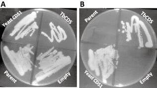
Plasmid shuffle shows TbCDS can complement the yeast cds1::KanMX knockout.
A. After transformation of haploid cds1::KanMX [pRS425‐MET25‐CDS1] with empty p405‐TEF1, p405‐TEF1‐TbCDS or p405‐TEF1‐CDS1 and selection for both plasmids, the three strains plus the cds1::KanMX [pRS425‐MET25‐CDS1] parent strain were plated on YPD, where all strains grew normally.
B. Strains were then plated onto YPD +5‐FOA in order to select against the pRS425‐MET25‐CDS1 plasmid. Parent = cds1::KanMX [pRS425‐MET25‐CDS1]; Empty = Parent transformed with p405‐TEF1; Yeast = Parent transformed with p405‐TEF1‐CDS1; TbCDS = parent transformed with p405‐TEF1‐TbCDS.
Is CDS essential in bloodstream form T. brucei?
According to the TriTrypDB, the TbCDS (Tb427.07.220) is expressed equally throughout the various life cycles stages, and this was confirmed in strain 427 by RT‐PCR in both the bloodstream and procyclic forms (Fig. S7). In addition, washed bloodstream T. brucei membranes exhibited CDS activity, as they were able to catalyse the conversion of [3H]CTP and PA into [3H]CDP‐DAG product (Fig. 1B).
To ascertain if TbCDS is an essential gene in T. brucei, a conditional TbCDS knockout was created. As the ‘wild type’ cell line used here constitutively expresses the T7 RNA polymerase and the tetracycline repressor protein, a tetracycline inducible (Ti) haemagglutinin (HA)‐tagged ectopic copy of the TbCDS was introduced (pLew 100) into the cell line prior to deletion of both TbCDS alleles in the presence of tetracycline, allowing the creation of a conditional double knockout (Wirtz et al., 1999). The resulting TbCDS‐HATi ΔTbCDS::PAC/ΔTbCDS::HYG clone was obtained and the genotype of the cell‐line confirmed by PCR (Fig. S8A), while expression of the ectopic TbCDS copy was confirmed as being tetracycline inducible by anti‐HA Western blot (Fig. S8B). Please note, the apparent mobility of CDS anti‐HA band is significantly higher than the predicted ∼ 47 kDa, as is commonly seen for multiple transmembrane domain proteins on SDS‐PAGE.
To establish whether CDS is an essential gene, the growth of the TbCDS‐HATi ΔTbCDS::PAC/ΔTbCDS::HYG (TbCDS CKO) cell line was monitored in tetracycline‐free HMI‐9 in the absence or presence of tetracycline (Fig. 3A and B). In the absence of tetracycline a growth defect was apparent after 24 h, and after 48 h cells started to die and no visible TbCDS‐HA protein was observed (Fig. 3C, lane 1). By 100 h the cell numbers were below the limits of detection by light microscopy, however, some live cells were visible after 120 h, following which growth resumed as normal. The growth pattern observed was highly reproducible. This revertant growth was likely due to a loss of tetracycline control, as has previously been shown in knockouts of essential genes (Chang et al., 2002; Martin and Smith, 2006a), and was confirmed by anti‐HA Western blot showing production of the HA‐tagged TbCDS in the absence of tetracycline (Fig. 3C, lane 3).
Figure 3.
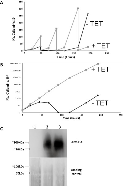
Growth curves and Western blot of the TbCDS conditional double knockout cell line. Cells were washed with tetracycline‐free HMI‐9, transferred to tetracycline‐free media and counted daily. Growth curves are shown for wild type cells in this media.
A. Growth curves of T. brucei CKO in the absence (black/−TET) or presence (grey/+TET) of tetracycline (A) actual number of cells per ml of culture × 104 after number of hours cultured in the absence (black/−TET) or presence (grey/+TET) of tetracycline.
B. Growth curves of log cumulative number of cells over time in the presence and absence of tetracycline. These growth curves were done numerous times and were highly reproducible.
C. Anti‐HA western of T. brucei CKO either after 48 h without tetracycline (lane 1), or with tetracycline (lane 2), or 150 h without tetracycline (lane 3). Reoccurrence of HA‐tagged TbCDS despite the absence of tetracycline suggests regrowth is due to cells reverting and breaking the tetracycline control of TbCDS expression. Loading control is ponceau stained Western blotted membrane.
Morphological phenotype of the CDS conditional knockout
There were morphological changes in the TbCDS CKO cell line after 42 h in the absence of tetracycline compared with wild type cells (Fig. 4). In many of the trypanosomes there was an apparent cell cycle arrest after replication of the flagellum and the formation of a furrow. A pre‐cytokinesis stall is not uncommon in T. brucei mutants, but the stall usually occurs before furrow formation (Hammarton et al., 2007). DAPI staining revealed that in most cases these cells had stalled at 1K2N, often with enlarged kinetoplast and nuclei. This 1K2N phenotype is surprising given the kinetoplast replicates prior to the nucleus and suggests a defect in kinetoplastid segregation. This downstream affect of depleted CDP‐DAG levels suggests key structural and/or signalling phospholipids may play an important part in correct kinteoplastid DNA replication/segregation. This could be at the tripartite attachment zone, where the basal body is physically attached to the kinetoplast, passing through both the cell and mitochondrial membranes. A very similar, but not identical phenotype was observed in the knock‐down of T. brucei MOB1, which caused a stall in cytokinesis through an inhibitory effect on kinetoplast segregation (Hammarton et al., 2005).
Figure 4.
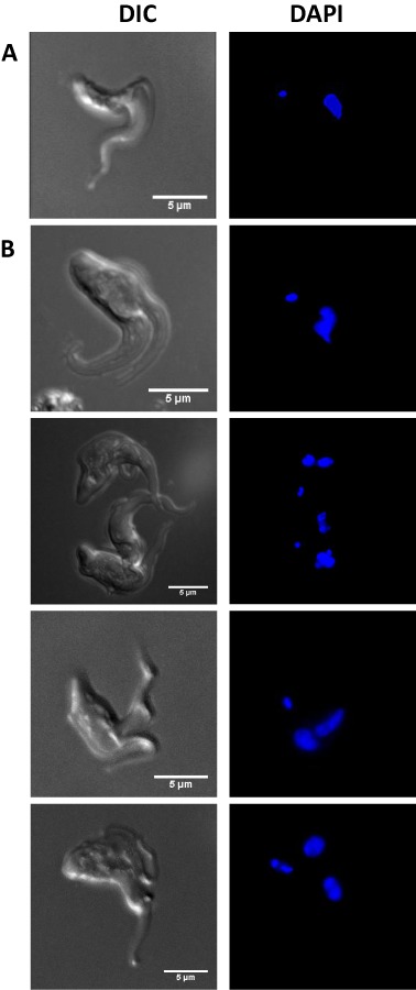
Comparison of wild‐type and TbCDS CKO cell morphology. Projections of Z‐series images taken by light microscopy. Left panel shows DIC and right panel shows DAPI staining and. Cells were fixed with 4% paraformaldehyde, allowed to adhere to poly‐lysine coated slides and mounted with anti‐fade GOLD reagent containing DAPI.
A. Wild‐type cells harvested from mid log culture and fixed in 4% paraformaldehyde.
B. TbCDS CKO cells harvested from culture 48 h after the removal of tetracycline.
Biochemical phenotype of the TbCDS conditional knockout
Total lipids were extracted from TbCDS CKO cells grown in the absence (42 h) or presence of tetracycline and analysed by electrospray‐mass spectrometry. As expected, lipid extracts from TbCDS CKO grown in the presence of tetracycline have a similar distribution of molecular species to that of wild‐type cells across the phospholipid classes, PI, PA, PG, PS, PE, PC and sphingolipids to that of wild‐type cells (data not shown). Comparison of the positive ion mode survey scans of TbCDS CKO in the presence and absence of tetracycline shows the relative proportion of the PC and SM remain relatively unchanged (Fig. 5A and B). Some notable differences were an increase in DAG 36:2–4 (615.5 m/z) and an increase in TAG C62:7–9 (987.6 m/z) (Fig. 5B). The negative ion mode survey scans of TbCDS CKO in the presence and absence of tetracycline show dominant ions at 727.1 and 774.1 m/z (Fig. 5C and D) corresponding to the major plasminogen PE (a‐36:1) and PS (a‐36:1) respectively. In the absence of tetracycline two species at 686.1 and 700.0 m/z identified by fragmentation as PA a‐C18:0/C18:2 (Fig. S9A), and its diacyl counterpart PA C18:0/C18:2 (Fig. S9B) are both absent compared with cells grown the presence of tetracycline. These reductions coincide with a significant increase in several PG species including those at 760.0 and 825.9 m/z identified as including a‐C18:0/C18:2 and C18:0/C22:4 respectively (Fig. S9C and D).
Figure 5.
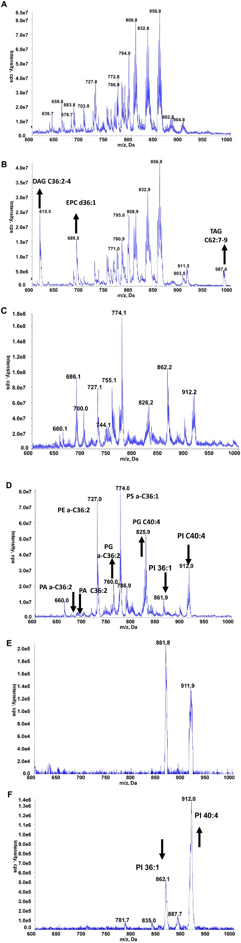
Lipidomic analyses by electrospray mass spectrometry.
A and B. Mass spectrometric analyses: Positive ion survey of total phospholipids from wild‐type (A) and TbCDS CKO in absence of tetracycline for 42 h (B).
C–F. Negative ion survey of total phospholipids from wild‐type (C) and TbCDS CKO cells in absence of tetracycline for 42 h (D). Parent‐ion scanning of the collision induced fragment at 241 m/z of wild‐type single marker cells (E) and TbCDS CKO cells in absence of tetracycline for 42 h (F). The data presented here are representative scans from one set of samples, but these analyses were carried out on several different samples along with their corresponding controls.
Finally, the absence of tetracycline gives rise to a significant decrease in the major PI species at 861 and 912 m/z in corresponding to C36:1 and C40:4 respectively (Fig. 5C and D), with a disproportional reduction between these major PI species. To investigate this further, parent‐ion scanning of m/z 241 (collision‐induced fragmentation, forming inositol‐1,2‐cyclic phosphate) in negative ion mode which only shows the PI species (Fig. 5E and F) allowed the relative ratio to be calculated, showing a decrease from 1.29 to 0.38 (861 to 912 m/z). Quantification of the total myo‐inositol containing phospholipids by GC‐MS showed that in the absence of tetracycline there was only 57 ± 6% myo‐inositol containing lipids compared with cells grown in the presence of tetracycline.
The decrease in CDS activity obviously causes a marked reduction in PI levels, however the disproportional reduction implies the C36:1 species turns over faster than the C40:4 species. No significant disproportional reduction was observed in our previous work on either INO1 (Martin and Smith, 2006a) or PI synthase (Martin and Smith, 2006b). This study may indicate that that the cellular location of either the synthesis or downstream use of the PI species may be different. Alternatively, it may highlight an issue with availability of certain CDP‐DAG species under conditions of limiting CDS activity.
The biochemical phenotype of the TbCDS CKO cells was also investigated by in vivo radiolabelling experiments with [3H]myristate, [3H]glycerol and [3H]inositol after the cells had been grown either in the presence or absence of tetracycline for 42 h and compared with wild‐type cells. After labelling, lipids were extracted, desalted, separated by high performance thin layer chromatography (HPTLC), and detected by fluorography.
Labelling of the TbCDS CKO cells grown in the absence of tetracycline with [3H]myristate (Fig. S10A) showed no significant decrease to those grown in the presence of tetracycline, although there was a slight increase in the labelling of neutral lipids (diacylglycerol and triacylglycerol), confirming the observed increase in the positive ion mode survey scan (Fig. 5B). This is also a clear indication that at this point under permissive conditions the cells are still metabolically active. The incorporation of [3H]glycerol into different glycerolipids (Fig. S10B) was considerably altered in the TbCDS CKO cells in the absence of tetracycline. There is significantly less labelling of PE and PI in the knockout, but PC seems unaffected, while PG seems to have increased. Surprisingly, the incorporation of [3H]inositol into PI and PIP seems relatively unaffected in the TbCDS CKO (Fig. S10C), despite the [3H]glycerol labelling showing a decrease in newly formed PI. However, this anomaly is likely due to head‐group swapping with pre‐existing unlabelled PI by the PI synthase as observed previously (Martin and Smith, 2006a).
To investigate the possible detrimental effects on GPI anchor formation caused by limited amounts of CDP‐DAG, washed trypanosomal membranes formed from wild‐type and TbCDS CKO in the absence of tetracycline (42 h) were used in a cell‐free‐system assay with GDP‐[3H]Man in the presence or absence of UDP‐GlcNAc. As expected, with wild‐type membranes the presence of GDP‐[3H]Man allows dolichol‐phosphate‐[3H]Mannose (DPM) to be formed (Fig. 6A, lane 3) (Smith et al., 1996). The subsequent addition of UDP‐GlcNAc allows the formation of [3H]mannosylated GPI intermediates Man1GlcN‐PI (M1), Man2GlcN‐PI (M2), Man3GlcN‐PI (M3), Man3GlcN‐(acyl)‐PI (aM3) (Fig. 6A, lane 1). The TbCDS CKO membranes in the presence of GDP‐[3H]Man formed DPM (Fig. 6A, lane 4). However, in the presence of UDP‐GlcNAc, there was no M1, M2 or aM3 bands with only a very faint M3 band visible (Fig. 6A, lane 2), indicating a significant reduction in flux through the GPI pathway.
Figure 6.
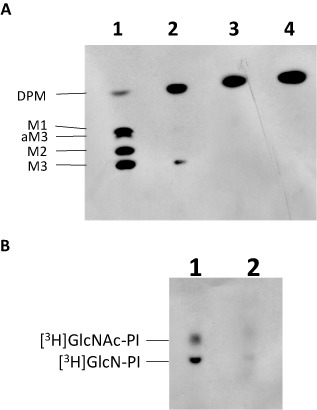
TbCDS is essential for formation of GPI intermediates.
A. Cell free system of wild‐type (lanes 1 and 3) and TbCDS CKO cells in absence of tetracycline for 42 h (lanes 2 and 4), membranes were incubated with GDP‐[3H]Man in the presence (lanes 1 and 2) and absence (lanes 3 and 4) of UDP‐GlcNAc.
B. Cell free system of wild‐type (lane 1) and TbCDS CKO cells in absence of tetracycline for 42 h (lane 2) membranes incubated with UDP‐[3H]GlcNAc. O = origin, F = front. Solvent: 180:140:9:9:23 CHCl3 : MeOH : 30% NH3:1 M NH4Ac : H2O. Positions of the GPI intermediates are shown on left hand side. The data presented here are representative TLCs, of labellings done on several biological replicates along with their corresponding controls.
In order to clarify that this lack of GPI biosynthetic flux was due to a lack of newly synthesized PI from the ER resident PI synthase (that utilizes CDP‐DAG), a separate assay with UDP‐[3H]GlcNAc in the absence of GDP‐Man was conducted. As expected wild‐type membranes formed two strong bands corresponding to GlcNAc‐PI and GlcN‐PI (Fig. 6B, lane 1), indicating ample endogenous PI acceptor was available to catalyse the initial steps of the GPI pathway. In contrast the TbCDS CKO membranes showed significantly fainter bands for both GlcNAc‐PI and GlcN‐PI (Fig. 6B, lane 2), suggesting a lack of endogenous PI in the ER membranes.
Collectively, these results clearly demonstrate the reduced flux through the GPI biosynthetic pathway is caused by a shortage of PI in the ER, due to depletion of CDS activity.
Subcellular localization of TbCDS in bloodstream form T. brucei
The observed differential effects on the two major PI species caused by reduced TbCDS expression in BSF T. brucei suggests two separate pools of CDP‐DAG may be synthesized by distinctly localized TbCDS enzymes.
To examine the subcellular localization of TbCDS, we used the TbCDS CKO cell line, as the viability conferred by the tetracycline induced copy of pLew 100 TbCDS‐HA confirms that the protein is correctly expressed and localized. The TbCDS‐HA protein was detected by Western blot of total protein (Fig. 3C, lane 2). The cells were stained against the HA‐tagged CDS and for ER localized BiP protein (Bangs et al., 1993; Fig. 7). The strongest signal from HA‐tagged TbCDS comes from the region between the nucleus and kinetoplast, while punctuate staining is visible throughout the cell. Since TbCDS is a membrane protein this pattern of staining indicates that it is localized to the membrane of subcellular organelles rather than the plasma membrane. Intense staining between the nucleus and kinetoplast is indicative of a Golgi localization, which has previously been shown to be the location of the downstream enzyme, PIS (Martin and Smith, 2006a) and is consistent with a requirement of CDP‐DAG for the synthesis of the bulk of PI in the cell. The perinuclear staining, and the staining throughout the cell indicate ER localization, where TbCDS could supply CDP‐DAG required for the ER localized PIS to make PI for GPI anchors.
Figure 7.
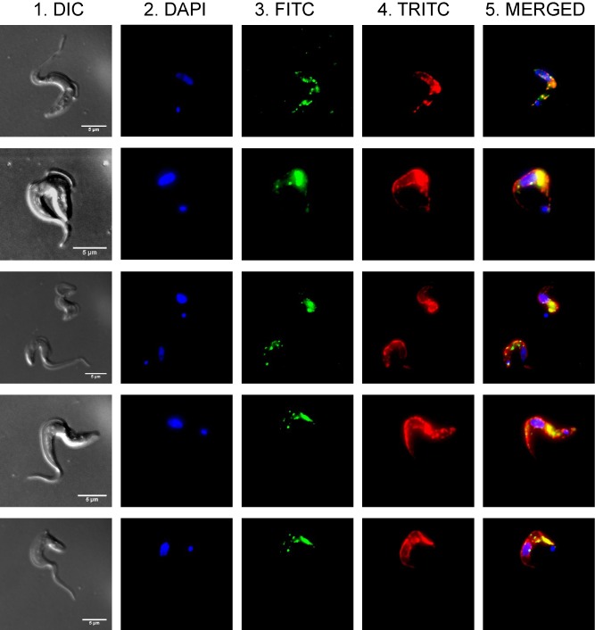
Localization of CDS‐HA in bloodstream T. brucei. T. brucei cells expressing CDS‐HA were co‐stained for the nuclear marker DAPI, the HA epitope and the ER marker BiP. 1. DIC image, 2. DAPI staining. 3. HA‐epitope staining and FITC detection. 4. BiP staining and TRITC detection. 5. Merged image.
Some co‐staining of TbCDS with the ER protein BiP does occur, particularly between the nucleus and the kinetoplast, although co‐staining is not complete and some punctuate staining of TbCDS appears to occur outwith the ER. Even within the ER, TbCDS is not evenly distributed and it may be present in specific areas of the ER (i.e. between the nucleus and the kinetoplast), possibly the mitochondrial associated membranes.
Discussion
This study has given us new insight into the glycerophospholipid biosynthesis in bloodstream form T. brucei. Figure 8 presents a model of glycerophospholipid synthesis revised to include the alternative mechanisms indicated by our results.
Figure 8.
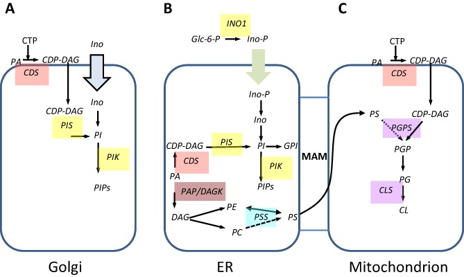
Model of revised pathways for glycerophospholipid synthesis in bloodstream form T. brucei. Enzymes for which candidate genes have been identified are indicated. Block arrows indicate transporters. MAM = mitochondrial associated membranes. Metabolite abbreviations: Glc‐6‐P = glucose 6‐phosphate; PA – phosphatidic acid; CDP‐DAG – cytidine diphosphate diacylglycerol; PGP – phosphatidylglycerophosphate; PG – phosphatidylglycerol, CL – cardiolipin, PS – phosphatidylserine, PE – phosphatidylethanolamine; PI – phosphatidylinositol; PIPs – phosphorylated derivatives of phosphatidyl inositol; DAG – diacylglycerol; PE – phosphatidylethanolamine; IPC – inositol phosphorylceramide. Enzyme abbreviations: CDS – cytidine diphosphate diacylglycerol synthase, PGPS – phosphatidylglycerolphosphate synthase, CLS – cardiolipin synthase, PIS phosphatidlyinositol synthase, PIK – phosphatidylinositol kinase, PSS – phosphatidylserine synthase, PSD – phosphatidylserine decarboxylase, PAP – phosphatidic acid phosphatase, DAGK – DAG kinase. Dotted lines indicate putative pathways.
CDP‐DAG synthase and PI synthase proteins are each encoded by single genes but CDP‐DAG and PI are synthesized as separate pools in the Golgi and the ER, possibly for different functions (Martin and Smith, 2006b).
A myo‐inositol transporter takes cytosolic inositol into the lumen of the Golgi where it is utilized to make PI via the Golgi resident PI synthase, whose active site faces the lumen of the Golgi (Gonzalez‐Salgado et al., 2012). The Golgi resident CDS may provide the CDP‐DAG for this PI synthase; however, the CDP‐DAG must be made on the cytosolic face of the Golgi as it uses cytosolic CTP, and therefore CDP‐DAG must be translocated across the membrane into the lumen of the Golgi.
The PI made in the Golgi is the source of bulk cellular PI, and is not used for GPI anchor synthesis, but is probably used for PIP formation by the resident PI‐4‐kinase required for post‐Golgi sorting (Martin and Smith, 2006b).
Inositol‐3‐phosphate formed from glucose 6‐phosphate by the cytosolic INO1 is taken in to the ER by an unknown transporter/mechanism, where it is dephosphorylated by inositol monophosphatase activity (Eisenberg and Parthasrathy, 1987; Martin and Smith, 2006a). This de novo synthesized inositol, presumably along with CDP‐DAG synthesized by a second pool of TbCDS in the ER is used by the pool of ER resident PI synthase (Martin and Smith, 2006b) In the ER, as in the Golgi, the CDP‐DAG must be translocated to the inner leaflet. The resulting PI is subsequently used for essential GPI biosynthesis (Martin and Smith, 2006a). The high flux through the PI to GPI pathway is likely be on a par with or higher than that of the PI synthesized in the Golgi, meaning that the dynamic pool of PI in the ER is very small as it is rapidly used. The ER synthesized PI is likely also used for the production of PIPs, which may occur in a subcompartment of the ER; recently described in humans (Kim et al., 2011).
A cell cycle stall caused by the knockout of TbCDS could indicate that PIP signalling has a role in cell cycle regulation/control in bloodstream form T. brucei. Additionally, depletion of GPI anchor biosynthesis as observed in this study has previously been shown to cause a cell cycle stall due to the cells being unwilling to dilute their GPI anchored VSG on the cell surface (Smith et al., 2009).
Previous work has indicated that the synthesis of PS via CDP‐DAG does not occur in T. brucei (Signorell et al., 2008) and in support of this, the T. brucei PSS enzyme shows sequence similarity to the mammalian PSS2 which utilizes PE in a head‐group exchange mechanism, rather than the yeast or prokaryotic PSS proteins which are CDP‐alcohol phosphatidyltransferases (Nikawa et al., 1987; Johnson et al., 1997). However, since knockdown of de novo synthesis of PE via the Kennedy pathway did not affect the amount of PS in bloodstream form T. brucei (Gibellini et al., 2009), it is possible that the T. brucei PSS is also capable of utilizing PC as a substrate. The production of PE through PS decarboxylation occurs in prokaryotes, yeast, and to a limited extent in mammals (reviewed in Schuiki and Daum, 2009), but this activity does not occur in bloodstream form T. brucei, even under conditions of depleted PE levels (Gibellini et al., 2009). Surprisingly, the T. brucei does encode a functional PS decarboxylase as demonstrated by recombinant assays and preliminary work indicates that it is essential in bloodstream form (T.K. Smith, unpublished).
For the first time, evidence is presented here that in T. brucei PG must be synthesized via an alternative mechanism neither directly or indirectly involving CDP‐DAG as a lipid donor. The phosphatidylglycerol‐phosphate synthase (PGPS) in procyclic T. brucei has been shown to be responsible for PG production, and its downregulation causes a growth defect (Serricchio and Buetikofer, 2013). However, the enzyme's substrate preference has not been experimentally confirmed. Given the increase in PG species seen in this study under limiting CDS activity it seems unlikely that CDP‐DAG is the sole substrate for PGPS. Many enzymes of the phospholipase‐D like superfamily, which includes PGPS, indicate a lack of substrate specificity (Shibuya et al., 1985; Nishijima et al., 1988; Chang et al., 1998; Tan et al., 2012). It is therefore possible that PGPS may be able to utilize an alternative to CDP‐DAG, for example PS as the lipid donor to produce PG. PS synthesis from PE is thought to occur in the mitochondrial associated membrane of the ER (Vance, 1990; Shiao and Vance, 1995; Stone and Vance, 2000) and so it would be ideally localized to feed into the mitochondrial phospholipid pathway where both T. brucei PGPS and CLS are found (Serricchio and Buetikofer, 2012; 2013).
The non‐reliance upon CDG‐DAG by this branch of lipid metabolism may be a strategy by the parasites to limit CTP use, as the CTP level is already dangerously low in bloodstream form T. brucei.
The T. brucei cellular response to glycerophospholipid disruption results in excess PA or DAG, which is fed into the Kennedy Pathway and converted to PG via PS, possibly in order to maintain a balance of cellular lipid as PG is considered a relatively harmless lipid to produce in excess. Whether PS is the major source of PG and CL under normal circumstances or whether synthesis of PGP from CDP‐DAG normally produces the bulk of PG remains to be determined. In many organisms CDS is localized to the mitochondria, and there is a chance that this is also the case in bloodstream form T. brucei, although further colocalization experiments and subcellular fractionation would be required to confirm this.
In summary this study indicates that the CDP‐DAG synthesized is primarily utilized by PI synthases in both the ER and Golgi. This new knowledge may lead to the discovery and validation of other novel drug targets against African sleeping sickness.
Experimental procedures
Cloning of the T. brucei TbCDS gene
A putative CDS gene was identified in the T. brucei genome database (Sanger Centre) using BlastP search with the S. cerevisiae CDS1 genomic sequence as the query. The open reading frame was amplified from T. brucei genomic DNA using the forward and reverse primers 5′‐GAGAAGCTTATGAAAACAAGTCCAGCAAG‐3′ and 5′‐TGCTTAATTAACTTTTCTGTGAGAGACCTATTAAG‐3′ containing HindIII and PacI restriction sites respectively (underlined). A band of the expected size of c. 1.2 kb was amplified using KOD polymerase, purified using a QIAquick PCR purification kit (Qiagen) and cloned into pCR‐Blunt II TOPO (Invitrogen).
Bioinformatics
Phylogenetic analysis was performed using Mobyle@Pasteur platform from the Institut Pasteur ‘Projets et Développements en Bioinformatique’ Team and the Ressource Parisienne en Bioinformatique Structurale (http://mobyle.pasteur.fr). Sequences were aligned using Clustal O 1.0.2: Multiple Alignment (Sievers et al., 2011). Alignments were manually checked and gap regions and ambiguously aligned regions were excluded from further analysis. Phylogeny was inferred using the protein sequence parsimony method from the PHYLIP package (Phlipi 3.67 ProtPars) (Felsenstein, 1989).
Saccharomyces cerevisae culture
The Saccharomyces cerevisiae strain heterozygous for the cds1::KanMX knockout was created by the Saccharomyces Genome Deletion Project (Winzeler et al., 1999) and obtained from Euroscarf collection. Basic yeast methods, growth media, and routine recombinant DNA methodology were performed as previously described (Gietz et al., 1992; Amberg et al., 2005). For general growth and maintenance of cultures Saccharomyces cerevesiae was either grown on liquid YPD containing 1% bacto‐yeast extract, 2% bacto‐peptone and 2% dextrose, or YPD plates containing 2% bacto‐agar. Otherwise, auxotrophs were grown on complete minimal glucose medium (SC) containing 0.67% bacto‐yeast nitrogen base without amino acids (Sigma) and 2% dextrose. This was supplemented with appropriate amino acid mix to select for plasmids with auxotrophic markers. Initially the amino acids were made up as sterile stocks and added as required, but later the appropriate amino acid mixes were bought from Sigma‐Aldrich. Yeast cultures were grown at 30°C, with constant shaking when in liquid media.
Construction of TbCDS and CDS1 pRS426‐MET25 and p405‐TEF1
TbCDS was isolated from T. brucei strain 427 genomic DNA using the forward and reverse primers 5′‐GAGGGATCCGATGAAAAACAAGTCCAGCAAG‐3′ and 5′‐CGGCTCGAGCCTATTCTGTGAGAGACCTATTAAGAGTCGAG‐3′ containing the HindIII and XhoI restriction sites respectively. Yeast CDS1 was isolated by colony PCR from S. cerevisiae using the forward and reverse primers 5′‐CCCAAGCTTATGTCTTGACAACCCTGGAGATGAA‐3′ and 5′‐CGGCTCGAGTTCAAGAGTGATTGGTCAATGATTTCTTGGTCAC‐3′ containing the HindIII and XhoI restriction sites respectively (underlined). Bands of the expected size of c. 1.2 kb (TbCDS) or 1.4 kb (yeast CDS1) were amplified using KOD polymerase, purified using a QIAquick PCR purification kit (Qiagen) and cloned into the yeast expression vector pRS426 MET25 (2μ URA3) or p405‐TEF1 [gift from Nicholas Buchler (Addgene plasmid #15968)] using the HindIII and XhoI restriction sites.
Complementation of cds1::KanMX with TbCDS and yeast CDS1 by tetrad analysis and plasmid shuffling
The diploid S. cerevisiae cds1::KanMX heterozygous strain was transformed with pRS426‐MET25 containing either yeast CDS1, T. brucei CDS, or with the empty vector, selecting transformants on SCD–Ura medium. These transformants, together with the original heterozygous CDS1/cds1::KanMX null strain were sporulated and tetrads dissected in the absence of methionine in order to allow expression of the CDS gene on the pRS426‐MET25 plasmid. While haploid cds1::KanMX segregants containing yeast CDS1on pRS426‐MET25 could be readily isolated, this was not the case with pRS426‐MET25 carrying T. brucei CDS. In case this reflected poor expression from the MET25promoter on pRS426‐MET25, the haploid cds1::KanMX knockouts containing yeast CDS1 on pRS416 MET25 (MET15 his3 leu2 ura3 cds1::KanMX [pRS425‐MET25‐CDS1]) were used to carry out a ‘plasmid shuffle’ with TbCDS or yeast CDS1 expressed instead from p405‐TEF1, which carries the strong TEF1 promoter. The yeast cds1::KanMX [pRS425‐MET25‐CDS1] was transformed with p405‐TEF1‐TbCDS, p405‐TEF1‐CDS1 or empty p405‐TEF1, selecting for both plasmids on SC–Leu–Met–Ura. Cells were then cultured on SC–Leu–Met (i.e. + Ura) followed by plating on SC–Leu + 5‐fluoro‐orotic acid (5‐FOA) to select against the URA3 containing pRS426‐MET25‐CDS1, thereby testing for complementation of cds1::KanMX by the p405‐TEF1‐based plasmids.
Construction of TbCDS pLew vectors
The TbCDS1 ORF was excised from Topo using HindIII and PacI and subsequently ligated into the tetracycline inducible expression vectors pLew82 and pLew100 (Wirtz et al., 1999) via the HindIII and PacI restriction sites.
Construction of T. brucei gene replacement cassettes
The 5′ and 3′ UTRs immediately adjacent to the TbCDS open reading frame were amplified from T. brucei genomic DNA using KOD polymerase. The primers 5′‐ATAAGTAAGCGGCCGCTGATTAACGCACCC‐3′ and 5′‐CGTTTAAACTTACGGACCGTCAAGCTTCTAGACGCTTTCACTGAAAAATGC‐3′ were used for the 5′ untranslated region (UTR), amplifying the expected 516 bp product and primers 5′‐TGACGGTCCGTAAGTTTAAACGGATCCTCCATGTTGGAAGGGCACG‐3′ and 5′‐AGTAAGCGGCCGCCCTTGAAAACAATGGTGTATAC‐3′ resulted in the correct 179 bp 3′ UTR product.
These amplified products were used in a knitting PCR, resulting in a 695 bp product in which the 5′ UTR was joined to the 3′ UTR via a short BamHI–HindIII linker region contained within the described primers (italics) and a NotI site (underlined) at each end. This PCR product was ligated into pGEM‐5Zf(+) (Promega) using NotI sites, and the hygromycin (HYG) or puromycin (PAC) resistance genes were ligated between the BamHI and HindIII restriction sites. Plasmid DNA was prepared using a QIAprep Miniprep Plasmid Kit (Qiagen). After digestion with NotI the DNA was precipitated with sodium acetate/ethanol and dissolved in sterile water to a final concentration of 1 μg μl−1.
Cultivation and genetic modification of T. brucei
Bloodstream form T. brucei strain 427, which had been previously modified to express both T7 polymerase and the tetracycline repressor protein (Wirtz et al., 1999), are referred to here as wild‐type cells for convenience. Cells were grown HMI‐9 media supplemented with G418 (2.5 μg ml−1) at 37°C with 5% CO2 as described elsewhere (Wirtz et al., 1999; Chang et al., 2002; Roper et al., 2002). Transformation conditions and subsequent drug selection were also described elsewhere (Wirtz et al., 1999; Chang et al., 2002; Roper et al., 2002).
After drug selection with phleomycin, several TbCDS‐HATi clones were obtained and integration of the pLew100 ectopic copy was confirmed by PCR using primers specific to the pLew100 vector (data not shown). The first TbCDS allele was deleted from the TbCDS‐HATi cell line using a PAC knockout cassette. TbCDS‐HATi ΔTbCDS::PAC clones were obtained by selection with puromycin. The second TbCDS allele was replaced with the hygromycin resistance gene.
For experiments requiring tetracycline‐free conditions, Tet‐system approved fetal calf serum (Clonetech) was used. When required, tetracycline was added to the media at a final concentration of 1 μg ml−1. For tetracycline‐free experiments, cells were washed three times in tetracycline‐free HMI‐9 and resuspended in tetracycline‐free media at 5 × 10⁴ cells ml−1. Cells were counted each day and were passaged only when the density was between 2 and 3 × 10⁶ cells ml−1 (normally every second day).
PCR confirmation of TbCDS knockout
The genotype of the conditional mutant was verified by PCR analysis using primer combinations to amplify sections of the knockout construct and primer combinations to amplify the TbCDS in its genomic context. If both genomic copies of the gene had indeed been deleted, there should be no product with the latter. The following forward primers were used: 5′‐AAGCTTATGACCGAGTACAAGCCCACGGTGCGCC‐3′, 5′‐AAGCTTATGAAAAAGCCTGAACTCACCGCGAC‐3′ or 5′‐GAGAAGCTTATGAAAACAAGTCCAGCAAG‐3′ along with the reverse primer 5′‐AGTAAGCGGCCGCCCTTGAAAACAATGGTGTATAC‐3′.
RT‐PCR
To confirm TbCDS transcription, RT‐PCR was performed using the 5′‐ATAAGTAAGCGGCCGCTGATTAACGCACCC‐3′ and CDS pLew R primers to amplify the TbCDS ORF. Reverse transcription PCR was performed using the SuperScript™ One‐Step RT‐PCR kit with Platinum® Taq (Invitrogen) following the manufacturers recommendation. Reaction components were 2× Reaction Mix, 10 pg–1 μg template RNA, 10 μM sense primer, 10 μM anti‐sense primer, 1 μl 0.2 μM RT/Platinum® Taq Mix, DEPC treated water to 50 μl.
CDS enzyme assays
The assay used for TbCDS activity was based on those described by Sparrow and Raetz (1985) and Wu et al. (1995). Washed T. brucei cells were harvested by centrifugation, washed twice in trypanosome dilution buffer (TDB, 5 mM KCl, 80 mM NaCl, 1 mM MgSO4, 20 mM NaH2PO4, 2 mM NaH2PO4, 20 mM glucose, pH 7.7) and lysed with 800 μl ice cold water. Two hundred microlitres of 5× incorporation buffer (250 mM Tris‐maleate pH 6.5; 15 mM MgCl2; 5 mg ml−1 BSA, 1.25 mM DTT) was added to the lysed cells, followed by centrifugation at 1000 rpm to remove whole cells. The cell membrane pellet was washed twice in incorporation buffer and the cell pellet resuspended in 85 μl incorporation buffer followed by sonication. Resuspended membranes were added to reaction tubes to make a final assay volume of 100 μl. The standard assay reaction consisted of washed crude membrane extract in 50 mM Tris‐maleate pH 6.5; 3 mM MgCl2; 1 mg ml−1 BSA; 0.25 mM DTT; 1 mM [3H]CTP at 9 μCi mM−1; 1.5 mM phosphatidic acid, 15 mM TX‐100 in a total volume of 150 μL. The assay was incubated at 30°C for 20 min, and stopped by addition of 666 μl 1:1 (v/v) CHCl3:MeOH. Radiolabelled glycolipids were extracted into the resulting 10:10:3 CHCl3 : MeOH : H2O (v/v) by shaking at 4°C for at least 1 h. Assay mixtures were spun at full speed and the supernatant evaporated to dryness. Two hundred microlitres of CHCl3 and 200 μl of 0.1 M HCl were then added to the tubes which were vortexed and briefly spun. The chloroform layer was removed and washed twice with 0.1 M HCl followed by evaporation to dryness. The resultant radiolabelled glycolipids were analysed by silica 60‐HPTLC in solvent system 25:15:4:2 (v/v) CHCl3 : MeOH : CH3COOH : H2O. Dried HPTLC plates were sprayed with En3hanceTM Spray and radiolabelled glycolipids visualized by fluorography.
In vivo T. brucei metabolic labelling
For each metabolic labelling, 1–2 × 10⁷ mid‐log cells were centrifuged (800 g, 10 min), washed in glucose free minimal essential media, before being resuspended in the same media at a final concentration of 1 × 10⁷ cells ml−1. Cells were labelled for 1 h at 37°C with 50 μCi ml−1 of either [9,10‐3H(N)]‐Tetradecanoic acid (myristic acid) (47 Ci mmol−1) (Perkin Elmer); [2‐3H]‐inositol (20 Ci mmol−1) or [1,2,3‐[3H]]‐glycerol (20 Ci mmol−1) (both American Radiolabelled Chemicals) in a shaking water bath. The cells were collected by centrifugation (800 g for 10 min) and samples taken for analysis. Lipids were extracted using chloroform : methanol : water (10:10:3 v/v) for 1 h, the supernatant removed and the pellet re‐extracted with chloroform : methanol (2:1 v/v) for 1 h. The supernatants were pooled and dried before samples were desalted using butanol/water partitioning. Lipids were separated by HPTLC using silica 60 HPTLC plates and chloroform : methanol : water (10:10:3 v/v) as the solvent. Radiolabelled lipids were detected by fluorography at −80°C, after spraying with En3hanceTM and using Kodak XAR‐5 film with an intensifying screen.
Lipidomic analysis
Lipids were extracted from T. brucei as described previously (Richmond et al., 2010). Briefly, mid‐log cells are collected by centrifugation (800 g, 10 min) washed with PBS and suspended in 100 μl PBS and transferred to a glass tube. A total of 375 μl of 1:2 (v/v) CHCl3:MeOH added and vortexed. The sample is agitated vigorously for a further 10–15 min. The sample is now made biphasic by the addition of 125 μl of CHCl3, vortex and then 125 μl of H2O and vortexed again and centrifuged at 1000 g at RT for 5 min. The lower chloroform phase is transferred to a new glass vial, dried under nitrogen and stored in the fridge.
Total lipid extracts were dissolved in 15 μl of choloroform : methanol (1:2) and 15 μl of acetonitrile : isopropanol : water (6:7:2) and analysed with a Absceix 4000 QTrap – a triple quadrupole mass spectrometer equipped with a nanoelectrospray source.
Samples were delivered using either thin‐wall nanoflow capillary tips or a Nanomate interface in direct infusion mode (∼ 125 nl min−1). The lipid extracts were analysed in both positive and negative ion modes using a capillary voltage of 1.25 kV. MS/MS scanning (daughter, precursor and neutral loss scans) were performed using nitrogen as the collision gas with collision energies between 35–90 V. Each spectrum encompasses at least 50 repetitive scans.
Tandem mass spectra (MS/MS) were obtained with collision energies as follows: 35–45 V, PC in positive ion mode, parent‐ion scanning of m/z 184; 35–55 V, PI in negative ion mode, parent‐ion scanning of m/z 241; 35–65 V, PE in negative ion mode, parent‐ion scanning of m/z 196; 20–35 V, PS in negative ion mode, neutral loss scanning of m/z 87; and 40–90 V, for all glycerophospholipids (including PA, PG and CL) detected by precursor scanning for m/z 153 in negative ion mode. MS/MS fragmentation/daughter ion scanning was performed with collision energies between 35–90 V. Assignment of phospholipid species is based upon a combination of survey, daughter, precursor and neutral loss scans, as well as previous assignments (Richmond et al., 2010). The identity of phospholipid peaks was verified using the LIPID MAPS: Nature Lipidomics Gateway (http://www.lipidmaps.org). In order to compare the peak heights relative to each other, peaks were measured by ion intensities and their relative ratios were calculated.
Cell‐free system assay of GPI biosynthesis
Membranes of T. brucei wild‐type and TbCDS conditional null mutants grown in the presence or absence of tetracycline for 30, 36 and 42 h were isolated and prepared as described previously (without the addition of tunicamycin prior to lysis) (Masterson et al., 1989), snap frozen in liquid nitrogen and stored at −80°C until required. A total of 1 × 10⁷ cell equivalents were used per assay: Briefly, membranes were washed twice in 10 ml of wash buffer (50 mM NaHEPES, pH 7.4, 250 mM KCl, 5 mM MgCl2, 100 μM tosyl‐l‐lysine chloromethyl ketone and 1 μg ml−1 leupeptin). The membranes were then resuspended by sonication in 2× incorporation buffer (2 times concentrated wash buffer containing 10 mM MnCl2, 1 μg ml−1 of tunicamycin and 1 mM DTT) and added to a reaction tube containing an equal volume of GDP‐[3H]Mannose (0.3 μCi per 107 cell equivalents), with and without 1 mM UDP‐GlcNAc, or just [3H] UDP‐GlcNAc (1.0 μCi per 107 cell equivalents). The cell‐free system was then sonicated briefly, incubated at 30°C for 1 h and the reaction stopped by the addition of 267 μl of chloroform: methanol (1:1 v/v). Radiolabelled glycolipid products were recovered by extraction into a chloroform/methanol/water mixture (10:10:3), evaporated to dryness, partitioned between butan‐1‐ol and water (Smith et al., 1996), and analysed by HPTLC in solvent system 180:140:9:9:23 (chloroform: methanol: 1 M ammonium acetate: concentrated ammonia: water). Dried HPTLC plates were sprayed with En3HanceTM (Perkin Elmer), and radiolabelled components were visualized by fluorography at −80°C using Kodak Biomax MS films with an intensifying screen.
Inositol analysis
Mid‐log cells were collected by centrifugation (800 g, 10 min) washed with TDB and stored at −20°C. Lipids were extracted from these samples by the addition of 500 μl of chloroform methanol mixture (2:1 v/v) and incubated at room temperature for 1 h. The supernatant was removed and the pellet re‐extracted with chloroform : methanol : water mixture (1:2:0.8 v/v). Pooled supernatants were dried under nitrogen prior to desalting by biphasic partitioning using 2:1 butanol : water (v/v). An internal standard of D6 myo‐inositol was added to samples prior to hydrolysis by strong acid (6 M HCl, 110°C), derivitization with TMS and analysis by GC‐MS, according to the method of Ferguson (Ferguson, 1993). myo‐Inositol was quantified and the mean of three separate analyses were determined.
Immunofluorescence
At least 1 × 10⁶ cells were harvested from culture by centrifugation at 800 g for 10 min at room temperature and washed with TDB. Cells were fixed in PBS plus 4% paraformaldehyde and processed on slides as described previously (Martin and Smith, 2006a). Anti‐BiP was a kind gift from Jay Bangs, Buffalo. After the final washes in PBS the slides were mounted in SlowFade® Gold Antifade Reagent with DAPI (Invitrogen) and a coverslip was placed over the slide. Slides were allowed to cure for 24 h at room temperature before sealing the sides with nail varnish.
All immunofluoresence microscopy was performed on a DeltaVision microscope (Applied Precision Inc.) and processed using ImageJ software (Rasband, 1997–2012).
Supplementary Material
Supporting Information
Acknowledgements
This work was supported in part by a Wellcome Trust Senior Research Fellowship 067441 and Wellcome Trust grants 082596 and 093228, with a BBSRC PhD studentship for A.C.L.
References
- Agabian, N. (1990) Transsplicing of nuclear pre‐messenger‐RNAs. Cell 61: 1157–1160 [DOI] [PubMed] [Google Scholar]
- Amberg, D., Burke, D., and Strathern, J. (2005) Methods in Yeast Genetics: A Cold Spring Harbor Laboratory Course Manual, 2005 edn (Cold Spring) Woodbury, NY: Cold Spring Harbor Laboratory Press [Google Scholar]
- Baelee, M.S., and Carman, G.M. (1984) Phosphatidylserine synthesis in Saccharomyces cerevisiae – purification and characterization of membrane‐associated phosphatidylserine synthase. J Biol Chem 259: 857–862 [PubMed] [Google Scholar]
- Bangs, J.D., Uyetake, L., Brickman, M.J., Balber, A.E., and Boothroyd, J.C. (1993) Molecular‐cloning and cellular‐localization of a BiP homolog in Trypanosoma brucei – divergent er retention signals in a lower eukaryote. J Cell Sci 105: 1101–1113 [DOI] [PubMed] [Google Scholar]
- Bell, R.M., Mavis, R.D., Osborn, M.J., and Vagelos, P.R. (1971) Enzyymes of phospholipid metabolism – localization in cytoplasmic and outer membrane of cell envelope of Escherichia coli and Salmonella typhimurium. Biochim Biophys Acta 249: 628–636 [DOI] [PubMed] [Google Scholar]
- Bishop, H.H., and Strickland, K.P. (1976) Studies on formation by rat‐brain preparations of CDP‐diglyceride from CTP and phosphatidic acids of varying fatty‐acid compositions. Can J Biochem 54: 249–260 [DOI] [PubMed] [Google Scholar]
- Carman, G.M., and Fischl, A.S. (1992) Phosphatidylinositol synthase from yeast. Methods Enzymol 209: 305–312 [DOI] [PubMed] [Google Scholar]
- Carman, G.M., and Han, G.‐S. (2011) Regulation of phospholipid synthesis in the yeast Saccharomyces cerevisiae. Annu Rev Biochem 80: 856–883 [DOI] [PMC free article] [PubMed] [Google Scholar]
- Carman, G.M., and Baelee, M. (1992) Phosphatidylserine synthase from yeast. Methods Enzymol 209: 298–305 [DOI] [PubMed] [Google Scholar]
- Carman, G.M., and Kelley, M.J. (1992) Cdpdiacylglycerol synthase from yeast. Methods Enzymol 209: 242–247 [DOI] [PubMed] [Google Scholar]
- Carter, J.R., and Kennedy, E.P. (1966) Enzymatic synthesis of cytidine diphosphate diglyceride. J Lipid Res 7: 678–683 [PubMed] [Google Scholar]
- Chang, S.C., Heacock, P.N., Clancey, C.J., and Dowhan, W. (1998) The pel1 gene (Renamed pgs1) encodes the phosphatidylglycerophosphate synthase of Saccharomyces cerevisiae. J Biol Chem 273: 9829–9836 [DOI] [PubMed] [Google Scholar]
- Chang, T., Milne, K.G., Guther, M.L.S., Smith, T.K., and Ferguson, M.A.J. (2002) Cloning of the Trypansoma brucei and Leishmania major genes encoding the glcnac‐phoshatidylinositol DE‐N‐acetylase of glycosylphosphatidylinositol biosynthesis that is essential to the african sleeping sickness parasite. J Biol Chem 277: 50176–50182 [DOI] [PubMed] [Google Scholar]
- Cross, G.A.M. (1996) Antigenic variation in trypanosomes: secrets surface slowly. Bioessays 18: 283–291 [DOI] [PubMed] [Google Scholar]
- Eisenberg, F.J., and Parthasarathy, R. (1987) Measurement of biosynthesis of myo‐inositol from glucose‐6‐phosphate. Methods Enzymol 141: 127–143 [DOI] [PubMed] [Google Scholar]
- Felsenstein, J. (1989) Mathematical evolutionary theory. Science 246: 941–942 [DOI] [PubMed] [Google Scholar]
- Ferguson, M.A.J. (1993) GPI membrane anchors: isolation and analysis In Glycobiology: A Practical Approach. Fukuda, M. , and Kobata, A. (eds). Oxford: Irl Press, pp. 349–383 [Google Scholar]
- Ganong, B.R., and Raetz, C.R.H. (1982) Massive accumulation of phosphatidic‐acid in conditionally lethal CDP‐diglyceride synthetase mutants and cytidine auxotrophs of Escherichia coli. J Biol Chem 257: 389–394 [PubMed] [Google Scholar]
- Ganong, B.R., Leonard, J.M., and Raetz, C.R.H. (1980) Phosphatidic‐acid accumulation in the membranes of Escherichia coli mutants defective in CDP‐diglyceride synthetase. J Biol Chem 255: 1623–1629 [PubMed] [Google Scholar]
- Gibellini, F., Hunter, W.N., and Smith, T.K. (2009) The ethanolamine branch of the kennedy pathway is essential in the bloodstream form of Trypanosoma brucei. Mol Microbiol 73: 826–843 [DOI] [PMC free article] [PubMed] [Google Scholar]
- Gietz, D., Stjean, A., Woods, R.A., and Schiestl, R.H. (1992) Improved method for high‐efficiency transformation of intact yeast‐cells. Nucleic Acids Res 20: 1425–1425 [DOI] [PMC free article] [PubMed] [Google Scholar]
- Gonzalez‐Salgado, A., Steinmann, M., Greganova, E., Mäser, P., Sigel, E., and Bütikofer, P. (2012) Myo‐inositol uptake is essential for bulk inositol phospholipid but not glycosylphosphatidylinositol synthesis in Trypanosoma brucei. J Biol Chem 287: 13313–13323 [DOI] [PMC free article] [PubMed] [Google Scholar]
- Halford, S., Dulai, K.S., Daw, S.C., Fitzgibbon, J., and Hunt, D.M. (1998) Isolation and chromosomal localization of two human CDP‐diacylglycerol synthase (CDS) genes. Genomics 54: 140–144 [DOI] [PubMed] [Google Scholar]
- Hammarton, T.C., Lillico, S.G., Welburn, S.C., and Mottram, J.C. (2005) Trypanosoma brucei MOB1 is required for accurate and efficient cytokinesis but not for exit from mitosis. Mol Microbiol 56: 104–116 [DOI] [PMC free article] [PubMed] [Google Scholar]
- Hammarton, T.C., Monnerat, S., and Mottram, J.C. (2007) Cytokinesis in trypanosomatilds. Curr Opin Microbiol 10: 520–527 [DOI] [PubMed] [Google Scholar]
- Haselier, A., Akbari, H., Weth, A., Baumgartner, W., and Frentzen, M. (2010) Two closely related genes of arabidopsis encode plastidial cytidinediphosphate diacylglycerol synthases essential for photoautotrophic growth. Plant Physiol 153: 1372–1384 [DOI] [PMC free article] [PubMed] [Google Scholar]
- Heacock, A.M., and Agranoff, B.W. (1997) CDP‐diacylglycerol synthase from mammalian tissues. Biochim Biophys Acta 1348: 166–172 [DOI] [PubMed] [Google Scholar]
- Heacock, A.M., Uhler, M.D., and Agranoff, B.W. (1996) Cloning of CDP‐diacylglycerol synthase from a human neuronal cell line. J Neurochem 67: 2200–2203 [DOI] [PubMed] [Google Scholar]
- Icho, T., Sparrow, C.P., and Raetz, C.R.H. (1985) Molecular‐cloning and sequencing of the gene for CDP‐diglyceride synthetase of Escherichia coli. J Biol Chem 260: 2078–2083 [PubMed] [Google Scholar]
- Inglis‐Broadgate, S.L., Ocaka, L., Banerjee, R., Gaasenbeek, M., Chapple, J.P., Cheetham, M.E., et al (2005) Isolation and characterization of murine CDS (CDP‐diacylglycerol synthase) 1 and 2. Gene 356: 19–31 [DOI] [PubMed] [Google Scholar]
- Johnson, J.E., Edwards, A.S., and Newton, A.C. (1997) A putative phosphatidylserine binding motif is not involved in the lipid regulation of protein kinase C. J Biol Chem 272: 30787–30792 [DOI] [PubMed] [Google Scholar]
- Kelley, M.J., and Carman, G.M. (1987) Purification and characterization of CDP‐diacylglycerol synthase from Saccharomyces cerevisiae. J Biol Chem 262: 14563–14570 [PubMed] [Google Scholar]
- Kim, Y.J., Guzman‐Hernandez, M.L., and Balla, T. (2011) A highly dynamic ER‐derived phosphatidylinositol‐synthesizing organelle supplies phosphoinositides to cellular membranes. Dev Cell 21: 813–824 [DOI] [PMC free article] [PubMed] [Google Scholar]
- Koonin, E.V. (1996) A duplicated catalytic motif in a new superfamily of phosphohydrolases and phospholipid synthases that includes poxvirus envelope proteins. Trends Biochem Sci 21: 242–243 [PubMed] [Google Scholar]
- Kuchler, K., Daum, G., and Paltauf, F. (1986) Subcellular and submitochondrial localization of phospholipid‐synthesizing enzymes in Saccharomyces cerevisiae. J Bacteriol 165: 901–910 [DOI] [PMC free article] [PubMed] [Google Scholar]
- Kyte, J., and Doolittle, R.F. (1982) A simple method for displaying the hydropathic character of a protein. J Mol Biol 157: 105–132 [DOI] [PubMed] [Google Scholar]
- Langley, K.E., and Kennedy, E.P. (1978) Partial‐purification and properties of CTP – phosphatidic‐acid cytidylyltransferase from membranes of Escherichia coli. J Bacteriol 136: 85–95 [DOI] [PMC free article] [PubMed] [Google Scholar]
- Lykidis, A. (2007) Comparative genomics and evolution of eukaryotic phospholipid biosynthesis. Prog Lipid Res 46: 171–199 [DOI] [PubMed] [Google Scholar]
- Lykidis, A., Jackson, P.D., Rock, C.O., and Jackowski, S. (1997) The role of CDP‐diacylglycerol synthetase and phosphatidylinositol synthase activity levels in the regulation of cellular phosphatidylinositol content. J Biol Chem 272: 33402–33409 [DOI] [PubMed] [Google Scholar]
- Martin, D., Gannoun‐Zaki, L., Bonnefoy, S., Eldin, P., Wengelnik, K., and Vial, H. (2000) Characterization of Plasmodium falciparum CDP‐diacylglycerol synthase, a proteolytically cleaved enzyme. Mol Biochem Parasitol 110: 93–105 [DOI] [PubMed] [Google Scholar]
- Martin, K.L., and Smith, T.K. (2006a) The glycosylphosphatidylinositol (GPI) biosynthetic pathway of bloodstream‐form Trypanosoma brucei is dependent on the de novo synthesis of inositol. Mol Microbiol 61: 89–105 [DOI] [PMC free article] [PubMed] [Google Scholar]
- Martin, K.L., and Smith, T.K. (2006b) Phosphatidylinositol synthesis is essential in bloodstream form Trypanosoma brucei. Biochem J 396: 287–295 [DOI] [PMC free article] [PubMed] [Google Scholar]
- Masterson, W.J., Doering, T.L., Hart, G.W., and Englund, P.T. (1989) A novel pathway for glycan assembly – biosynthesis of the glycosyl‐phosphatidylinositol anchor of the trypanosome variant surface glycoprotein. Cell 56: 793–800 [DOI] [PubMed] [Google Scholar]
- Mok, A.Y.P., McDougall, G.E., and McMurray, W.C. (1992) CDP‐diacylglycerol synthesis in rat‐liver mitochondria. FEBS Lett 312: 236–240 [DOI] [PubMed] [Google Scholar]
- Nagamune, K., Nozaki, T., Maeda, Y., Ohishi, K., Fukuma, T., Hara, T., et al (2000) Critical roles of glycosylphosphatidylinositol for Trypanosoma brucei. Proc Natl Acad Sci USA 97: 10336–10341 [DOI] [PMC free article] [PubMed] [Google Scholar]
- Nickels, J.T., Buxeda, R.J., and Carman, G.M. (1994) Regulation of phosphatidylinositol 4‐kinase from the yeast Saccharomyces cerevisiae by CDP‐diacylglycerol. J Biol Chem 269: 11018–11024 [PubMed] [Google Scholar]
- Nigou, J., and Besra, G.S. (2002) Cytidine diphosphate‐diacylglycerol synthesis in Mycobacterium smegmatis. Biochem J 367: 157–162 [DOI] [PMC free article] [PubMed] [Google Scholar]
- Nikawa, J., Kodaki, T., and Yamashita, S. (1987) Primary structure and disruption of the phosphatidylinositol synthase gene of Saccharomyces cerevisiae. J Biol Chem 262: 4876–4881 [PubMed] [Google Scholar]
- Nishijima, S., Asami, Y., Uetake, N., Yamagoe, S., Ohta, A., and Shibuya, I. (1988) Disruption of the Escherichia coli cls gene responsible for cardiolipin synthesis. J Bacteriol 170: 775–780 [DOI] [PMC free article] [PubMed] [Google Scholar]
- Ponting, C.P., and Kerr, I.D. (1996) A novel family of phospholipase D homologues that includes phospholipid synthases and putative endonucleases: identification of duplicated repeats and potential active site residues. Protein Sci 5: 914–922 [DOI] [PMC free article] [PubMed] [Google Scholar]
- Raetz, C.R.H., and Kennedy, E.P. (1973) Function of cytidine diphosphate‐diglyceride and deoxycytidine diphosphate‐diglyceride in biogenesis of membrane lipids in Escherichia coli. J Biol Chem 248: 1098–1105 [PubMed] [Google Scholar]
- Rasband, W.S. (1997–2012) ImageJ. Bethesda, MD: US National Institutes of Health [Google Scholar]
- Richmond, G.S., Gibellini, F., Young, S.A., Major, L., Denton, H., Lilley, A., and Smith, T.K. (2010) Lipidomic analysis of bloodstream and procyclic form Trypanosoma brucei. Parasitology 137: 1357–1392 [DOI] [PMC free article] [PubMed] [Google Scholar]
- Rodgers, M.J., Albanesi, J.P., and Phillips, M.A. (2007) Phosphatidylinositol 4‐kinase III‐Β is required for golgi maintenance and cytokinesis in Trypanosoma brucei. Eukaryot Cell 6: 1108–1118 [DOI] [PMC free article] [PubMed] [Google Scholar]
- Roper, J.R., Guther, M.L.S., Milne, K.G., and Ferguson, M.A.J. (2002) Galactose metabolism is essential for the African sleeping sickness parasite Trypanosoma brucei. Proc Natl Acad Sci USA 99: 5884–5889 [DOI] [PMC free article] [PubMed] [Google Scholar]
- Saito, S., Goto, K., Tonosaki, A., and Kondo, H. (1997) Gene cloning and characterization of CDP‐diacylglycerol synthase from rat brain. J Biol Chem 272: 9503–9509 [DOI] [PubMed] [Google Scholar]
- Sato, N., Hagio, M., Wada, H., and Tsuzuki, M. (2000) Requirement of phosphatidylglycerol for photosynthetic function in thylakoid membranes. Proc Natl Acad Sci USA 97: 10655–10660 [DOI] [PMC free article] [PubMed] [Google Scholar]
- Schuiki, I., and Daum, G. (2009) Phosphatidylserine decarboxylases, key enzymes of lipid metabolism. IUBMB Life 61: 151–162 [DOI] [PubMed] [Google Scholar]
- Serricchio, M., and Buetikofer, P. (2012) An essential bacterial‐type cardiolipin synthase mediates cardiolipin formation in a eukaryote. Proc Natl Acad Sci USA 109: E954–E961 [DOI] [PMC free article] [PubMed] [Google Scholar]
- Serricchio, M., and Buetikofer, P. (2013) Phosphatidylglycerophosphate synthase associates with a mitochondrial inner membrane complex and is essential for growth of Trypanosoma brucei. Mol Microbiol 87: 569–579 [DOI] [PubMed] [Google Scholar]
- Shen, H., and Dowhan, W. (1996) Reduction of CDP‐diacylglycerol synthase activity results in the excretion of inositol by Saccharomyces cerevisiae. J Biol Chem 271: 29043–29048 [DOI] [PubMed] [Google Scholar]
- Shen, H.F., Heacock, P.N., Clancey, C.J., and Dowhan, W. (1996) The cds1 gene encoding CDP‐diacylglycerol synthase in Saccharomyces cerevisiae is essential for cell growth. J Biol Chem 271: 789–795 [DOI] [PubMed] [Google Scholar]
- Shiao, Y.J., and Vance, J.E. (1995) Evidence for an ethanolamine cycle – differential recycling of the ethanolamine moiety of phosphatidylethanolamine derived from phosphatidylserine and ethanolamine. Biochem J 310: 673–679 [DOI] [PMC free article] [PubMed] [Google Scholar]
- Shibuya, I., Yamagoe, S., Miyazaki, C., Matsuzaki, H., and Ohta, A. (1985) Biosynthesis of novel acidic phospholipid analogs in Escherichia coli. J Bacteriol 161: 473–477 [DOI] [PMC free article] [PubMed] [Google Scholar]
- Sievers, F., Wilm, A., Dineen, D., Gibson, T.J., Karplus, K., Li, W., et al (2011) Fast, scalable generation of high‐quality protein multiple sequence alignments using Clustal Omega. Mol Syst Biol 7: 539. [DOI] [PMC free article] [PubMed] [Google Scholar]
- Signorell, A., Rauch, M., Jelk, J., Ferguson, M.A.J., and Buetikofer, P. (2008) Phosphatidylethanolamine in Trypanosoma brucei is organized in two separate pools and is synthesized exclusively by the kennedy pathway. J Biol Chem 283: 23636–23644 [DOI] [PMC free article] [PubMed] [Google Scholar]
- Smith, T.K., and Buetikofer, P. (2010) Lipid metabolism in Trypanosoma brucei. Mol Biochem Parasitol 172: 66–79 [DOI] [PMC free article] [PubMed] [Google Scholar]
- Smith, T.K., Cottaz, S., Brimacombe, J.S., and Ferguson, M.A.J. (1996) Substrate specificity of the dolichol phosphate mannose: glucosaminyl phosphatidylinositol alpha 1–4‐mannosyltransferase of the glycosylphosphatidylinositol biosynthetic pathway of African trypanosomes. J Biol Chem 271: 6476–6482 [DOI] [PubMed] [Google Scholar]
- Smith, T.K., Crossman, A., Brimacombe, J.S., and Ferguson, M.A.J. (2004) Chemical validation of gpi biosynthesis as a drug target against African sleeping sickness. EMBO J 23: 4701–4708 [DOI] [PMC free article] [PubMed] [Google Scholar]
- Smith, T.K., Vasileva, N., Gluenz, E., Terry, S., Portman, N., Kramer, S., et al (2009) Blocking variant surface glycoprotein synthesis in Trypanosoma brucei triggers a general arrest in translation initiation. PLoS ONE 4: e7532. [DOI] [PMC free article] [PubMed] [Google Scholar]
- Sparrow, C.P., and Raetz, C.R.H. (1985) Purification and properties of the membrane‐bound CDP‐diglyceride synthetase from Escherichia coli. J Biol Chem 260: 2084–2091 [PubMed] [Google Scholar]
- Sribney, M., Dove, J.L., and Lyman, E.M. (1977) Studies on synthesis of CDP‐diacylglycerol – stimulation by GTP and inhibition by ATP and fluoride. Biochem Biophys Res Commun 79: 749–755 [DOI] [PubMed] [Google Scholar]
- Stone, S.J., and Vance, J.E. (2000) Phosphatidylserine synthase‐1 and ‐2 are localized to mitochondria‐associated membranes. J Biol Chem 275: 34534–34540 [DOI] [PubMed] [Google Scholar]
- Tan, B.K., Bogdanov, M., Zhao, J., Dowhan, W., Raetz, C.R.H., and Guan, Z. (2012) Discovery of a cardiolipin synthase utilizing phosphatidylethanolamine and phosphatidylglycerol as substrates. Proc Natl Acad Sci USA 109: 16504–16509 [DOI] [PMC free article] [PubMed] [Google Scholar]
- Thompson, W., and MacDonald, G. (1975) Isolation and characterization of cytidine diphosphate diglyceride from beef liver. J Biol Chem 250: 6779–6785 [PubMed] [Google Scholar]
- Vance, J.E. (1990) Phospholipid‐synthesis in a membrane‐fraction associated with mitochondria. J Biol Chem 265: 7248–7256 [PubMed] [Google Scholar]
- Volta, M., Bulfone, A., Gattuso, C., Rossi, E., Mariani, M., Consalez, G.G., et al (1999) Identification and characterization of CDS2, a mammalian homolog of the drosophila CDP‐diacylglycerol synthase gene. Genomics 55: 68–77 [DOI] [PubMed] [Google Scholar]
- White, D.A., Albright, F.R., Lennarz, W.J., and Schnaitm, C.A. (1971) Distribution of phospholipid‐synthesizing enzymes in wall and membrane subfractions of envelope of Escherichia coli. Biochim Biophys Acta 249: 636–642 [DOI] [PubMed] [Google Scholar]
- Winzeler, E.A., Shoemaker, D.D., Astromoff, A., Liang, H., Anderson, K., Andre, B., et al (1999) Functional characterization of the S. cerevisiae genome by gene deletion and parallel analysis. Science 285: 901–906 [DOI] [PubMed] [Google Scholar]
- Wirtz, E., Leal, S., Ochatt, C., and Cross, G.A.M. (1999) A tightly regulated inducible expression system for conditional gene knock‐outs and dominant‐negative genetics in Trypanosoma brucei. Mol Biochem Parasitol 99: 89–101 [DOI] [PubMed] [Google Scholar]
- Wu, L., Niemeyer, B., Colley, N., Socolich, M., and Zuker, C.S. (1995) Regulation of PLC‐mediated signaling in‐vivo by CDP‐diacylglycerol synthase. Nature 373: 216–222 [DOI] [PubMed] [Google Scholar]
Associated Data
This section collects any data citations, data availability statements, or supplementary materials included in this article.
Supplementary Materials
Supporting Information