Abstract
Cell differentiation, tissue formation, and organogenesis are fundamental patterns during the development of multicellular animals from the dividing cells of fertilized eggs. Hence, the complete morphogenesis of any developing organism of the animal kingdom is based on a complex series of interactions that is always associated with the development of a blastula, a one-layered hollow sphere. Here we document an alternative pathway of differentiation, organogenesis, and morphogenesis occurring in an adult protochordate colonial organism. In this system, any minute fragment of peripheral blood vessel containing a limited number of blood cells isolated from Botrylloides, a colonial sea squirt, has the potential to give rise to a fully functional organism possessing all three embryonic layers. Regeneration probably results from a small number of totipotent stem cells circulating in the blood system. The developmental process starts from disorganized, chaotic masses of blood cells. At first an opaque cell mass is formed. Through intensive cell divisions, a hollow, blastula-like structure results, which may produce a whole organism within a short period of a week. This regenerative power of the protochordates may be compared with some of the characteristics associated with the formation of mammalian embryonal carcinomous bodies. It may also serve as an in vivo model system for studying morphogenesis and differentiation by shedding more light on the controversy of the "stem cell" vs. the "dedifferentiation" theories of regeneration and pattern formation.
Full text
PDF
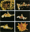
Images in this article
Selected References
These references are in PubMed. This may not be the complete list of references from this article.
- De Loof A. All animals develop from a blastula: consequences of an undervalued definition for thinking on development. Bioessays. 1992 Aug;14(8):573–575. doi: 10.1002/bies.950140815. [DOI] [PubMed] [Google Scholar]
- FREEMAN G. THE ROLE OF BLOOD CELLS IN THE PROCESS OF ASEXUAL REPORODUCTION IN THE TUNICATE PEROPHORA VIRIDIS. J Exp Zool. 1964 Jul;156:157–183. doi: 10.1002/jez.1401560204. [DOI] [PubMed] [Google Scholar]
- KLEINSMITH L. J., PIERCE G. B., Jr MULTIPOTENTIALITY OF SINGLE EMBRYONAL CARCINOMA CELLS. Cancer Res. 1964 Oct;24:1544–1551. [PubMed] [Google Scholar]
- Mackillop W. J., Ciampi A., Till J. E., Buick R. N. A stem cell model of human tumor growth: implications for tumor cell clonogenic assays. J Natl Cancer Inst. 1983 Jan;70(1):9–16. [PubMed] [Google Scholar]
- Martin G. R. Teratocarcinomas and mammalian embryogenesis. Science. 1980 Aug 15;209(4458):768–776. doi: 10.1126/science.6250214. [DOI] [PubMed] [Google Scholar]
- Martin G. R. Teratocarcinomas as a model system for the study of embryogenesis and neoplasia. Cell. 1975 Jul;5(3):229–243. doi: 10.1016/0092-8674(75)90098-7. [DOI] [PubMed] [Google Scholar]
- PIERCE G. B., DIXON F. J., Jr Testicular teratomas. I. Demonstration of teratogenesis by metamorphosis of multipotential cells. Cancer. 1959 May-Jun;12(3):573–583. doi: 10.1002/1097-0142(195905/06)12:3<573::aid-cncr2820120316>3.0.co;2-m. [DOI] [PubMed] [Google Scholar]
- Pierce G. B., Speers W. C. Tumors as caricatures of the process of tissue renewal: prospects for therapy by directing differentiation. Cancer Res. 1988 Apr 15;48(8):1996–2004. [PubMed] [Google Scholar]
- Shimizu H., Sawada Y., Sugiyama T. Minimum tissue size required for hydra regeneration. Dev Biol. 1993 Feb;155(2):287–296. doi: 10.1006/dbio.1993.1028. [DOI] [PubMed] [Google Scholar]
- Slack J. M. The source of cells for regeneration. Nature. 1980 Aug 21;286(5775):760–760. doi: 10.1038/286760a0. [DOI] [PubMed] [Google Scholar]
- Sleigh M. J. Differentiation and proliferation in mouse embryonal carcinoma cells. Bioessays. 1992 Nov;14(11):769–775. doi: 10.1002/bies.950141109. [DOI] [PubMed] [Google Scholar]
- Venuti J. M., Jeffery W. R. Cell lineage and determination of cell fate in ascidian embryos. Int J Dev Biol. 1989 Jun;33(2):197–212. [PubMed] [Google Scholar]
- Watt F. M. Cell culture models of differentiation. FASEB J. 1991 Mar 1;5(3):287–294. doi: 10.1096/fasebj.5.3.2001788. [DOI] [PubMed] [Google Scholar]


