Abstract
We have tested the hypothesis that circadian oscillations in the concentration of cytosolic free calcium ([Ca2+]cyt) can encode information. We imaged oscillations of [Ca2+]cyt in the cotyledons and leaves of Arabidopsis (Arabidopsis thaliana) that have a 24-h period in light/dark cycles and also constant light. The amplitude, phase, and shape of the oscillations of [Ca2+]cyt and [Ca2+]cyt at critical daily time points were controlled by the light/dark regimes in which the plants were grown. These data provide evidence that 24-h oscillations in [Ca2+]cyt encode information concerning daylength and light intensity, which are two major regulators of plant growth and development.
INTRODUCTION
Calcium is a ubiquitous second messenger involved in the transduction of many environmental and developmental stimuli in plants and animals (Trewavas, 1999; Sanders et al., 2002; Schuster et al., 2002). In response to diverse stimuli, cells generate transient increases in the concentration of cytosolic free calcium ([Ca2+]cyt) that vary in amplitude, frequency, duration, cellular location, and timing (McAinsh et al., 1995; McAinsh and Hetherington, 1998; Trewavas, 1999; Berridge et al., 2000; Evans et al., 2001; Allen et al., 2001). Important information regarding the nature of the stimulus may therefore be encoded in the different spatiotemporal profiles of [Ca2+]cyt increases (McAinsh and Hetherington, 1998; Sanders et al., 2002; Schuster et al., 2002). The majority of stimuli tested to date result in single, rapid [Ca2+]cyt spikes (also called bursts) or in complex [Ca2+]cyt oscillations recurring with a period of 1 to 20 min (Sanders et al., 2002; Schuster et al., 2002). However, longer-term [Ca2+]cyt oscillations, characterized by a period of ∼24 h, occur in the cytosol and chloroplast of Nicotiana plumbaginifolia, in the cytosol of Arabidopsis (Arabidopsis thaliana; Johnson et al., 1995), and in the cytosol of neurons of the mouse suprachiasmatic nucleus, which is the primary circadian pacemaker in mammals (Ikeda et al., 2003). These circadian rhythms of [Ca2+]cyt are believed to be involved in signaling to or from the endogenous circadian clock (Gómez and Simón, 1995; Trewavas, 1999; Webb, 2003), although their precise role remains unclear (Sai and Johnson, 1999). One possibility is that circadian oscillations of [Ca2+]cyt encode information in a manner analogous to the short-period oscillations of [Ca2+]cyt induced by extracellular signals. Known regulators of circadian behavior include light duration and intensity. Thus, circadian oscillations of [Ca2+]cyt might have the potential to encode temporal information about the daily duration and intensity of light, which are two major regulators of plant growth and development.
Eukaryotic circadian clocks have similar basic structures; input pathways integrate environmental cues such as the daily (24-h) cycle of alternating light and dark intervals and temperature to entrain a core molecular oscillator that, in turn, regulates multiple output pathways and transduces temporal information (Harmer et al., 2001). In plants, the circadian clock controls fundamental aspects of plant physiology and development, including gene expression (Harmer et al., 2000) and the movements of stomata (Webb, 1998, 2003) and leaves (Gómez and Simón, 1995). The circadian clock is also the internal chronometer by which photoperiodic or daylength sensitive responses, such as seasonal growth, the induction and breaking of bud dormancy, vegetative reproduction, and floral development, can occur (Yanovsky and Kay, 2002; Hayama and Coupland, 2003). Despite the wealth of knowledge concerning the input pathways that entrain the circadian clock (Somers et al., 1998; Devlin, 2002; Fankhauser and Staiger, 2002), the molecular nature of the core circadian oscillator (Alabadi et al., 2001; Somers 2001; Hall et al., 2002; Hayama and Coupland, 2003) and the circadian and photoperiodic responses that are regulated by the oscillator (Lumsden, 1998; Hayama and Coupland, 2003; Webb 2003), little is known about the signaling pathways by which the circadian oscillator regulates cellular events (Morre et al., 2002; Webb 2003). We hypothesized that circadian Ca2+ oscillations act as second messengers in circadian signaling, in a similar manner to the more rapid [Ca2+]cyt oscillations induced by extracellular stimuli (McAinsh et al., 1995; McAinsh and Hetherington, 1998; Trewavas, 1999; Berridge et al., 2000; Allen et al., 2001; Evans et al., 2001). To test this hypothesis, we have examined information encoding in 24-h oscillations of [Ca2+]cyt to determine whether these could act as outputs in the circadian signaling pathway carrying temporal and other information. We show that circadian (measurements performed in constant light [LL]) and diurnal (measurements performed in light/dark [LD] cycles) oscillations in [Ca2+]cyt in the cotyledons and leaves of Arabidopsis have the potential to encode essential information about the LD cycle in which plants are grown.
RESULTS
Circadian Oscillations of [Ca2+]cyt in the Leaves of Arabidopsis
Aequorin bioluminescence was measured in vivo, using photon-counting imaging (Figure 1). Seedlings in LL had circadian oscillations in aequorin luminescence that increased during the subjective day to a peak value and decreased during the subjective night to a minimum. The circadian alteration in the bioluminescent signal was localized to the cotyledons and the emerging leaves (Figure 1). The changes in aequorin bioluminescence reflected alterations in [Ca2+]cyt because oscillations of luminescence were not detected when we imaged untransformed plants treated with coelenterazine or transformed plants without coelenterazine. The oscillations were not a consequence of alterations in the abundance of cytosolic aequorin because the total aequorin pool measured using antibodies (Johnson et al., 1995) and photon counting of the luminescence of the entire pool following discharge in excess Ca2+ (data not shown) did not change in a circadian manner. No differences in growth rate, flowering time, and morphology were detected between wild-type and transgenic plants, demonstrating that transgene insertion and expression had no effect on circadian behavior (data not shown). The period of the [Ca2+]cyt oscillations was ∼24 h and predicted both subjective dawn and subjective dusk, which are characteristics of circadian rhythms (Millar and Kay, 1996).
Figure 1.
Imaging Circadian Oscillations of [Ca2+]cyt in Leaves and Cotyledons of Arabidopsis.
(A) Bright-field (BF) and pseudocolored photon-counting image of aequorin luminescence (AL; [C]) of Arabidopsis seedlings expressing aequorin.
(B) Aequorin luminescence of Arabidopsis seedlings in circadian free-run (LL) at a PFD of 110 μmol m−2 s−1. Seedlings were entrained to a 12L/12D photoperiod at 60 μmol m−2 s−1 PFD for 11 d before LL. Closed black circles represent the mean luminescence of 12 seedling clusters with standard error bars. Closed red circles represent the luminescence from a single seedling cluster. The numbers beside the points during the second circadian cycle indicate the number of hours into LL that each photon-counting image was acquired and correspond to the images presented in (C). The theoretical best-fit curve for FFT at 95% confidence probability is shown in blue. The bar above the abscissa indicates the subjective light regime during LL. Open areas (in the bar) represent subjective day, and hatched areas represent subjective night. The closed black area represents the final dark period before LL.
(C) Pseudocolored photon-counting images of the luminescence emitted by a single cluster of Arabidopsis seedlings expressing aequorin. Numbers indicate the time in LL when each image was recorded. Cold colors (blue and green) represent regions of low luminescence counts, corresponding to low [Ca2+]cyt. Warm colors (yellow and orange) represent regions of more intense luminescence, indicating higher [Ca2+]cyt. The closed red circles in (B) represent the integrated luminescence emitted by this seedling cluster at the indicated times.
The mean whole-plant [Ca2+]cyt typically oscillated between ∼100 nmol L−1 at the trough and 300 nmol L−1 at the peak. However, this may be an underestimate of the peak of [Ca2+]cyt occurring in individual cells in response to circadian signals because (1) the [Ca2+]cyt of different populations of cells oscillate with different circadian phases (Wood et al., 2001), and (2) individual cells in a population can have different circadian phases (see Webb, 2003). The multiple phases of individual cells in the plant and any cells not responding will result in the mean peak whole-plant [Ca2+]cyt being lower than the peak of [Ca2+]cyt attained in the cells.
The Amplitude of Circadian Oscillations of [Ca2+]cyt Is Modulated by Photon Flux Density
We hypothesized that if circadian [Ca2+]cyt oscillations act in the circadian signaling network, then these oscillations should encode information that regulates circadian behavior, such as light intensity and daylength. First, we examined whether circadian oscillations in [Ca2+]cyt were modulated by light intensity. Seedlings were entrained for 11 d in a 12-h-light/12-h-dark (12L/12D) regime with a photon flux density (PFD) of 60 μmol m−2 s−1. Seedlings were transferred to LL with a PFD of 60 μmol m−2 s−1 or 110 μmol m−2 s−1, and aequorin luminescence imaged. The amplitude of the circadian rhythm of aequorin luminescence for seedlings exposed to a LL PFD of 60 μmol m−2 s−1 was consistently lower than for seedlings exposed to 110 μmol m−2 s−1 (Figure 2). The peak height of the circadian oscillations of mean whole-plant [Ca2+]cyt was ∼50 nmol L−1 higher in 110 μmol m−2 s−1 than in 60 μmol m−2 s−1. However, other characteristics of the circadian [Ca2+]cyt rhythms were not altered by PFD. The period of the circadian oscillations was not significantly different in the two PFD. The periods were 21.93 ± 1.85 h (n = 16) in 60 μmol m−2 s−1 LL and 23.8 ± 2.25 h (n = 14) in 110 μmol m−2 s−1 LL. In addition, there was no difference in the phase of the oscillations in the two light intensities. These data suggest that the amplitude of circadian [Ca2+]cyt oscillations was sensitive to the light intensity of LL and may encode information about the PFD.
Figure 2.
Light Intensity Modulates the Amplitude of Circadian Oscillations of [Ca2+]cyt.
Points represent the mean luminescence from seedling clusters, with standard error bars shown. Seedlings were entrained to a 12L/12D photoperiod at 60 μmol m−2 s−1 for 11 d before transfer to LL at a PFD of 110 μmol m−2 s−1 (closed circles; n = 10) or 60 μmol m−2 s−1 (closed triangles; n = 8). Open areas indicate subjective day, and shaded areas indicate subjective night. The closed area represents the final dark period before LL.
The Phase of Circadian Oscillations of [Ca2+]cyt Is Modulated by the Photoperiod of Entrainment
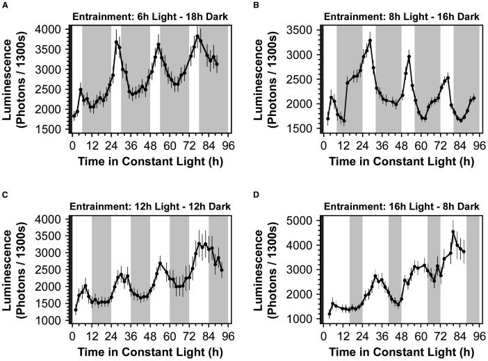
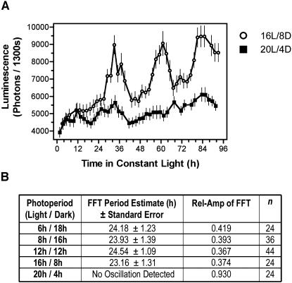
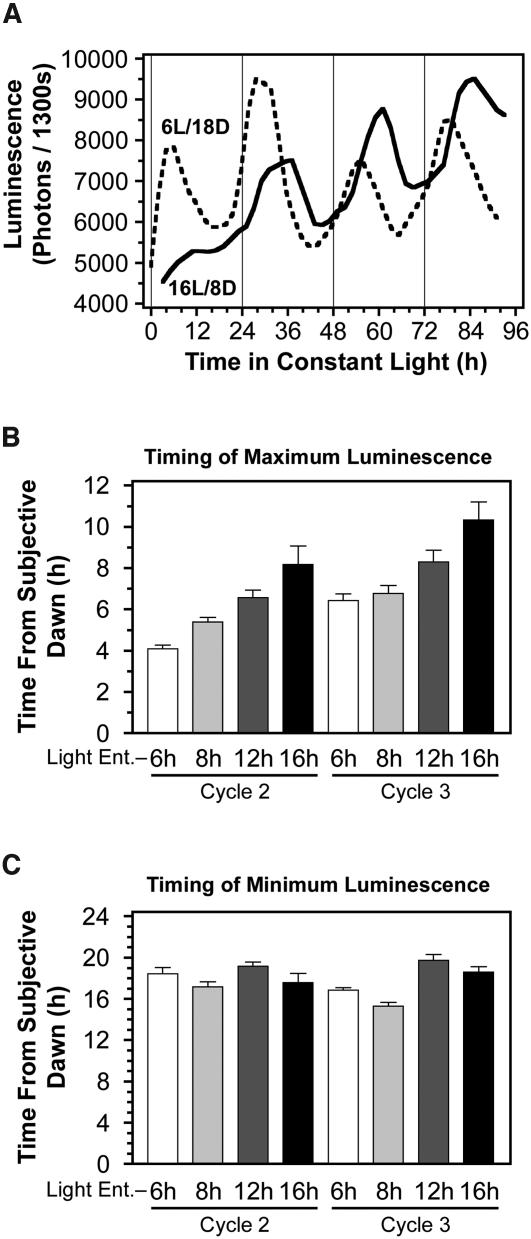
We tested whether circadian [Ca2+]cyt oscillations were modulated by changes in daylength (photoperiod). Seedlings were entrained in 6L/18D, 8L/16D, 12L/12D, 16L/8D, and 20L/4D. Seedlings entrained in 6L/18D, 8L/16D, 12L/12D, and 16L/8D had free-running rhythms in aequorin luminescence (Figure 3) with an approximate period of 24 h (Figure 4B). By contrast, seedlings entrained in 20L/4D had no rhythmic component to the bioluminescence signal (Figures 4A and 4B). The phase of the circadian [Ca2+]cyt oscillations was dependent on the entrainment photoperiod, with peak [Ca2+]cyt occurring later per 24-h cycle in plants entrained in long photoperiods than in plants entrained in short photoperiods (Figure 5A). The luminescence of seedlings entrained in 6L/18D peaked at a mean of 4.09 ± 0.19 h (n = 24) after subjective dawn for circadian cycle 2 (24 to 48 h after transfer to LL). Seedlings entrained in 8L/16D had peak luminescence at 5.39 ± 0.22 h (n = 36) after subjective dawn, seedlings entrained in 12L/12D had peak luminescence at 6.59 ± 0.37 h (n = 44) after subjective dawn, and seedlings entrained in 16L/8D were maximally luminescent 8.16 ± 0.90 h (n = 24) after subjective dawn, for circadian cycle 2. Thus, the longer the photoperiod during entrainment, the later the peak luminescence after subjective dawn in LL. This trend was repeated in the subsequent cycles of circadian free-run (e.g., cycle 3, from 48 to 72 h, and cycle 4, from 72 to 96 h after transfer to LL). The timing of minimum luminescence in LL was unaffected by the entrainment daylength and occurred ∼19 to 20 h after subjective dawn in seedlings from all entrainment regimes (Figure 5C). Consequently, the circadian [Ca2+]cyt oscillations were asymmetric, and the asymmetry depended on the entrainment photoperiod because the longer the photoperiod, the shorter the time difference between maximum and minimum [Ca2+]cyt. Therefore, the daylength during entrainment controlled both the phase and the shape (symmetry) of the [Ca2+]cyt oscillations during circadian free-run in LL.
Figure 3.
Circadian Oscillations of [Ca2+]cyt in Arabidopsis Seedlings Entrained to Different Photoperiods.
Aequorin luminescence emitted by seedlings in 110 μmol m−2 s−1 LL. Seedlings were entrained in 6L/18D (A), 8L/16D (B), 12L/12D (C), and 16L/8D (D) for 11 d before LL. During the entrainment light period, the PFD was 60 μmol m−2 s−1. Points represent the mean bioluminescence of 12 seedling clusters ±se. Open areas indicate the subjective day, and shaded areas indicate the subjective night. The closed area represents the final dark period of the entrainment.
Figure 4.
Circadian Oscillations of [Ca2+]cyt Were Arrhythmic in Very Long Photoperiods.
(A) Aequorin luminescence emitted by Arabidopsis seedlings entrained in 60 μmol m−2 s−1 16L/8D (open circles) or 20L/4D (closed squares) for 11 d before transfer to 110 μmol m−2 s−1 LL. Points represent the mean of 12 seedling clusters ±se. The period of the mean oscillation in aequorin bioluminescence was 26 h (relative amplitude error [Rel-Amp] = 0.31) for seedlings entrained in 16L/8D. However, no rhythmic component to the mean bioluminescence of seedlings entrained in 20L/4D was determined by FFT-NLLS.
(B) Table showing the mean ± se of the period of bioluminescence for seedling clusters expressing aequorin in LL. The period of bioluminescence was calculated for each cluster using FFT-NLLS. Seedlings were entrained in the photoperiods indicated at 60 μmol m−2 s−1 for 11 d before transfer to 110 μmol m−2 s−1 LL.
Figure 5.
The Photoperiod of Entrainment Shifts the Phase of Circadian Oscillations of [Ca2+]cyt in Constant Light.
(A) Aequorin luminescence of Arabidopsis seedlings in 110 μmol m−2 s−1 LL. Seedlings were entrained in 6L/18D (dashed line) or in 16L/8D (solid line) at a PFD of 60 μmol m−2 s−1 for 11 d before LL. For clarity, the points representing the mean values do not appear. The thin, vertical lines that intersect the abscissa at 0, 24, 48, and 72 h indicate subjective dawn for all seedlings.
(B) Timing of maximum aequorin luminescence, relative to subjective dawn, of Arabidopsis seedlings in LL, entrained to 6L/18D (open bars), 8L/16D (light-shaded bars), 12L/12D (dark-shaded bars), and 16L/8D (closed bars). Bars represent the mean luminescence of 24 to 36 seedling clusters, with standard errors shown. Circadian cycles are indicated below each set of bars: Cycle 2 corresponds to 24 to 48 h in LL, and cycle 3 corresponds to 48 to 72 h in LL.
(C) Timing of minimum aequorin luminescence, relative to subjective dawn, emitted by Arabidopsis seedlings in LL, entrained to 6L/18D (open bars), 8L/16D (light-shaded bars), 12L/12D (dark-shaded bars), and 16L/8D (closed bars). As in (B), bars represent the mean luminescence of 24 to 36 seedling clusters, with standard errors and the circadian cycles indicated below each set of bars.
The photoperiod-dependent phase shift of the circadian [Ca2+]cyt rhythms had significant consequences for [Ca2+]cyt at subjective dusk. At subjective dusk, relative luminescence (as a percentage of the maximum luminescence for each circadian cycle) was dependent on the duration of the entrainment photoperiod. The relative luminescence at subjective dusk of seedlings entrained to 6L/18D was 72.59 ± 10.92% (n = 24), that of seedlings entrained in 8L/16D was 59.82 ± 11.95% (n = 36), that of seedlings entrained in 12L/12D was 55.06 ± 11.10% (n = 44), and for seedlings entrained to 16L/8D, relative luminescence was 24.17 ± 8.91% (n = 24). Thus, at subjective dusk, [Ca2+]cyt was relatively high in plants entrained in short photoperiods and low in plants entrained in long photoperiods. These differences in [Ca2+]cyt at subjective dusk are a consequence of photoperiodic entrainment. Conversely, at subjective dawn, there was no significant difference in relative luminescence for seedlings entrained in the different LD regimes.
We next tested whether the phase and shape of circadian [Ca2+]cyt oscillations were set by the length of the dark period by transferring the seedlings to LL after a dark period equal to that they were entrained in (12 h) or following a final dark period that had been extended by 5 h (17 h total; Figure 6A). Extending the length of the final dark period did not alter the circadian period. The period estimate of the circadian rhythm of seedlings transferred to LL after a 5-h delay was 22.6 ± 1.47 h (n = 4), which was not significantly different to that of seedlings transferred to LL without delay; 21.9 ± 1.85 h (n = 10). Extending the length of the final dark period resulted in a shift in phase of the oscillations relative to each other, but the phase of the rhythms relative to the final subjective dawn was unaffected (Figure 6B). The timing of maximum luminescence relative to subjective dawn in the first three circadian cycles was not significantly different between the treatments (Figure 6B). The characteristics of the circadian luminescence oscillations were therefore determined by the entrainment regime and not by the length of the final dark period.
Figure 6.
The Effect of the Dark Period Length on Circadian Oscillations of [Ca2+]cyt.
(A) Aequorin luminescence of Arabidopsis seedlings in 110 μmol m−2 s−1 LL. Seedlings expressing apoaequorin were entrained in 60 μmol m−2 s−1 12L/12D for 11 d. Closed circles represent the mean luminescence with standard errors emitted by 10 seedling clusters that experienced the normal 12L/12D on day 11, before transfer to LL. Open circles represent the mean luminescence with standard errors emitted by four seedling clusters that received an additional 5 h of darkness before transfer to LL, hence a 12-h-light period followed by a 17-h-dark period on day 11. The bars on the abscissa indicate the subjective light regime during LL and are color-coded to match the graph. Open areas represent subjective day, hatched areas subjective night, and closed areas the final dark period before LL. For all seedlings, the time of transfer to LL is defined as the start (t = 0) of subjective dawn.
(B) Timing from subjective dawn of maximum aequorin luminescence emitted by Arabidopsis seedlings entrained in 60 μmol m−2 s−1 12L/12D. Bars represent the mean luminescence emitted by seedling clusters ±se. Closed bars (n = 10) indicate that seedlings received a 12-h-dark period before transfer to LL, and shaded bars (n = 4) denote seedlings for which the transfer from darkness to LL was delayed by 5 h. As for (A), the time of transfer to LL is defined as the t = 0 of subjective dawn.
Leaf [Ca2+]cyt Oscillations in LD Cycles Are Modulated by Photoperiod and Light Intensity
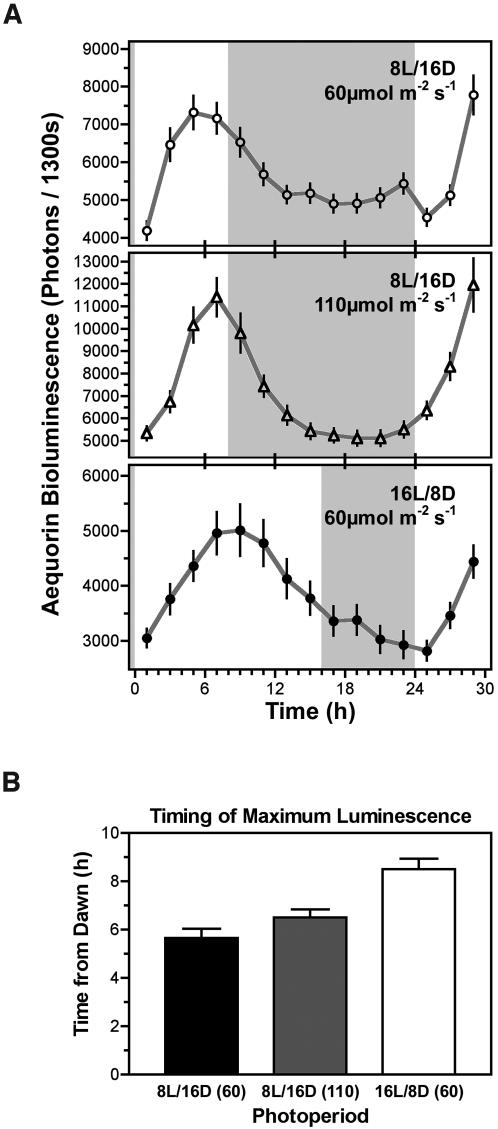
Although important information regarding biological clocks is obtained from studying organisms under constant conditions, such conditions are largely artificial. We therefore examined [Ca2+]cyt dynamics in more natural LD cycles to determine, firstly, whether [Ca2+]cyt oscillations with a 24-h period exist under these conditions and, secondly, whether the [Ca2+]cyt oscillations encode environmental information. Arabidopsis expressing aequorin was grown in 8L/16D or 16L/8D and luminescence measured in these same LD regimes rather than in LL (Figure 7). Plants grown in 16L/8D were exposed to a PFD of 60 μmol m−2 s−1. Plants grown in the 8L/16D regime were exposed to 60 μmol m−2 s−1 or to 110 μmol m−2 s−1, thus the latter received similar daily integrated photons as plants in 16L/8D.
Figure 7.
Light Intensity and Duration Modulates [Ca2+]cyt Oscillations in Light and Dark Cycles.
(A) Oscillations of aequorin luminescence from Arabidopsis seedlings in LD. Seedlings expressing apoaequorin were grown in 60 μmol m−2 s−1 8L/16D (open circles, top graph), in 110 μmol m−2 s−1 8L/16D (open triangles, middle graph), or in 60 μmol m−2 s−1 16L/8D (closed circles, bottom graph) for 14 d. Points represent the mean of 12 seedling clusters ±se. Open areas represent the light periods, and shaded areas represent the dark periods experienced by the seedlings during bioluminescence imaging.
(B) Timing from dawn of maximum aequorin luminescence emitted by Arabidopsis in 60 μmol m−2 s−1 8L/16D (closed bars), in 110 μmol m−2 s−1 8L/16D (shaded bars), or in 60 μmol m−2 s−1 16L/8D (open bars). Bars represent the mean luminescence of 12 seedling clusters ±se.
Seedlings in LD had [Ca2+]cyt oscillations that were strikingly similar to those observed in circadian free-run (LL). In LD, [Ca2+]cyt increased during the day to a peak value, peaked before dusk, and then decreased to a minimum during the dark period. The period of the [Ca2+]cyt oscillations in LD also was ∼24 h. Importantly, the timing of peak luminescence (phase) in LD was consistent with that in LL and dependent on the photoperiod (Figure 7). At dusk, the relative luminescence of seedlings in 8L/16D was 59.57 ± 15.02% (n = 22) compared with 24.89 ± 4.34% (n = 22) in 16L/8D. The relative intensity at subjective dusk was very similar whether measured in LD or LL. Seedlings entrained to 8L/16D had dusk relative luminescence values of 59% in both LD and in LL. Moreover, the relative luminescence at subjective or true dusk of seedlings entrained to 16L/8D was 24% in both LL and LD. Additionally, the amplitude of the [Ca2+]cyt oscillations in LD was consistently larger in the higher PFD (Figure 7A). These data demonstrate that leaf [Ca2+]cyt oscillates with a period of 24 h in light and dark cycles that are characteristic of natural environments and that the pattern of these oscillations was dependent on the photoperiod and the intensity of light.
DISCUSSION
We have examined both the location and potential for information encoding in circadian oscillations of [Ca2+]cyt. The spatial resolution provided by photon-counting imaging enabled localization of circadian [Ca2+]cyt oscillations to the leaves and cotyledons of Arabidopsis in both LL and LD. It is likely that both the epidermal and mesophyll cells contribute to the circadian oscillations of [Ca2+]cyt in the leaves (Wood et al., 2001). However, the imaging system we used did not allow us to investigate the localization of the [Ca2+]cyt oscillations at a cellular level. These data demonstrate that the circadian oscillations of [Ca2+]cyt reported previously for whole plants (Johnson et al., 1995) are present in the photosynthetic organs, where light stimuli are perceived and integrated.
The localization of 24-h oscillations of [Ca2+]cyt to the cotyledons and leaves suggests that they might have a role in transducing circadian and photoperiodic information (Lumsden, 1998). Our estimate of the amplitude of the circadian [Ca2+]cyt oscillation (∼300 nmol L−1) and that of Johnson et al. (1995; 600 nmol L−1) are comparable to the [Ca2+]cyt increases after excitation by extracellular stimuli in plant cells and are sufficient to activate many Ca2+-dependent signaling mechanisms (Webb et al., 2001; Hetherington and Woodward, 2003). More accurate measurement of the circadian control of [Ca2+]cyt in single cells requires experimental and technical advances. [Ca2+]cyt oscillations might serve in the transduction of light input to the circadian oscillator through the phytochrome and cryptochrome photoreceptors (Shacklock et al., 1992; Bowler, et al., 1994; Somers et al., 1998; Baum et al., 1999; Devlin and Kay, 2000; Guo et al., 2001; Fankhauser and Staiger, 2002) or as a regulatory output from the clock (Johnson et al., 1995; Sai and Johnson, 1999; Webb, 2003). Candidates for regulation by 24-h oscillations of [Ca2+]cyt include physiological and developmental processes in the leaf that are known to be regulated by both the circadian clock and alterations in [Ca2+]cyt (Bakrim et al., 2001; Jung et al., 2002; Webb, 2003). However, the role of [Ca2+]cyt oscillations has limitations: Circadian [Ca2+]cyt rhythms are not involved in the circadian regulation of the LIGHT-HARVESTING COMPLEX B (LHCB) promoter. The rhythms of LHCB promoter activity and [Ca2+]cyt are asynchronous in LL and uncoupled such that in callus, rhythms of LHCB promoter activity can be detected in the absence of [Ca2+]cyt rhythms (Sai and Johnson, 1999).
Previously, circadian oscillations in [Ca2+]cyt have been detected in N. plumbaginifolia and in Arabidopsis (Johnson et al., 1995; Wood et al., 2001). In those studies, plants were entrained in 16L/8D, and [Ca2+]cyt peaked just before subjective dusk. The data presented here indicated an earlier peak in [Ca2+]cyt for plants entrained in 16L/8D that occurred between 8 h and 10 h after subjective dawn. Our study differs in the light intensity supplied to the plants (22 μmol m−2 s−1 in the previous studies [Johnson et al., 1995; Wood et al., 2001] compared with 60 or 110 μmol m−2 s−1 for our measurements), suggesting one possible explanation for the different phasing of the circadian [Ca2+]cyt oscillations in the two studies. However, when we tested the effect of varying PFD, we found that PFD affected the amplitude but not the period or the phase of the circadian bioluminescence oscillations (Figure 2). The discrepancy between this investigation and the earlier studies may instead be as a result of the different methods of bioluminescence detection used. Aequorin luminescence was previously quantified by luminometry, as an integrated signal from whole seedlings (Johnson et al., 1995; Wood et al., 2001). However, different tissues have differently phased circadian [Ca2+]cyt oscillations (Wood et al., 2001) that may summate to produce the integrated signal detected using the luminometer. Conversely, in this study, aequorin bioluminescence was imaged from only the cotyledons and emerging leaves of the seedlings. Thus, the enhanced resolution of photon-counting imaging may explain the differences between these and earlier data.
Our data provide new evidence that circadian and photoperiodic information can be encoded in the pattern of [Ca2+]cyt oscillations in both circadian free-run in LL and in LD cycles. This has important implications for understanding signaling in plants. We have demonstrated that the phase (or timing of the peak of [Ca2+]cyt) and shape of free-running circadian oscillations of [Ca2+]cyt in LL were dependent on the length of the light and dark cycles during entrainment. The peak of [Ca2+]cyt after subjective dawn in LL occurred later in those plants that had been entrained to long light periods than the peak in plants entrained to short light periods. These data demonstrate that the phase of the circadian oscillations of [Ca2+]cyt was sensitive to the length of the light and/or dark cycles during entrainment. However, the timing of the trough of [Ca2+]cyt relative to subjective dawn was not altered by the photoperiodic entrainment.
Another effect of the phase shift of the [Ca2+]cyt rhythms was that the timing of the peak was altered in relation to subjective dusk. The peak of [Ca2+]cyt in LL in plants entrained to 6L/18D occurred at the end of the subjective light period, 0 to 2 h before subjective dusk. By comparison, the peak in plants entrained to 16L/8D occurred in the middle of the subjective light period, 6 to 8 h before subjective dusk. Plants entrained in 6L/18D therefore emitted ∼80% of the peak luminescence level at subjective dusk, whereas plants entrained in 16L/8D emitted only 25% of the peak luminescence level at subjective dusk.
These data demonstrate that the entrainment photoperiod regulates the phase and shape of the circadian [Ca2+]cyt oscillations and importantly, the [Ca2+]cyt at subjective dusk. It is therefore possible that these characteristics of circadian [Ca2+]cyt oscillations encode information that affects the integration and outcomes of Ca2+-based signaling networks (Berridge et al., 2000; Evans et al., 2001; Sanders et al., 2002; Schuster et al., 2002; Hetherington and Woodward, 2003).
Two aspects of circadian [Ca2+]cyt oscillations in Arabidopsis seedlings, the phase and the amplitude of the oscillations, are modulated by light quantity and entrainment photoperiod. The modulation of the phase and amplitude of circadian oscillations of [Ca2+]cyt by environmental stimuli is consistent with the hypothesis that information is encoded in the dynamics of circadian [Ca2+]cyt oscillations (Jaffe, 1991; McAinsh et al., 1995; Ehrhardt et al., 1996; McAinsh and Hetherington, 1998; Allen et al., 2001; Evans et al., 2001; Shaw and Long, 2003). If information is encoded, there must also be mechanisms for decoding circadian [Ca2+]cyt oscillations. In plants these mechanisms remain obscure, but models exist based on counteracting and differentially sensitive regulatory enzymes and posttranslational and biochemical modification of Ca2+-sensitive proteins in phase with [Ca2+]cyt oscillations (McAinsh and Hetherington, 1998; Schuster et al., 2002; Cullen, 2003).
We also imaged the daily [Ca2+]cyt dynamics of the leaves and cotyledons of seedlings in light and dark cycles more representative of the natural environment. The [Ca2+]cyt oscillations in LD were strikingly similar to those observed in LL: [Ca2+]cyt rose during the day, peaked several hours after dawn, and then decreased, attaining a minimal value during the night. Additionally, the photoperiod-dependent phase shift of [Ca2+]cyt oscillations and amplitude modulation observed in LL occurred also in LD. In 8L/16D, the peak [Ca2+]cyt occurred at 6 h after dawn but in 16L/8D, the peak [Ca2+]cyt occurred at 8 h after dawn. Higher PFD in 8L/16D increased the amplitude of the oscillations in [Ca2+]cyt, in agreement with our observations of circadian [Ca2+]cyt oscillations in LL, but PFD did not affect the phase of the [Ca2+]cyt oscillations in LD. Thus, the effects of daylength on [Ca2+]cyt in LD were because of the duration of the light and dark cycles and not the amount of light to which the seedlings were exposed. These data demonstrate that [Ca2+]cyt in cotyledon and leaf cells oscillated with a 24-h period in LD cycles and that the phase of these oscillations was sensitive to the entrainment regime and the amplitude was sensitive to PFD. Importantly, the level of [Ca2+]cyt at dusk was relatively high in plants in short days and low in plants in long days. This observation strongly suggests that [Ca2+]cyt oscillations in LD are not directly regulated by light but are instead associated with the circadian clock and may play a role in circadian signaling in response to LD cycles.
There was one major difference between the oscillations of [Ca2+]cyt in LL and those in LD. The luminescence of seedlings in LD did not increase significantly before dawn but only rose after illumination. This difference between the Ca2+ dynamics of plants in LD or in LL can be explained by the observation that [Ca2+]cyt is arrhythmic in darkness (Johnson et al., 1995). Therefore, it appears that [Ca2+]cyt can rise in anticipation of dawn only in the light, which is why the circadian anticipation of dawn was only detected in LL but not LD cycles.
The slow rates of increase and decrease of [Ca2+]cyt in LD suggest that the daily [Ca2+]cyt oscillations were not generated as an immediate, acute response to illumination but instead by a complex signaling network cued to LD cycles and the circadian clock (Millar and Kay, 1996; Hall et al., 2002). Nevertheless, short-term or acute responses of [Ca2+]cyt have been reported. In Triticum aestivum (wheat) protoplasts, dark-light transitions generate brief elevations in [Ca2+]cyt lasting <5 min in response to red light (Shacklock et al., 1992). In Arabidopsis and Nicotiana tabacum (tobacco), blue light elevates [Ca2+]cyt via PHOT1 and PHOT2 phototropin-mediated signaling (Baum et al., 1999; Stoelzle et al., 2003). Organellar [Ca2+] also has an acute response to illumination. In the chloroplast, the dark-light transition generates a rapid increase in stromal [Ca2+] that reaches a peak value between 25 to 30 min after dawn (Sai and Johnson, 1999). Our assay was not designed to measure short-term changes in [Ca2+]cyt, and our measurements began 30 min after the dark-to-light transition to avoid the possibility of re-entraining the plants to a new rhythm. It is therefore possible that short, transient increases in [Ca2+]cyt immediately after dawn were superimposed on the 24-h oscillation of [Ca2+]cyt that occurred in LD and were not detected by our method.
Perhaps one of the most important plant responses regulated by daylength is the photoperiodic transition from vegetative growth to the development of flowers (Yanovsky and Kay, 2002; Hayama and Coupland, 2003). In LD, [Ca2+]cyt oscillates with amplitude and phase that are sensitive to light quantity and entrainment photoperiod. Most importantly, these diurnal [Ca2+]cyt oscillations result in substantially different [Ca2+]cyt at critical periods of the day, depending on the length of the light period. Consequently, it is compelling to consider a role for [Ca2+]cyt in the signal transduction pathways of photoperiodic responses. Pharmacological studies (Friedman et al., 1989) and direct measurement of [Ca2+]cyt (Walczysko et al., 2000) support a role for alterations in [Ca2+]cyt in the transduction pathways regulating floral induction in response to changes in photoperiod. It has also been proposed that Ca2+-dependent protein kinase activity acts as a Ca2+ sensor in photoperiodism (Jaworski et al., 2003). Additionally, it has recently been suggested that extracellular Ca2+ may have a role in regulating the switch to flowering through the action of a cell-surface Ca2+ sensor (Han et al., 2003). Strikingly, this cell-surface Ca2+ sensor also has a role in generating oscillations of [Ca2+]cyt in the guard cell in response to elevations of external [Ca2+]cyt (Han et al., 2003). Although our data demonstrate that [Ca2+]cyt oscillations encode circadian and photoperiodic information, further work is required to determine whether this information is decoded and contributes to physiological processes, such as the control of floral induction.
METHODS
Seedling Growth and Photoperiodic Entrainment
Transgenic Arabidopsis seeds (ecotype Wassilewskija) that constitutively express the apoaequorin cDNA under the control of the 35S promoter of Cauliflower mosaic virus were surface-sterilized and germinated on solid medium (0.5× Murashige and Skoog medium and 0.8% (w/v) agar, pH = 6.8) in clusters of 15 to 20 plants. Surface sterilization was performed by rinsing seeds for 1 min in 100% ethanol, then by incubating seeds for 10 min in 50% (v/v) sodium hypochlorite, followed by two washes in sterile water. Seed germination was synchronized by vernalization at 4°C for 2 d in the dark. Seedlings were grown in white light at a PFD of 60 μmol m−2 s−1 at 19°C for 11 d. During the growth period, seedlings were entrained to several photoperiods: 6L/18D, 8L/16D, 12L/12D, 16L/8D, or 20L/4D. Plants were then transferred to 60 μmol m−2 s−1 or 110 μmol m−2 s−1 of LL for imaging circadian [Ca2+]cyt oscillations under circadian free-run conditions. In a separate protocol, plants remained under the appropriate entrainment regime for measurement of [Ca2+]cyt dynamics under LD conditions. During the dark period, plants were handled in a dark room under green safelight.
Imaging Aequorin Bioluminescence
[Ca2+]cyt was measured in vivo using the bioluminescent Ca2+-reporter aequorin. For plants in LL conditions, 100 μL of 5 μmol L−1 coelenterazine-free base (Prolume, Pinetop, AZ) was applied to each seedling cluster 24 h and 12 h before imaging, to ensure high levels of reconstituted aequorin. Photon counting was performed in a light-tight box using a Photek (Hastings, UK) ICCD225 photon-counting camera system mounted above the seedlings. The camera was controlled by Photek IFS32 software. Immediately before photon counting, bright-field images of the seedlings in position under the camera were captured. The bright-field illumination was from low-voltage, biologically safe, green light–emitting diodes, which provided a PFD <1 μmol m−2 s−1. The bioluminescence emitted by each seedling cluster was recorded for 1500 s, every 2 h, for 4 d. The first 200 s of each photon-counting integration contained luminescence generated by the autofluorescence decay of chlorophyll and was discarded. Eight-bit images of the photon density were generated from the remaining 1300 s of each integration and pseudocolored. Unless otherwise indicated, ∼10 μL of 5 μmol L−1 coelenterazine was applied to each seedling cluster after imaging, to maintain high levels of aequorin in the plants. The baseline of aequorin luminescence increased during the experiments as a result of a combination of regular coelenterazine application and seedling growth. Coelenterazine treatment had no adverse effects on the seedlings, which increased in size during the experiments, showed no visible signs of damage, and could be successfully grown to seed. For plants imaged under LD rather than LL conditions, seedlings were dosed twice daily with 100 μL of 10 μmol L−1 coelenterazine for 3 d before imaging. These seedlings received no further coelenterazine during imaging.
The integrated 8-bit photon-counting images were processed using the Photek IFS32 software. Regions of interest, based on the bright-field image, were demarcated around each seedling cluster, and total photon counts (luminescence) were quantified. Rhythms of aequorin luminescence, corresponding to rhythms in [Ca2+]cyt, were analyzed using two methods. First, the fast Fourier transform–nonlinear least-squares method (FFT-NLLS) described by Plautz et al. (1997) was used to determine the period of each circadian rhythm in luminescence of the mean signal from 12 seedling clusters. The degree of confidence in the FFT-NLLS is described by the relative amplitude error, which ranges from 0 to 1, with 0 indicating a rhythm known to infinite precision and 1 indicating a rhythm that was not statistically significant (Plautz et al., 1997). Second, the timing of minimum and maximum bioluminescence during each 24-h period was determined for each seedling cluster and averaged. Cycle 1, from 0 to 24 h, immediately followed the last dark period of the entrainment regime and was therefore not considered a true circadian cycle. Cycles 2 and 3, from 24 to 48 h and from 48 to 72 h, respectively, represented genuine circadian cycles. Cycle 4, from 72 to 96 h, was also a circadian cycle but was not used because not all data sets continued until 96 h. n in the text refers to the number of seedling clusters analyzed.
Estimation of the Total Aequorin Pool and Magnitude of the Oscillations in [Ca2+]cyt
The size of the aequorin pool was quantified in 30 seedlings at opposite phases of the circadian cycle (t = 7.5 and 19.5) by discharging the entire pool with 1 M L−1 CaCl2 in 10% (v/v) ethanol and quantifying the luminescent signal using a photon-counting luminometer (photon multiplier tube 9899A cooled to −14 C with a FACT50 housing; Electron Tubes, Middlesex, UK).
The long time scale of the measurements compromise estimation of [Ca2+]cyt based on the rate of aequorin consumption as a proportion of the total pool (Johnson et al., 1995). Additionally, we could not measure the total aequorin pool using the photon-counting camera because the light emission after discharge of the entire pool (Fricker et al., 1999) saturated the detector. Instead, we used the fold increase in light to estimate the circadian changes in [Ca2+]cyt (Cobbold and Rink, 1987). Fold increases in light were converted to [Ca2+]cyt by comparison with the fold increase in light during imaging of transient excursions of [Ca2+]cyt in response to NaCl stimulation of seedlings.
To generate a calibration curve, we used data obtained from photon-counting imaging of 7-d-old aequorin-expressing seedlings irrigated with 100, 150, 200, or 300 mol m−3 NaCl in Murashige and Skoog medium (n = 20; F. Tracy and M. Tester, personal communication). Functional aequorin had been generated by overnight incubation with 10 μmol L−1 coelenterazine in the dark. The seedlings were imaged continuously before, during, and after perfusion with NaCl containing solution. A time-resolved integration graph of the integrated photon count s−1 was plotted, and the peak fold increase in light was calculated. Previously, the mean peak [Ca2+]cyt increase in response to 100, 150, 200, or 300 mol L−1 NaCl in Murashige and Skoog medium had been measured using photon-counting luminometry calibrated exactly as described in Fricker et al. (1999; n = 120; F. Tracy and M. Tester, personal communication). By comparing the mean peak height of the [Ca2+]cyt transients in response to NaCl stimulation measured using luminometry with the mean peak fold change in light detected by the imaging camera, we made a calibration curve of estimated [Ca2+]cyt increase against fold change in light detected by the camera. We estimated the changes in [Ca2+]cyt in response to circadian and light signals by comparing of the fold increases in light during the circadian experiments with the calibration curve generated from the NaCl experiments.
Acknowledgments
We thank Marc Knight of the University of Oxford (Oxford, UK) Department of Plant Sciences for his kind gift of Arabidopsis expressing apoaequorin, and Carl H. Johnson of Vanderbilt University (Nashville, TN) and Enid MacRobbie and Julia Davies (University of Cambridge, Cambridge, UK) for their helpful comments on the manuscript. We thank Francis Tracey and Mark Tester (University of Cambridge) for providing access to their unpublished data. We also acknowledge the financial support of the Broodbank Fellowship Trust of the University of Cambridge (to J.L.), the Royal Society of London, and the Biotechnology and Biological Sciences Research Council of the United Kingdom (to A.A.R.W).
The author responsible for distribution of materials integral to the findings presented in this article in accordance with the policy described in the Instructions for Authors (www.plantcell.org) is: Alex A.R. Webb (alex.webb@plantsci.cam.ac.uk).
Article, publication date, and citation information can be found at www.plantcell.org/cgi/doi/10.1105/tpc.020214.
References
- Alabadi, D., Oyama, T., Yanovsky, M.J., Harmon, F.G., Mas, P., and Kay, S.A. (2001). Reciprocal regulation between TOC1 and LHY/CCA1 within the Arabidopsis circadian clock. Science 293, 880–883. [DOI] [PubMed] [Google Scholar]
- Allen, G.J., Chu, S.P., Harrington, C.L., Schumacher, K., Hoffmann, T., Tang, Y.Y., Grill, E., and Schroeder, J.I. (2001). A defined range of guard cell calcium oscillation parameters encodes stomatal movements. Nature 411, 1053–1057. [DOI] [PubMed] [Google Scholar]
- Bakrim, N., Brulfert, J., Vidal, J., and Chollet, R. (2001). Phosphoenolpyruvate carboxylase kinase is controlled by a similar signaling cascade in CAM and C4 plants. Biochem. Biophys. Res. Commun. 286, 1158–1162. [DOI] [PubMed] [Google Scholar]
- Baum, G., Long, J.C., Jenkins, G.I., and Trewavas, A.J. (1999). Stimulation of the blue light phototropic receptor NPH1 causes a transient increase in cytosolic Ca2+. Proc. Natl. Acad. Sci. USA 96, 13554–13559. [DOI] [PMC free article] [PubMed] [Google Scholar]
- Berridge, M.J., Lipp, P., and Bootman, M.D. (2000). The versatility and universality of calcium signalling. Nat. Rev. Mol. Cell Biol. 1, 11–21. [DOI] [PubMed] [Google Scholar]
- Bowler, C., Neuhaus, G., Yamagata, H., and Chua, N.H. (1994). Cyclic GMP and calcium mediate phytochrome phototransduction. Cell 77, 73–81. Erratum. Cell 79, 743. [DOI] [PubMed] [Google Scholar]
- Cobbold, P.H., and Rink, T.J. (1987). Fluorescence and bioluminescence measurement of cytoplasmic free calcium. Biochem. J. 248, 313–328. [DOI] [PMC free article] [PubMed] [Google Scholar]
- Cullen, P.J. (2003). Calcium signalling: The ups and downs of protein kinase C. Curr. Biol. 13, R699–R701. [DOI] [PubMed] [Google Scholar]
- Devlin, P.F. (2002). Signs of the time: Environmental input to the circadian clock. J. Exp. Bot. 53, 1535–1550. [DOI] [PubMed] [Google Scholar]
- Devlin, P.F., and Kay, S.A. (2000). Cryptochromes and phytochromes are required for phytochrome signalling to the circadian clock but not for rhythmicity. Plant Cell 12, 2499–2509. [DOI] [PMC free article] [PubMed] [Google Scholar]
- Ehrhardt, D.W., Wais, R., and Long, S.R. (1996). Calcium spiking in plant root hairs responding to Rhizobium nodulation signals. Cell 85, 673–681. [DOI] [PubMed] [Google Scholar]
- Evans, N.H., McAinsh, M.R., and Hetherington, A.M. (2001). Calcium oscillations in higher plants. Curr. Opin. Plant Biol. 4, 415–420. [DOI] [PubMed] [Google Scholar]
- Fankhauser, C., and Staiger, D. (2002). Photoreceptors in Arabidopsis thaliana: Light perception, signal transduction and entrainment of the endogenous clock. Planta 216, 1–16. [DOI] [PubMed] [Google Scholar]
- Fricker, M.D., Plieth, C., Knight, H., Blancaflor, E., Knight, M.R., White, N.S., and Gilroy, S. (1999). Fluorescence and luminescence techniques to probe ion activities in living plant cells. In Fluorescent and Luminescent Probes for Biological Activity, 2nd ed., W.T. Mason, ed (San Diego, CA: Academic Press), pp. 569–596.
- Friedman, H., Goldschmidt, E.E., and Halevy, A.H. (1989). Involvement of calcium in the photoperiodic flower induction process of Pharbitis nil. Plant Physiol. 89, 530–534. [DOI] [PMC free article] [PubMed] [Google Scholar]
- Gómez, L.A., and Simón, E. (1995). Circadian rhythm of Robinia pseudoacacia leaflet movement: Role of calcium and phytochrome. Photochem. Photobiol. 61, 210–215. [Google Scholar]
- Guo, H., Mockler, T., Duong, H., and Lin, C. (2001). SUB1, an Arabidopsis Ca2+-binding protein involved in cryptochrome and phytochrome coaction. Science 291, 487–490. [DOI] [PubMed] [Google Scholar]
- Hall, A., Kozma-Bognár, L., Bastow, R.M., Nagy, F., and Millar, A.J. (2002). Distinct regulation of CAB and PHYB gene expression by similar circadian clocks. Plant J. 32, 529–537. [DOI] [PubMed] [Google Scholar]
- Han, S., Tang, R., Anderson, L.K., Woerner, T.E., and Pei, Z.-M. (2003). A cell surface receptor mediates extracellular Ca2+ sensing in guard cells. Nature 425, 196–200. [DOI] [PubMed] [Google Scholar]
- Harmer, S.L., Hogenesch, J.B., Straume, M., Chang, H.S., Han, B., Zhu, T., Wang, X., Kreps, J.A., and Kay, S.A. (2000). Orchestrated transcription of key pathways in Arabidopsis by the circadian clock. Science 290, 2110–2113. [DOI] [PubMed] [Google Scholar]
- Harmer, S.L., Panda, S., and Kay, S.A. (2001). Molecular basis of circadian rhythms. Annu. Rev. Cell Dev. Biol. 17, 215–253. [DOI] [PubMed] [Google Scholar]
- Hayama, R., and Coupland, G. (2003). Shedding light on the circadian clock and the photoperiodic control of flowering. Curr. Opin. Plant Biol. 6, 13–19. [DOI] [PubMed] [Google Scholar]
- Hetherington, A.M., and Woodward, F.I. (2003). The role of stomata in sensing and driving environmental change. Nature 424, 901–908. [DOI] [PubMed] [Google Scholar]
- Ikeda, M., Sugiyama, T., Wallace, C.S., Gompf, H.S., Yoshioka, T., Miyawaki, A., and Allen, C.N. (2003). Circadian dynamics of circadian and nuclear Ca2+ in single suprachiasmatic nucleus neurones. Neuron 38, 253–263. [DOI] [PubMed] [Google Scholar]
- Jaffe, L.F. (1991). The path of calcium in cytosolic calcium oscillations: A unifying hypothesis. Proc. Natl. Acad. Sci. USA 88, 9883–9887. [DOI] [PMC free article] [PubMed] [Google Scholar]
- Jaworski, K., Szmidt-Jaworska, A., Tretyn, A., and Kopcewicz, J. (2003). Biochemical evidence for a calcium-dependent protein kinase from Pharbitis nil and its involvement in photoperiodic flower induction. Phytochemistry 62, 1047–1055. [DOI] [PubMed] [Google Scholar]
- Johnson, C.H., Knight, M.R., Kondo, T., Masson, P., Sedbrook, J., Haley, A., and Trewavas, A.J. (1995). Circadian oscillations of cytosolic and chloroplastic free calcium in plants. Science 269, 1863–1865. [DOI] [PubMed] [Google Scholar]
- Jung, J.Y., Kim, Y.W., Kwak, J.M., Hwang, J.U., Young, J., Schroeder, J.I., Hwang, I., and Lee, Y. (2002). Phosphatidylinositol 3- and 4-phosphate are required for normal stomatal movements. Plant Cell 14, 2399–2412. [DOI] [PMC free article] [PubMed] [Google Scholar]
- Lumsden, P.J. (1998). Photoperiodic induction in short-day plants. In Biological Rhythms and Photoperiodism in Plants, P. Lumsden and A. Millar, eds (Oxford, UK: Bios Scientific Publishers), pp. 167–182.
- McAinsh, M.R., and Hetherington, A.M. (1998). Encoding specificity in Ca2+ signaling systems. Trends Plant Sci. 3, 32–36. [Google Scholar]
- McAinsh, M.R., Webb, A., Taylor, J.E., and Hetherington, A.M. (1995). Stimulus-induced oscillations in guard cell cytosolic free calcium. Plant Cell 7, 1207–1219. [DOI] [PMC free article] [PubMed] [Google Scholar]
- Millar, A.J., and Kay, S.A. (1996). Integration of circadian and phototransduction pathways in the network controlling CAB gene expression in Arabidopsis. Proc. Natl. Acad. Sci. USA 93, 15491–15496. [DOI] [PMC free article] [PubMed] [Google Scholar]
- Morre, D.J., Chueh, P.J., Pletcher, J., Tang, X., Wu, L.Y., and Morre, D.M. (2002). Biochemical basis for the biological clock. Biochemistry 41, 11941–11945. [DOI] [PubMed] [Google Scholar]
- Plautz, J.D., Straume, M., Stanewsky, R., Jamison, C.F., Brandes, C., Dowse, H.B., Hall, J.C., and Kay, S.A. (1997). Quantitative analysis of Drosophilla period gene transcription in living animals. J. Biol. Rhythms 12, 204–217. [DOI] [PubMed] [Google Scholar]
- Sai, J., and Johnson, C.H. (1999). Different circadian oscillators control Ca2+ fluxes and Lhcb gene expression. Proc. Natl. Acad. Sci. USA 96, 11659–11663. [DOI] [PMC free article] [PubMed] [Google Scholar]
- Sanders, D., Pelloux, J., Brownlee, C., and Harper, J.F. (2002). Calcium at the crossroads of signaling. Plant Cell 14, S401–S417. [DOI] [PMC free article] [PubMed] [Google Scholar]
- Schuster, S., Marhl, M., and Hofer, T. (2002). Modelling of simple and complex calcium oscillations. From single-cell responses to intercellular signalling. Eur. J. Biochem. 269, 1333–1355. [DOI] [PubMed] [Google Scholar]
- Shacklock, P.S., Read, N.D., and Trewavas, A.J. (1992). Cytosolic free calcium mediates red light-induced photomorphogenesis. Nature 358, 753–755. [Google Scholar]
- Shaw, S.L., and Long, S.R. (2003). Nod factor elicits two separable calcium responses in Medicago truncatula root hair cells. Plant Physiol. 131, 976–984. [DOI] [PMC free article] [PubMed] [Google Scholar]
- Somers, D.E. (2001). Clock-associated genes in Arabidopsis: A family affair. Philos. Trans. R. Soc. Lond. B Biol. Sci. 356, 1745–1753. [DOI] [PMC free article] [PubMed] [Google Scholar]
- Somers, D.E., Devlin, P.F., and Kay, S.A. (1998). Phytochromes and cryptochromes in the entrainment of the Arabidopsis circadian clock. Science 282, 1488–1490. [DOI] [PubMed] [Google Scholar]
- Stoelzle, S., Kagawa, T., Wada, M., Hedrich, R., and Dietrich, P. (2003). Blue light activates calcium-permeable channels in Arabidopsis mesophyll cells via the phototropin signaling pathway. Proc. Natl. Acad. Sci. USA 100, 1456–1461. [DOI] [PMC free article] [PubMed] [Google Scholar]
- Trewavas, A.J. (1999). Le calcium c'est la vie: Calcium makes waves. Plant Physiol. 120, 1–6. [DOI] [PMC free article] [PubMed] [Google Scholar]
- Walczysko, P., Wagner, E., and Albrechtova, J.T. (2000). Use of co-loaded Fluo-3 and Fura Red fluorescent indicators for studying the cytosolic Ca2+ concentrations distribution in living plant tissue. Cell Calcium 28, 23–32. [DOI] [PubMed] [Google Scholar]
- Webb, A.A.R. (1998). Stomatal rhythms. In Biological Rhythms and Photoperiodism in Plants, P. Lumsden and A. Millar, eds (Oxford, UK: Bios Scientific Publishers), pp. 69–80.
- Webb, A.A.R. (2003). The physiology of circadian rhythms in plants. New Phytol. 160, 281–303. [DOI] [PubMed] [Google Scholar]
- Webb, A.A.R., Larman, M.G., Montgomery, L.T., Taylor, J.E., and Hetherington, A.M. (2001). The role of calcium in ABA-induced gene expression and stomatal movements. Plant J. 26, 351–362. [DOI] [PubMed] [Google Scholar]
- Wood, N.T., Haley, A., Viry-Moussaïd, M., Johnson, C.H., van der Luit, A.H., and Trewavas, A.J. (2001). The calcium rhythms of different cell types oscillate with different circadian phases. Plant Physiol. 125, 787–796. [DOI] [PMC free article] [PubMed] [Google Scholar]
- Yanovsky, M.J., and Kay, S.A. (2002). Molecular basis of seasonal time measurement in Arabidopsis. Nature 419, 308–312. [DOI] [PubMed] [Google Scholar]


