

Abstract
The mosquito Aedes aegypti is the principal vector that transmits dengue virus (DENV) to humans. The primary factors that trigger a susceptible or refractory interaction of A. aegypti with DENV are not well understood. In this study, our aim is to characterize the influence of vector genotype on differential expression of susceptible vs. refractory A. aegypti strains to DENV infection. To accomplish that we identified differential expression of a set of cDNAs (n = 9,504) of the D2S3 (susceptible) and Moyo-D (refractory) strains of A. aegypti to dengue virus serotype 2 (JAM1409) and compared these results to the differential expression of cDNAs in a different susceptible vector genotype (Moyo-S) relative to the same refractory genotype (Moyo-D) identified from our previous study. We observed that, although the number of differentially expressed transcripts (DETs) were similar in both the studies, about ~95% of the DETs were distinct between Moyo-D/D2S3 vs. Moyo-D/Moyo-S. This suggested that A. aegypti response, to infection of a given genotype of dengue, is largely dependent upon the vector genotype. However, we observed a set of common DETs among the vector strains that were associated with predicted functions such as endocytosis, regulation of autophagy, peroxisome and lipid metabolism that may be relatively universal in conferring mosquito response to DENV infection.
Keywords: Vector competence, dengue virus, microarray, Aedes aegypti, vector-virus interaction
Introduction
Dengue, dengue with warning and severe dengue (WHO, 2009) collectively represent a major public health problem in more than 100 countries in subtropical and tropical regions. Dengue virus (DENV), the causative agent, is vector borne and is primarily transmitted by the mosquito Aedes aegypti. At present, there are no licensed vaccines or drugs for dengue prevention/cure and, therefore, vector control remains the major strategy to prevent dengue infection. Because A. aegypti co-habits with approximately 40% of the world population, the global risk of DENV infection is significant. According to the World Health Organization, more than 500,000 people are hospitalized each year due to dengue related diseases and many of them (~ 20,000) lead to severe complications resulting in death.
The resilient nature of A. aegypti has become a major subject of scrutiny in the recent years (Philips 2008, WHO 2009). Natural populations of A. aegypti mosquitoes show varying degrees of susceptibility to DENV (Gubler et al. 1979; Rosen et al. 1985; Bennett et al. 2002; Diallo et al. 2008). Although progress has been made in our understanding of functional genomics of A. aegypti (Severson and Behura 2012), precise mechanisms of how the mosquito hosts or defends against DENV infection are unclear.
Susceptibility or refractoriness of A. aegypti populations to DENV infection is controlled by robust activation of molecular factors in the infected mosquito. DENV enters the mosquito upon blood feeding on viremic human hosts and then must establish an infection in epithelial cells of the mosquito’s midgut. The success or failure of establishment of DENV infection in the midgut is one of the important factors that define vector competence of the mosquito. The intrinsic ability of A. aegypti to either host or defend against viral infection is generally referred to as ‘vector competence’. Genetic studies suggest that DENV vector competence in A. aegypti is determined by multiple quantitative trait loci (QTL) in the genome (Bosio et al. 2000; Gomez-Machorro et al., 2004; Bennett et al., 2005). The limitation of DENV infection in a refractory A. aegypti strain has been shown to involve genetic mechanisms that either prohibit the virus from establishing an infection in the mid-gut epithelium (mid-gut infection barrier, MIB) or prevent virus escape from the mid-gut (mid-gut escape barrier, MEB) to other tissues including the salivary glands, that is essential for subsequent transmission to another human host (Bosio et al. 1998). To date, genetic barriers to salivary gland infection or escape have not been identified in A. aegypti populations.
The interaction between A. aegypti and dengue is a dynamic co-evolutionary process wherein the vector seeks to defend against infection and the virus undergoes adaptive selection to facilitate its survival (Rico-Hesse 2007). Accordingly, the outcomes of vector-virus interactions are intricately dependent upon the genotypes of vector, virus and the environmental factors as well. Gene expression studies have identified genes and pathways in both mosquito host and human host that may be involved in dengue virus infection (Sessions et al. 2009). It has been suggested that Toll and JAK-STAT pathways have important roles in susceptibility of A. aegypti to DENV (Xi et al., 2008; Souza-Neto et al. 2009). A candidate protein of A. aegypti has also been identified that binds to dengue virus and contributes to dengue infectivity in the mosquito (Mercado-Curiel et al. 2008). Differential expression of mid-gut serine protease and trypsin genes have also been suggested as having role in DENV-2 infectivity in A. aegypti (Molina-Cruz et al. 2005, Brackney et al. 2008). However, the potential for differential response of these genes or pathways to DENV infection in refractory versus susceptible mosquito genotypes is unclear. In earlier studies, we employed microarrays to compare gene expression profiles in susceptible and refractory genotypes of A. aegypti and identified genes that are expressed in a highly networked manner to trigger a susceptible or refractory response to DENV infection (Behura et al. 2011; Chauhan et al. 2012). The results identified modular patterns of gene expression that were significantly different between the susceptible and refractory genetic backgrounds. Based on sequences of genes identified as responsive and non-responsive to the infection, it was found that different intrinsic features of A. aegypti genes are correlated with the transcriptional response to DENV infection (Behura and Severson 2012). Further, we observed that several responsive genes in a susceptible (Moyo-S) strain (Chauhan et al. 2012) were also differentially expressed in another susceptible (Rockefeller) strain in response to dengue infection (Colpitt et al. 2011). The primary goal of this study is to better understand the plasticity in transcriptome response to DENV infection among different A. aegypti genetic backgrounds. In an effort to achieve that objective, here we compare transcriptome profiles in the D2S3 (susceptible) and Moyo-D (refractory) strains of A. aegypti in response to DENV infection and compare results with that of Chauhan et al. (2012) that identified gene expression patterns in Moyo-S (susceptible) vs. Moyo-D (refractory) strains. The results of this investigation show that a core set of genes are commonly differentially expressed in both D2S3/Moyo-D, as well as genes which are expressed in a susceptible or refractory strain-specific manner in response to DENV infection indicating that vector genotype plays an important role in conferring susceptible/refractory responses of the mosquito to dengue virus infection.
Materials and Methods
Mosquito strains and rearing methods
Two laboratory strains of A. aegypti, Moyo-In-Dry (Moyo-D) and D2S3 were used in this study. Moyo-D was originally collected in eastern Kenya, is refractory to infection by the avian malaria parasite Plasmodium gallinaceum (Thathy et al. 1994), and was subsequently determined to also be refractory to DENV (average infection rate of ~13% under our conditions). The D2S3 strain was selected for high dengue virus susceptibility from A. aegypti aegypti and A. aegypti formosus parents (Bennet et al. 2005), with ~46% average infection rate under our conditions (Schneider et al., 2007). Mosquitoes were reared and maintained in an environmental chamber at 26°C, 85% relative humidity, with a 16 h light/8 h dark cycle that included a 1 h crepuscular period to simulate dusk and dawn following our standard conditions (Clemons et al., 2010).
Oral DENV infection
Conditions for cell culture and virus infections were as described in Schneider et al. (2007). When ~ 80% confluent, the cells were infected with DENV serotype 2 JAM1409 at a multiplicity of infection (MOI) of 0.1 and incubated in maintenance media supplemented with 2% FBS for 7 days. A standard artificial blood feeding protocol using a membrane feeder (Rutledge et al., 1964) was followed to perform oral infections of the mosquitoes and the blood feeding scheme used in the present study is illustrated in Fig. 1. Equal parts of virus-infected C6/36 cell (Aedes albopictus cell) suspension and warmed defibrinated sheep blood (Colorado Serum Company: http://www.colorado-serum.com/) were fed to mosquitoes as an infectious blood meal. The uninfected (control) blood meals consisted of a non-virus-infected C6/36 cell suspension and warmed defibrinated sheep blood. Fully engorged females were maintained at 28°C and 85% relative humidity, and provided with 5% sucrose solution ad libitum via a soaked cotton ball. Test and control samples were collected at 3 hr and 3 days after infection and midguts were dissected from 20 females for each sample. Three independent biological replicates were performed for each feeding experiment.
Figure 1.
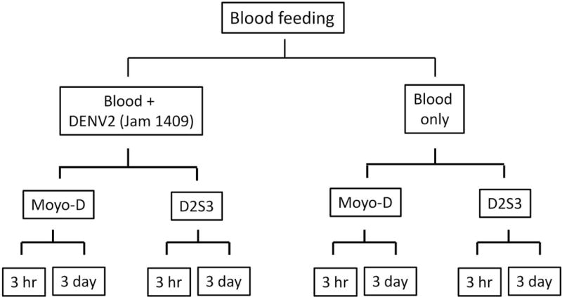
Experimental design of sample preparation for the microarray investigation. Adult females of the D2S3 strain (susceptible) and Moyo-D strain (refractory) were fed blood meals to generate orally infected test samples and uninfected control samples. At 3 hr and 3 days after infections, midgut samples were collected from individual females at each time point for both the strains. A total of 24 samples were generated from the three biological replicates for RNA preparations from pooled midguts and microarray hybridizations were performed using a dye swap method.
RNA isolation and sample preparation
RNA was isolated using TRIzol Reagent (Invitrogen: http://www.invitrogen.com/), according to manufacturer’s instruction from pooled midguts of each sample. Following RNA isolation, samples were treated with 1.0 U DNase I (Invitrogen). RNA concentration and quality was assessed using the NanoDrop ND-1000 (NanoDrop: http://www.nanodrop.com/) prior to labeling. First strand cDNA synthesis and labeling was performed with 500 ng of total RNA using the Genisphere 3DNA Array 900 kit (Genisphere: http://www.genisphere.com) for each dye, cyanine 3 (Cy3) and cyanine 5 (Cy5).
Microarray design and hybridizations
The contents, design of the custom cDNA microarray and the details of hybridization and scanning methods are described in Chauhan et al. (2012). Briefly, microarrays were generated from 9,504 unique cDNA amplicons obtained from a various tissue sources to assist in the A. aegypti genome annotation effort (Nene et al. 2007). Hybridizations were performed using the two step protocol as recommended by the manufacturer (Genisphere).
Data analysis
Statistical analysis of array data was conducted using Significance Analysis of Microarrays (SAM) version 2.0 (http://www-stat.stanford.edu/~tibs/SAM/). The SAM method performs cube root transformation to reduce the artifacts associated with the ratios involving small numbers and then assigns a score to each gene on the basis of changes in expression relative to the standard deviation of the replicates (Tusher et al. 2001). Genes with scores greater than a user defined threshold are considered significant. The software then uses permutations of the replicates to estimate the False Discovery Rate (FDR) associated with the selected gene set. For this exploratory investigation, we limited the FDR values to less than 5% to report significant expression differences in gene expression. We defined positive genes as those up-regulated in Moyo-D and/or down-regulated in D2S3, and negative genes as those down-regulated in Moyo-D and/or up-regulated in D2S3.
The software Cluster 3.0 (de Hoon et al. 2004) was used to generate hierarchal clusters of significant genes based on average correlation of Euclidian distances of expression of transcripts found significant among the four samples (2 strains × 2 time points). The cluster patterns were displayed using TreeView 1.60 (http://ranalbl.gov/EisenSoftware.htm). The gene ontology (GO) analyses of significant genes were carried out with A. aegypti gene annotation data available at Vectorbase (http://www.vectorbase.org) and BioMart (http://www.biomart.org). The pathway analysis was performed with the KEGG (Kyoto Encyclopedia of Genes and Genomes, Japan) pathways annotated from A. aegypti genes (www.kegg.jp). The hypergeometric tests of GO association differentially expressed genes were conducted with gene counts as described in Behura et al. (2011).
The data of the current study and a related study (Chauhan et al. 2012) were compared to assess the role of vector genotype in transcriptional response to DENV infection. Both studies were carried out after infecting mosquitoes with same strain of virus (DENV-2 JAM1409) and the same refractory mosquito strain (Moyo-D). However, the previous study utilized a different susceptible strain (MOYO-S). Here, we compared the differentially expressed transcripts (DETs) common to the 1 and 4 hr time points with the DETs identified at the 3 hr time point in the present study. Similarly, the 48 and 96 hr time points common DETs identified in the previous study were compared with the 3 day (72 hr) DETs of the present study. Our null hypothesis was that vector genotype has no role in modulating gene expression between susceptible/refractory strains upon dengue infection. After identifying the number of common and strain-specific DETs identified in both the studies, we performed hypergeometric tests, essentially as described in Fury et al. (2006) to determine if the null hypothesis can or cannot be rejected.
Quantitative real-time RT-PCR
To confirm microarray results, a total of 5 genes showing significant differential expression were examined by real-time qRT-PCR using SYBR green dye technology. The same RNA samples used for the current microarray experiment were used to perform these PCRs as described in Behura et al. (2011). Note that these genes were also identified and confirmed as differentially expressed in that study. Pearson correlation coefficients were calculated for the qRT-PCR data and the corresponding values from the current microarray data for D2S3 and Moyo-D strain expression levels of each of the five genes.
Results
Identification of differentially expressed transcripts
We identified differentially expressed transcripts (DETs) in the D2S3 and Moyo-D strains following oral infection with DENV-2 JAM1409 using custom microarrays containing 9,504 unique cDNA amplicons as previously described (Chauhan et al. 2012). A total of 24 samples [2 strains × 2 conditions (infection or control) × 2 time points × 3 biological replicates] were analyzed to identify DETs in infected samples in comparison to control samples. The D2S3 and Moyo-D strains show ~52% and ~15% infectivity, respectively, when challenged with DENV-2 JAM1409 under our conditions (Schneider et al. 2007). Two time points post-infection, 3 hr and 3 days, were chosen to represent an early- and a late post-infection period. All the hybridizations were conducted using the cye3/cye5 dye swap method where the ratio of dye intensity of D2S3 relative to Moyo-D was used to calculate relative expression changes in the infected and uninfected samples of the two strains. In other words, DETs which are up-regulated in the D2S3 strain are down-regulated in the Moyo-D strain and vice versa. The data of this microarray experiment were submitted to ArrayExpress (http://www.ebi.ac.uk/arrayexpress) under accession number E-MTAB-64.
The DETs were associated with significant changes in expression in the infected samples vs. uninfected samples as measured by the Significance Analysis of Microarray (SAM) method. The significance threshold value (delta) was kept greater than 0.31 and the average false discovery rate was kept less than 5% to identify the significant DETs. A total of 1,165 significant DETs were identified using these conditions. The fold change of expression of these genes varied from 1.2 to 7.0. On average, one in every four of the significant DETs was associated with greater than 2-fold changes in expression. By performing qRT-PCR with selected genes (n =5), it was found that each gene had highly similar expression patterns between the qRT-PCR assays and the microarray results (Online Resource 1). Additionally, we have compared expression pattern of common DETs between the current study and another independently performed microarray study (Chauhan et al. 2012; further details are described below) and observed highly similar patterns (Pearson correlation coefficient = 93%) further validating the microarray expression data of this study.
Time and strain specific expression
The number of significant DETs distributed among the two strains and two time points are shown in Figure 2. The complete list of these transcripts is provided in Online Resource 2 1. It shows that slightly higher numbers of transcripts are differentially expressed at the 3 hr post infection time than at the 3 day post infection time. This suggests that the early events after dengue infection may be transcriptionally more active than late events, such as at 3 days post-infection. This result is consistent with similar observations by Chauhan et al. (2012). Further, the number of DETs up-regulated specifically in the refractory strain is also greater than the number of DETs up-regulated in the susceptible strain indicating that defending against the infection evoked a more complex gene response than hosting DENV. Comparable observations were observed by Behura et al. (2011) for microarray studies with different refractory and susceptible genetic backgrounds.
Figure 2.
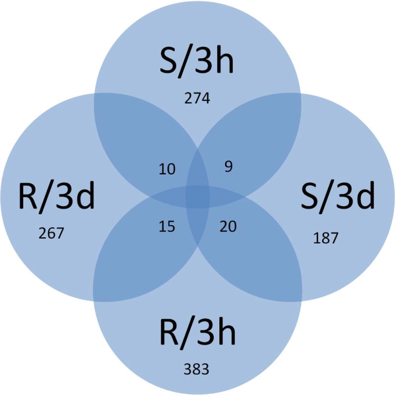
Venn diagram showing number and distribution of differentially expressed transcripts (DETs) between D2S3 and/or Moyo-D strains at 3 hr and/or 3 days after infection with dengue virus. The four samples are shown as S/3h, S/3d, R/3h and R/3d, where S and R represents the susceptible (D2S3) and the refractory (Moyo-D) strains respectively, and 3 h and 3 d represent the two post-infection time points (i.e. 3 h and 3 days), respectively. The numbers of DETs that are responsive to dengue infection exclusively in a specific strain at specific times are indicated. For example, 274 transcripts were up-regulated in the susceptible strain (or down-regulated in the refractory strain) at 3 hr after infection, whereas 267 transcripts were up-regulated in the refractory strain (or down-regulated in the susceptible strain) at 3 days after infection. The numbers shown as shared between samples represent DETs which are up-regulated in more than one strain or time point. For example, total 10 DETs were up-regulated in the susceptible strain at 3 h (or down-regulated in the refractory strain), but the same DETs were up-regulated in the refractory strain at the 3 day time point (or down-regulated in the susceptible strain).
In addition, several (n=54) transcripts also showed overlapping expression between the 3 hr as well as 3 day time points. The complete list of these transcripts is provided in Online Resource 3. They showed either similar or opposite expression patterns between the two strains. In the D2S3 strain, a total of nine transcripts were up-regulated at both time points. Another 10 transcripts were up-regulated in D2S3 at the 3hr time point, but were then down-regulated in Moyo-D at the 3 day time point. Similarly, 15 DETs were consistently up-regulated in Moyo-D at both time points, whereas a set of 20 transcripts were up-regulated in Moyo-D at 3hr, but down-regulated in D2S3 at 3 days post-infection (Figure 3). Because these 54 genes show overlapping expression between infection times and mosquito strains, it is highly likely that they be instrumental to global cross talk of genes in the mosquito host to produce either a permissive or non-permissive response to DENV infection.
Figure 3.
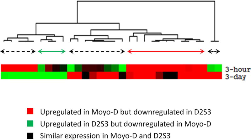
Hierarchical cluster of transcripts (Online Resource 3) based on differential expression at both post infection time points in either D2S3 or Moyo-D strains. The cluster tree shows different groups of transcripts (marked by colored horizontal arrows) that show similar expression patterns between the two strains and the two time points. Below the horizontal line are the self organizing maps of expression cluster of the transcripts and the color codes of the map are described below that. The color of horizontal arrows indicates the groups for the different expression patterns.
Gene ontology and pathway analysis of DENV responsive genes
The gene ontology (GO) terms of the DENV responsive genes were determined to predict functionality of differential expression of transcripts in the D2S3 and Moyo-D strains. Based on GO terms associated with A. aegypti genes (archived at Biomart database at VectorBase), it was found that the responsive genes show significant enrichment (hypergeometric p-value < 0.05) to gene ontology terms such as binding, catalytic, metabolic, hydrolase, membrane and kinase activities (Table 1). However, the individual GO terms were differentially associated with each strain and post-infection time (Online Resource 4). It was found that specific GO terms are associated with genes which are differentially expressed in a strain-specific manner, and also with genes which are commonly responsive to DENV infection in both the strains (Table 2). We also found that the differentially expressed genes represented several KEGG pathway genes (n =155) of A. aegypti (Online Resource 5). Many of these genes are related to endocytosis, transportation, signaling and metabolic activities that may be relevant to entry and early infection processes of DENV in the epithelium cells of mosquito midguts.
Table 1.
List of enriched gene ontology terms associated with DENV responsive genes. The number of response genes associated with each GO term and the total number of genes associated with the same term are listed. The p-value represents the hypergeometric distribution p-value calculated for each GO term as described in Behura et al. (2011).
| GO Term Name | No. of responsive genes | Total no. of genes | p-value |
|---|---|---|---|
| Protein binding | 37 | 2429 | 0.040 |
| Nucleotide binding | 17 | 877 | 0.027 |
| Binding | 14 | 749 | 0.044 |
| Metabolic process | 14 | 689 | 0.030 |
| Integral to membrane | 9 | 1143 | 0.035 |
| Nucleic acid binding | 9 | 1133 | 0.037 |
| Kinase activity | 8 | 355 | 0.049 |
| Nucleus | 8 | 1033 | 0.039 |
| Protein kinase activity | 7 | 247 | 0.027 |
| Protein phosphorylation | 7 | 256 | 0.030 |
| Protein serine/threonine kinase activity | 7 | 250 | 0.028 |
| GTP binding | 5 | 159 | 0.038 |
| GTPase activity | 5 | 96 | 0.007 |
| RNA binding | 5 | 164 | 0.042 |
| Acid-amino acid ligase activity | 4 | 49 | 0.003 |
| Protein modification process | 3 | 39 | 0.012 |
| Protein ubiquitination | 3 | 28 | 0.005 |
| Translation elongation factor activity | 3 | 20 | 0.002 |
| Ubiquitin-protein ligase activity | 3 | 33 | 0.008 |
| DNA recombination | 2 | 17 | 0.019 |
| Glycolysis | 2 | 20 | 0.025 |
| Regulation of protein metabolic process | 2 | 28 | 0.045 |
| Small conjugating protein ligase activity | 2 | 28 | 0.045 |
| Translational elongation | 2 | 21 | 0.027 |
Table 2.
Major GO terms associated with D2S3 and Moyo-D and that are commonly up-regulated in the two strains. The numbers show the number of differentially expressed genes associated with the GO term.
| D2S3 | |
|---|---|
| Metabolic process | 14 |
| Zinc ion binding | 14 |
| Cytoplasm | 8 |
| Transferase activity | 13 |
| Moyo-D | |
| Hydrolase activity | 18 |
| Transport | 9 |
| Common | |
| Binding activity | 37 (D2S3)/53 (Moyo-D) |
| Catalytic activity | 33 (D2S3)/25 (Moyo-D) |
| Intracellular activity | 31 (D2S3)/46 (Moyo-D) |
Role of mosquito genotype on transcriptional response to DENV infection
The current investigation (with susceptible D2S3 versus refractory Moyo-D strains) utilized the same custom microarray platform that was used for identifying differentially expressed genes in the Moyo-S (susceptible) and Moyo-D strains (Chauhan et al. 2012). Moyo-S is a sub-strain of Moyo-D that was selected for high susceptibility to P. gallinaceum (Thathy et al. 1994), and subsequently found to show ~57% infectivity with DENV-2 JAM1409 (Schneider et al. 2007). Because Moyo-D was used as a common refractory strain in the present as well as in the Chauhan et al. (2012) study, we compared both datasets to explore any signatures of gene expression in both or patterns that may be specific to the susceptible strains i.e. D2S3 (this study) or Moyo-S (Chauhan et al. 2012). From comparison of these data, we identified that nearly 95% of the DETs are different in D2S3/Moyo-D vs. Moyo-S/Moyo-D indicating that A. aegypti response to infection of a given dengue virus is largely dependent upon the vector genotype. However, we identified a set of 52 transcripts that were differentially regulated in both D2S3/Moyo-D vs. Moyo-S/Moyo-D strains (Online Resource 6). Based on the hypergeometric test (Fury et al. 2006) of the distribution of the common and the strain-specific DETs, we observed significant association (hypergeometric p-value < 0.01) between expression patterns and genotype differences (susceptible vs refractory) of A. aegypti to DENV suggesting that the null assumption (see Methods) can be rejected. Of these, 31 transcripts were up-regulated in the common refractory strain Moyo-D and the remaining 21 transcripts were up-regulated in Moyo-S and D2S3 (Figure 4). By analyzing the annotated function of these common genes, it was found that these genes are enriched (hypergeometric p-value < 0.05) in gene function such endocytosis, autophagy, peroxisome, lipid metabolism, signaling and translation. As these genes (Online Resource 6) were commonly responsive to dengue infection between mosquito strains, they may act as core genes of A. aegypti necessary to trigger both hosting and defensive reaction to dengue infection.
Figure 4.
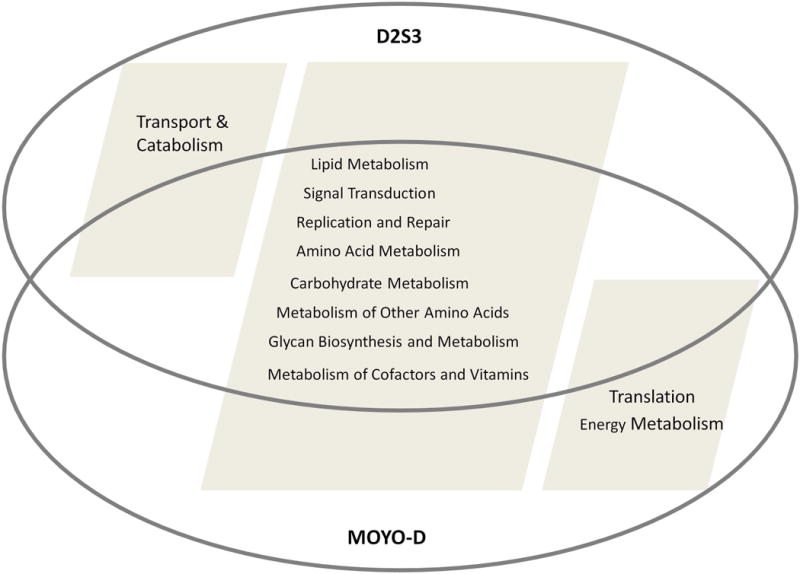
Hypergeometric distribution of genes which are differentially expressed between Moyo-D vs. D2S3 (current investigation) and Moyo-D vs. Moyo-S (Chauhan et al. 2012). The Venn diagram shows the number of up-regulated transcripts in susceptible (blue color) versus refractory strain (red color).
Discussion
It is well known that different laboratory strains and field isolates of A. aegypti show variable susceptibility to dengue virus infection, as well as variability in response to different DENV isolates (reviewed in Guzman et al. 2010, Rico-Hesse 2007). The current investigation is a part of our continuing effort toward understanding the plasticity of gene expression of the vector mosquito A. aegypti to dengue virus infection. It is clear that infection status is dynamic, complex, and subject to interactions between individual mosquito genotypes and environmental influences. Several laboratory strains of this mosquito show either low or high infectivity to DENV2 serotype (Schneider et al. 2007) that can be subjected to transcriptome studies for better understanding of susceptible/refractory response of A. aegypti genes to dengue infection. The D2S3 strain was selected for high susceptibility to DENV-2 (JAM1409) infection (Bennet et al. 2005). We chose this strain to compare gene expression with Moyo-D strain that shows low infectivity to dengue infection (Schneider et al. 2007). Moyo-D was chosen primarily for the availability of gene expression data (Chauhan et al. 2012) between Moyo-D and Moyo-S in response to dengue serotype 2 infection that allowed us to compare gene expression patterns of both the microarray studies.
The primary aim of the present study is to identify expression of a collection of cDNAs (n = 9,054), investigated by Chauhan et al. (2012), and to compare the differentially expressed transcripts between Moyo-S/Moyo-D vs. D2S3/Moyo-D vector genotypes. Our purpose is not to identify differentially expressed genes in a comprehensive manner in A. aegypti genome as we have already reported in a previous work (Behura et al. 2011). The cDNA array that we used in this study is the same array format that was used by Chauhan et al. (2012); complete details for cDNA amplicon selection for these arrays is presented therein. Briefly, the cDNA amplicons were chosen as they represented putative unique gene sequences from consensus sequence assemblies generated from expressed sequence tag (EST) collections from various tissue sources generated to assist in the A. aegypti genome annotation effort (Nene et al. 2007). The A. aegypti genes that were missing among these 9,045 cDNAs were not specific to any to particular function or pathway (data not shown). Hence it is unlikely that we are systematically losing any specific gene function from our cDNA analysis. Although usage of cDNA microarray is considered as a limitation for comprehensive analysis of gene expression, our approach is however appropriate for the specific aim of this study which is to compare expression patterns of a collection of A. aegypti cDNAs across genotypes. To meet that specific objective, we resorted to the same cDNA microarray instead of using another microarray or RNA-seq platform. Nevertheless, we have identified several known pathway genes which are significantly upregulated in D2S3 or Moyo-D strain at specific post-infection time points (Online Resource 5). Several genes associated with transport and catabolism activities such as endocytosis (AAEL002469, AAEL004319, AAEL007288, AAEL007845, AAEL008184 and AAEL009754), peroxisome and phagosome (AAEL002723, AAEL009112, AAEL013260 and AAEL007845), autophagy (AAEL002286) and lysozomal activities (AAEL001235 and AAEL005460) were identified among the significant genes. These genes are expected to have functional role in internalization and other early cellular processes of the virus in the midgut epithelium cells. Based on the expression patterns of these genes between D2S3 and Moyo-D (Online Resource 5), it appears that these genes are critical in establishing susceptible interaction between the virus and mosquito (Mosso et al. 2008).
Several, genes relating to energy metabolism, particularly oxidative phosphorylation (AAEL007777, AAEL008848, AAEL010330, AAEL011871 and AAEL013009) and different translational related processes including mRNA surveillance (AAEL010940, AAEL011742 and AAEL011817) and RNA transport (AAEL004378, AAEL005635, AAEL007078, AAEL009646, AAEL011817 and AAEL014733) were differentially expressed between the two strains (Online Resource 5). This is consistent with our earlier report (Behura et al. 2011) that defending against DENV infection is energetically more expensive to A. aegypti than hosting the virus. The refractory response requires a greater number of genes to be activated to defend against DENV than the number of genes required to host the virus (Figure 2). Moreover, several genes related to serine protease (AAEL011622), innate immunity (AAEL007642), apoptosis (AAEL012143) and proteolytic activities (AAEL011016 and AAEL005112) were activated in the Moyo-D strain but not in the D2S3 strain, suggesting a possible role of these genes in defense response to DENV in A. aegypti. Proteases, Toll pathway genes and apoptosis inducing genes have been indicated to regulate dengue susceptibility in A. aegypti from previous studies (Molina-Cruz et al. 2005, Brackney et al. 2008, Xi et al. 2008, Behura et al. 2011, Liu et al. 2013).
Genes related to signal transduction such as Jak-STAT signaling (AAEL013786), Phosphatidylinositol signaling (AAEL009294), TGF-beta signaling (AAEL009110), Hedgehog signaling (AAEL004351), mTOR signaling (AAEL008179) and Notch signaling (AAEL002389) were up-regulated in both the D2S3 and Moyo-D strains. These genes possibly play key roles in establishing signal transductions among the responsive genes for triggering either a susceptible or refractory response to dengue infection. Signal transduction is an important feature of molecular interactions between dengue and A. aegypti as also found from previous investigations (Souza-Neto et al. 2009, Behura et al. 2011, Chauhan et al. 2012). Based on the results obtained from this study and from others mentioned above, we provide a simplified intuitive model to explain the overall mechanism of susceptible and refractory response of A. aegypti to DENV infection (Figure 5). It is likely that DENV is controlled by a vacuolar trafficking process which is distinct between susceptible and refractory mosquitoes. The endoplasmic reticulum may play an important role in this process (Hsieh et al. 2008). The innate immunity of the mosquito plays the key role in defending against DENV infection which is likely to be orchestrated by multiple factors including apoptotic and proteolytic processes (Brackney et al. 2008, Liu et al. 2013).
Figure 5.

An intuitive model of cellular events that are activated in susceptible and refractory A. aegypti mosquitoes in response to dengue virus infection. The vertically downward arrows indicate the likely events in a susceptible mosquito versus a refractory mosquito. The question marks (?) indicate unknown factors or mechanisms that may be involved in triggering the refractory response.
The mechanism of susceptible and refractory responses of A. aegypti to DENV is subject to trade-offs between mosquito and DENV evolution, as the virus is obligated to adapt alternatively to the mosquito vector and the human host. Evidence suggests that when such trade-off effect is released by growing the virus in one cell line (either mosquito or human), the virus gains fitness in infectivity compared to DENV undergoing alternative passages in both mosquito and human cells (Vasilakis et al. 2009). The trade-off effect is also manifested within a host, wherein genotype variation in hosts acts as a retarding force on evolvable capacity of the virus (Holmes 2003). But, it is not known (to the best of our knowledge) if intra-clonal DENV (circulating within same genotype of mosquito) have higher rates of evolution and adaptive fitness than inter-clonal (circulating between different genotypes of mosquito). However, the present investigation also allowed us to compare gene expression patterns between D2S3/Moyo-D with that of Moyo-S/Moyo-D (Chauhan et al. 2012). From this comparative analysis, we found that different susceptible genetic backgrounds of A. aegypti show different responses to dengue virus infection. Thus, vector genotype has an effect in modulating transcriptional response of A. aegypti to DENV infection which may be associated with differential adaptability of DENV in different strains of the mosquito.
Supplementary Material
Acknowledgments
This research was funded by grant RO1-AI059342 from the National Institute of Allergy and Infectious Diseases, National Institutes of Health (NIAID/NIH), USA.
Footnotes
Ethical standards
This study was performed in accordance with the recommendations in the Guide for the Care and Use of Laboratory Animals of the National Institutes of Health. The animal use protocol was approved by the University of Notre Dame Institutional Animal Care and Use Committee (Study # 11-036).
Conflict of interest
The authors declare that they have no conflict of interest.
References
- Behura SK, Gomez-Machorro C, Harker BW, deBruyn B, Lovin DD, Hemme RR, Mori A, Romero-Severson J, Severson DW. Global cross-talk of genes of the mosquito Aedes aegypti in response to dengue virus infection. PLoS Negl Trop Dis. 2011;5:e1385. doi: 10.1371/journal.pntd.0001385. [DOI] [PMC free article] [PubMed] [Google Scholar]
- Behura SK, Severson DW. Intrinsic features of Aedes aegypti genes affect transcriptional responsiveness of mosquito genes to dengue virus infection. Infect Genet Evol. 2012;12:1413–1418. doi: 10.1016/j.meegid.2012.04.027. [DOI] [PMC free article] [PubMed] [Google Scholar]
- Bennett KE, Beaty BJ, Black WC., 4th Selection of D2S3, an Aedes aegypti (Diptera: Culicidae) strain with high oral susceptibility to Dengue 2 virus and D2MEB, a strain with a midgut barrier to Dengue 2 escape. J Med Entomol. 2005;42:110–119. doi: 10.1603/0022-2585(2005)042[0110:SODAAA]2.0.CO;2. [DOI] [PubMed] [Google Scholar]
- Bennett KE, Olson KE, Muñoz Mde L, Fernandez-Salas I, Farfan-Ale JA, Higgs S, Black WC, 4th, Beaty BJ. Variation in vector competence for dengue 2 virus among 24 collections of Aedes aegypti from Mexico and the United States. Am J Trop Med Hyg. 2002;67:85–92. doi: 10.4269/ajtmh.2002.67.85. [DOI] [PubMed] [Google Scholar]
- Bosio CF, Beaty BJ, Black WC., 4th Quantitative genetics of vector competence for dengue-2 virus in Aedes aegypti. Am J Trop Med Hyg. 1998;59:965–970. doi: 10.4269/ajtmh.1998.59.965. [DOI] [PubMed] [Google Scholar]
- Bosio CF, Fulton RE, Salasek ML, Beaty BJ, Black WC., 4th Quantitative trait loci that control vector competence for dengue-2 virus in the mosquito Aedes aegypti. Genetics. 2000;156:687–698. doi: 10.1093/genetics/156.2.687. [DOI] [PMC free article] [PubMed] [Google Scholar]
- Brackney DE, Foy BD, Olson KE. The effects of midgut serine proteases on dengue virus type 2 infectivity of Aedes aegypti. Am J Trop Med Hyg. 2008;79:267–274. [PMC free article] [PubMed] [Google Scholar]
- Bubner B, Gase K, Baldwin IT. Two-fold differences are the detection limit for determining transgene copy numbers in plants by real-time PCR. BMC Biotechnol. 2004;4:14. doi: 10.1186/1472-6750-4-14. [DOI] [PMC free article] [PubMed] [Google Scholar]
- Chauhan C, Behura SK, Debruyn B, Lovin DD, Harker BW, Gomez-Machorro C, Mori A, Romero-Severson J, Severson DW. Comparative expression profiles of midgut genes in dengue virus refractory and susceptible Aedes aegypti across critical period for virus infection. PLoS One. 2012;7:e47350. doi: 10.1371/journal.pone.0047350. [DOI] [PMC free article] [PubMed] [Google Scholar]
- Clemons AC, Mori A, Haugen M, Severson D, Duman-Scheel M. Aedes aegypti: culturing and egg collection. Cold Spring Harbor Protoc. 2010;2010 doi: 10.1101/pdb.prot5507. pdb.prot5507. [DOI] [PMC free article] [PubMed] [Google Scholar]
- Colpitts TM, Cox J, Vanlandingham DL, Feitosa FM, Cheng G, Kurscheid S, Wang P, Krishnan MN, Higgs S, Fikrig E. Alterations in the Aedes aegypti transcriptome during infection with West Nile, dengue and yellow fever viruses. PLoS Pathog. 2011;7:e1002189. doi: 10.1371/journal.ppat.1002189. [DOI] [PMC free article] [PubMed] [Google Scholar]
- de Hoon MJL, Imoto S, Nolan J, Miyano S. Open Source Clustering Software. Bioinformatics. 2004;20:1453–1454. doi: 10.1093/bioinformatics/bth078. [DOI] [PubMed] [Google Scholar]
- Diallo M, Ba Y, Faye O, Soumare ML, Dia I, Sall AA. Vector competence of Aedes aegypti populations from Senegal for sylvatic and epidemic dengue 2 virus isolated in West Africa. Trans R Soc Trop Med Hyg. 2008;102:493–498. doi: 10.1016/j.trstmh.2008.02.010. [DOI] [PubMed] [Google Scholar]
- Fury W, Batliwalla F, Gregersen PK, Li W. Overlapping probabilities of top ranking gene lists, hypergeometric distribution, and stringency of gene selection criterion. Conf Proc IEEE Eng Med Biol Soc. 2006;1:5531–5534. doi: 10.1109/IEMBS.2006.260828. [DOI] [PubMed] [Google Scholar]
- Gomez-Machorro C, Bennett KE, del Lourdes Munoz M, Black WC., 4th Quantitative trait loci affecting dengue midgut infection barriers in an advanced intercross line of Aedes aegypti. Insect Mol Biol. 2004;13:637–648. doi: 10.1111/j.0962-1075.2004.00522.x. [DOI] [PubMed] [Google Scholar]
- Gubler DJ, Nalim S, Tan R, Saipan H, Saroso JS. Variation in susceptibility to oral infection with dengue viruses among geographic strains of Aedes aegypti. Am J Trop Med Hyg. 1979;28:1045–1052. doi: 10.4269/ajtmh.1979.28.1045. [DOI] [PubMed] [Google Scholar]
- Guzman MG, Halstead SB, Artsob H, Buchy P, Farrar J, Gubler DJ, Hunsperger E, Kroeger A, Margolis HS, Martínez E, Nathan MB, Pelegrino JL, Simmons C, Yoksan S, Peeling RW. Dengue: a continuing global threat. Nat Rev Microbiol. 2010;8(Suppl 12):7–16. doi: 10.1038/nrmicro2460. [DOI] [PMC free article] [PubMed] [Google Scholar]
- Holmes EC. Patterns of intra- and interhost nonsynonymous variation reveal strong purifying selection in dengue virus. J Virol. 2003;77:11296–11308. doi: 10.1128/JVI.77.20.11296-11298.2003. [DOI] [PMC free article] [PubMed] [Google Scholar]
- Hsieh SC, Liu IJ, King CC, Chang GJ, Wang WK. A strong endoplasmic reticulum retention signal in the stem-anchor region of envelope glycoprotein of dengue virus type 2 affects the production of virus-like particles. Virology. 2008;374:338–350. doi: 10.1016/j.virol.2007.12.041. [DOI] [PubMed] [Google Scholar]
- Liu B, Behura SK, Clem RJ, Schneemann A, Becnel J, Severson DW, Zhou L. P53-mediated rapid induction of apoptosis conveys resistance to viral infection in insects. PLoS Pathog. 2013;9:e1003137. doi: 10.1371/journal.ppat.1003137. [DOI] [PMC free article] [PubMed] [Google Scholar]
- Molina-Cruz A, Gupta L, Richardson J, Bennett K, Black W, 4th, Barillas-Mury C. Effect of mosquito midgut trypsin activity on dengue-2 virus infection and dissemination in Aedes aegypti. Am J Trop Med Hyg. 2005;72:631–637. [PubMed] [Google Scholar]
- Mosso C, Galván-Mendoza IJ, Ludert JE, del Angel RM. Endocytic pathway followed by dengue virus to infect the mosquito cell line C6/36 HT. Virology. 2008;378:193–199. doi: 10.1016/j.virol.2008.05.012. [DOI] [PubMed] [Google Scholar]
- Phillips ML. Dengue reborn: widespread resurgence of a resilient vector. Environ Health Perspectives. 2008;116:382–389. doi: 10.1289/ehp.116-a382. [DOI] [PMC free article] [PubMed] [Google Scholar]
- Rico-Hesse R. Dengue virus evolution and virulence models. Clin Infect Dis. 2007;44:1462–1466. doi: 10.1086/517587. [DOI] [PMC free article] [PubMed] [Google Scholar]
- Rosen L, Roseboom LE, Gubler DJ, Lein JC, Chaniotis BN. Comparative susceptibility of mosquito species and strains to oral and parenteral infection with dengue and Japanese encephalitis viruses. Am J Trop Med Hyg. 1985;34:603–615. doi: 10.4269/ajtmh.1985.34.603. [DOI] [PubMed] [Google Scholar]
- Schneider JR, Mori A, Romero-Severson J, Chadee DD, Severson DW. Investigations of dengue-2 susceptibility and body size among Aedes aegypti populations. Med Vet Entomol. 2007;21:370–376. doi: 10.1111/j.1365-2915.2007.00699.x. [DOI] [PubMed] [Google Scholar]
- Sessions OM, Barrows NJ, Souza-Neto JA, Robinson TJ, Hershey CL, Rodgers MA, Ramirez JL, Dimopoulos G, Yang PL, Pearson JL, Garcia-Blanco MA. Discovery of insect and human dengue virus host factors. Nature. 2009;458:1047–10450. doi: 10.1038/nature07967. [DOI] [PMC free article] [PubMed] [Google Scholar]
- Severson DW, Behura SK. Mosquito genomics: progress and challenges. Annu Rev Entomol. 2012;57:143–166. doi: 10.1146/annurev-ento-120710-100651. [DOI] [PubMed] [Google Scholar]
- Souza-Neto JA, Sim S, Dimopoulos G. An evolutionary conserved function of the JAK-STAT pathway in anti-dengue defense. Proc Natl Acad Sci U S A. 2009;106:17841–17846. doi: 10.1073/pnas.0905006106. [DOI] [PMC free article] [PubMed] [Google Scholar]
- Thathy V, Severson DW, Christensen BM. Reinterpretation of the genetics of susceptibility of Aedes aegypti to Plasmodium gallinaceum. J Parasitol. 1994;80:705–712. [PubMed] [Google Scholar]
- Tusher VG, Tibshirani R, Chu G. Significance analysis of microarrays applied to the ionizing radiation response. Proc Natl Acad Sci U S A. 2001;98:5116–5121. doi: 10.1073/pnas.091062498. [DOI] [PMC free article] [PubMed] [Google Scholar]
- Vasilakis N, Deardorff ER, Kenney JL, Rossi S, Hanley K, Weaver S. Mosquitoes Put the Brake on Arbovirus Evolution:Experimental Evolution Reveals Slower Mutation Accumulation in Mosquito Than Vertebrate Cells. PLoS Pathog. 2009;5:1–18. doi: 10.1371/journal.ppat.1000467. [DOI] [PMC free article] [PubMed] [Google Scholar]
- WHO. Dengue guidelines for diagnosis, treatment, prevention and control. Third. Geneva: World Health Organization; 2009. [PubMed] [Google Scholar]
- Xi Z, Ramirez JL, Dimopoulos G. The Aedes aegypti Toll pathway controls dengue virus infection. PLoS Pathog. 2008;4:e1000098. doi: 10.1371/journal.ppat.1000098. [DOI] [PMC free article] [PubMed] [Google Scholar]
Associated Data
This section collects any data citations, data availability statements, or supplementary materials included in this article.