

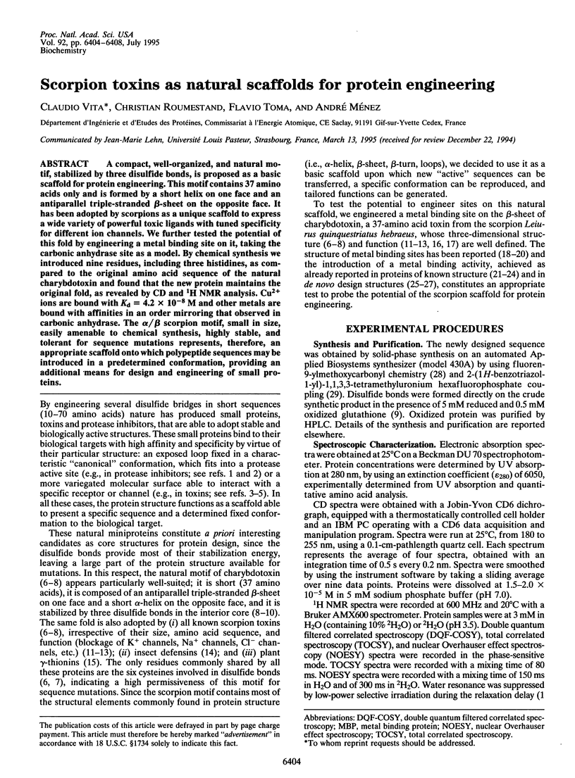
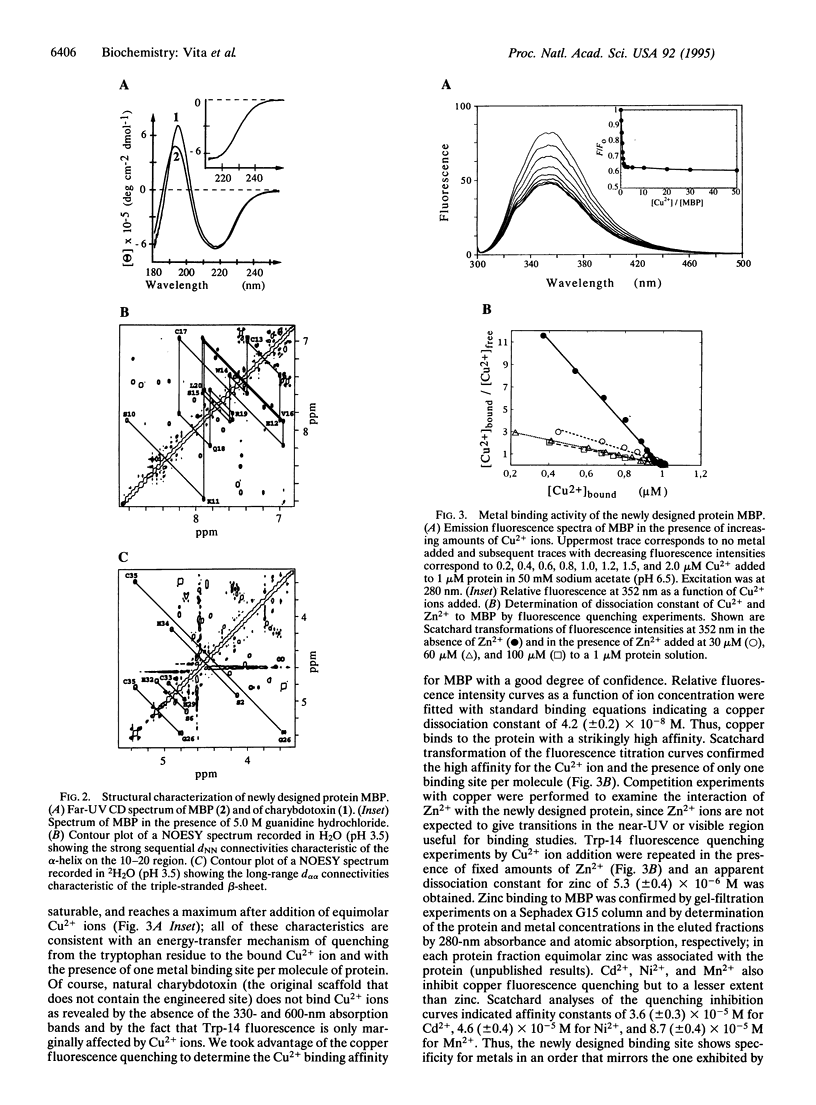
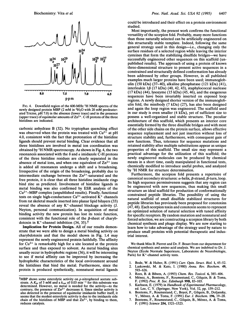
Abstract
A compact, well-organized, and natural motif, stabilized by three disulfide bonds, is proposed as a basic scaffold for protein engineering. This motif contains 37 amino acids only and is formed by a short helix on one face and an antiparallel triple-stranded beta-sheet on the opposite face. It has been adopted by scorpions as a unique scaffold to express a wide variety of powerful toxic ligands with tuned specificity for different ion channels. We further tested the potential of this fold by engineering a metal binding site on it, taking the carbonic anhydrase site as a model. By chemical synthesis we introduced nine residues, including three histidines, as compared to the original amino acid sequence of the natural charybdotoxin and found that the new protein maintains the original fold, as revealed by CD and 1H NMR analysis. Cu2+ ions are bound with Kd = 4.2 x 10(-8) M and other metals are bound with affinities in an order mirroring that observed in carbonic anhydrase. The alpha/beta scorpion motif, small in size, easily amenable to chemical synthesis, highly stable, and tolerant for sequence mutations represents, therefore, an appropriate scaffold onto which polypeptide sequences may be introduced in a predetermined conformation, providing an additional means for design and engineering of small proteins.
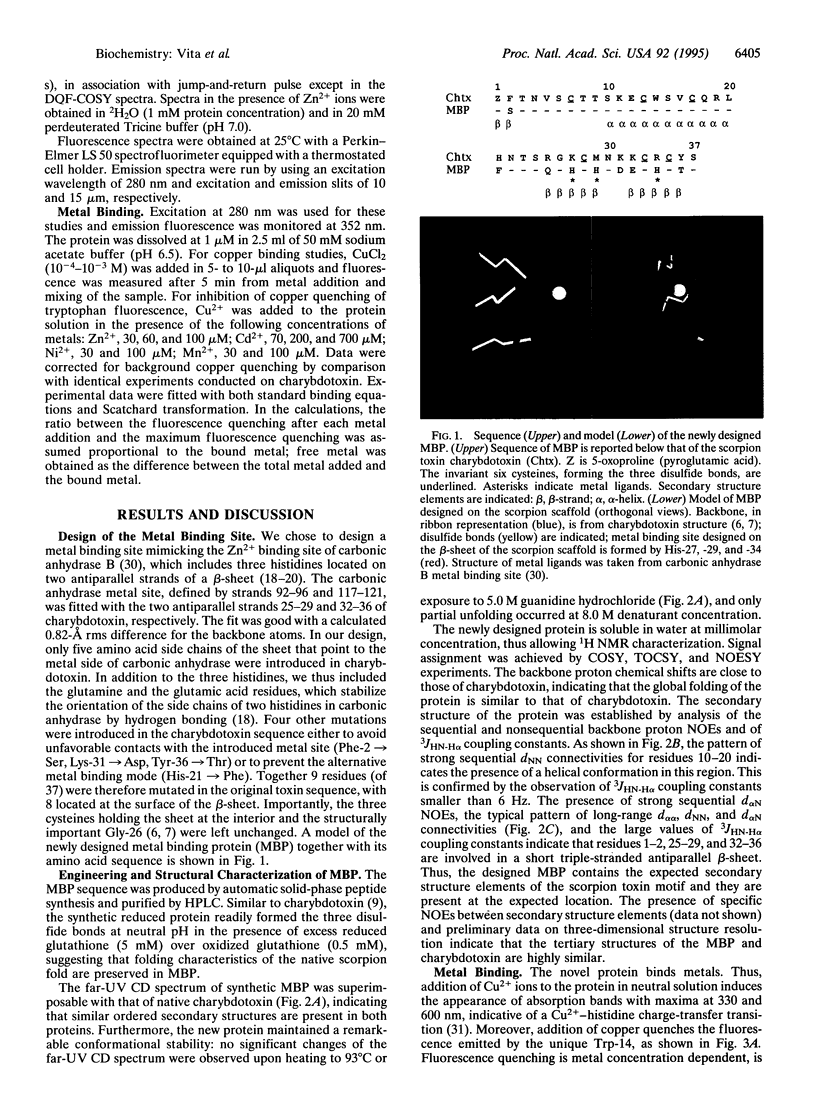
Full text
PDF
Images in this article
Selected References
These references are in PubMed. This may not be the complete list of references from this article.
- Arnold F. H., Haymore B. L. Engineered metal-binding proteins: purification to protein folding. Science. 1991 Jun 28;252(5014):1796–1797. doi: 10.1126/science.1648261. [DOI] [PubMed] [Google Scholar]
- Bonmatin J. M., Bonnat J. L., Gallet X., Vovelle F., Ptak M., Reichhart J. M., Hoffmann J. A., Keppi E., Legrain M., Achstetter T. Two-dimensional 1H NMR study of recombinant insect defensin A in water: resonance assignments, secondary structure and global folding. J Biomol NMR. 1992 May;2(3):235–256. doi: 10.1007/BF01875319. [DOI] [PubMed] [Google Scholar]
- Bontems F., Gilquin B., Roumestand C., Ménez A., Toma F. Analysis of side-chain organization on a refined model of charybdotoxin: structural and functional implications. Biochemistry. 1992 Sep 1;31(34):7756–7764. doi: 10.1021/bi00149a003. [DOI] [PubMed] [Google Scholar]
- Bontems F., Roumestand C., Boyot P., Gilquin B., Doljansky Y., Menez A., Toma F. Three-dimensional structure of natural charybdotoxin in aqueous solution by 1H-NMR. Charybdotoxin possesses a structural motif found in other scorpion toxins. Eur J Biochem. 1991 Feb 26;196(1):19–28. doi: 10.1111/j.1432-1033.1991.tb15780.x. [DOI] [PubMed] [Google Scholar]
- Bontems F., Roumestand C., Gilquin B., Ménez A., Toma F. Refined structure of charybdotoxin: common motifs in scorpion toxins and insect defensins. Science. 1991 Dec 6;254(5037):1521–1523. doi: 10.1126/science.1720574. [DOI] [PubMed] [Google Scholar]
- Bruix M., Jiménez M. A., Santoro J., González C., Colilla F. J., Méndez E., Rico M. Solution structure of gamma 1-H and gamma 1-P thionins from barley and wheat endosperm determined by 1H-NMR: a structural motif common to toxic arthropod proteins. Biochemistry. 1993 Jan 19;32(2):715–724. doi: 10.1021/bi00053a041. [DOI] [PubMed] [Google Scholar]
- Chakrabarti P. Geometry of interaction of metal ions with histidine residues in protein structures. Protein Eng. 1990 Oct;4(1):57–63. doi: 10.1093/protein/4.1.57. [DOI] [PubMed] [Google Scholar]
- Dreyer F. Peptide toxins and potassium channels. Rev Physiol Biochem Pharmacol. 1990;115:93–136. [PubMed] [Google Scholar]
- Garcia M. L., Galvez A., Garcia-Calvo M., King V. F., Vazquez J., Kaczorowski G. J. Use of toxins to study potassium channels. J Bioenerg Biomembr. 1991 Aug;23(4):615–646. doi: 10.1007/BF00785814. [DOI] [PubMed] [Google Scholar]
- Gimenez-Gallego G., Navia M. A., Reuben J. P., Katz G. M., Kaczorowski G. J., Garcia M. L. Purification, sequence, and model structure of charybdotoxin, a potent selective inhibitor of calcium-activated potassium channels. Proc Natl Acad Sci U S A. 1988 May;85(10):3329–3333. doi: 10.1073/pnas.85.10.3329. [DOI] [PMC free article] [PubMed] [Google Scholar]
- Hellinga H. W., Caradonna J. P., Richards F. M. Construction of new ligand binding sites in proteins of known structure. II. Grafting of a buried transition metal binding site into Escherichia coli thioredoxin. J Mol Biol. 1991 Dec 5;222(3):787–803. doi: 10.1016/0022-2836(91)90511-4. [DOI] [PubMed] [Google Scholar]
- Higaki J. N., Haymore B. L., Chen S., Fletterick R. J., Craik C. S. Regulation of serine protease activity by an engineered metal switch. Biochemistry. 1990 Sep 18;29(37):8582–8586. doi: 10.1021/bi00489a012. [DOI] [PubMed] [Google Scholar]
- Hynes T. R., Kautz R. A., Goodman M. A., Gill J. F., Fox R. O. Transfer of a beta-turn structure to a new protein context. Nature. 1989 May 4;339(6219):73–76. doi: 10.1038/339073a0. [DOI] [PubMed] [Google Scholar]
- Iverson B. L., Iverson S. A., Roberts V. A., Getzoff E. D., Tainer J. A., Benkovic S. J., Lerner R. A. Metalloantibodies. Science. 1990 Aug 10;249(4969):659–662. doi: 10.1126/science.2116666. [DOI] [PubMed] [Google Scholar]
- Kannan K. K., Notstrand B., Fridborg K., Lövgren S., Ohlsson A., Petef M. Crystal structure of human erythrocyte carbonic anhydrase B. Three-dimensional structure at a nominal 2.2-A resolution. Proc Natl Acad Sci U S A. 1975 Jan;72(1):51–55. doi: 10.1073/pnas.72.1.51. [DOI] [PMC free article] [PubMed] [Google Scholar]
- LINDSKOG S., NYMAN P. O. METAL-BINDING PROPERTIES OF HUMAN ERYTHROCYTE CARBONIC ANHYDRASES. Biochim Biophys Acta. 1964 Jun 1;85:462–474. doi: 10.1016/0926-6569(64)90310-4. [DOI] [PubMed] [Google Scholar]
- Langen H. T., Taylor J. W. Alkaline phosphatase-somatostatin hybrid proteins as probes for somatostatin-14 receptors. Proteins. 1992 Sep;14(1):1–9. doi: 10.1002/prot.340140103. [DOI] [PubMed] [Google Scholar]
- Lanza P., Billetta R., Antonenko S., Zanetti M. Active immunity against the CD4 receptor by using an antibody antigenized with residues 41-55 of the first extracellular domain. Proc Natl Acad Sci U S A. 1993 Dec 15;90(24):11683–11687. doi: 10.1073/pnas.90.24.11683. [DOI] [PMC free article] [PubMed] [Google Scholar]
- Laskowski M., Jr, Kato I. Protein inhibitors of proteinases. Annu Rev Biochem. 1980;49:593–626. doi: 10.1146/annurev.bi.49.070180.003113. [DOI] [PubMed] [Google Scholar]
- Lee G., Chan W., Hurle M. R., DesJarlais R. L., Watson F., Sathe G. M., Wetzel R. Strong inhibition of fibrinogen binding to platelet receptor alpha IIb beta 3 by RGD sequences installed into a presentation scaffold. Protein Eng. 1993 Sep;6(7):745–754. doi: 10.1093/protein/6.7.745. [DOI] [PubMed] [Google Scholar]
- Miller C., Moczydlowski E., Latorre R., Phillips M. Charybdotoxin, a protein inhibitor of single Ca2+-activated K+ channels from mammalian skeletal muscle. Nature. 1985 Jan 24;313(6000):316–318. doi: 10.1038/313316a0. [DOI] [PubMed] [Google Scholar]
- Neyton J., Pelleschi M. Multi-ion occupancy alters gating in high-conductance, Ca(2+)-activated K+ channels. J Gen Physiol. 1991 Apr;97(4):641–665. doi: 10.1085/jgp.97.4.641. [DOI] [PMC free article] [PubMed] [Google Scholar]
- Olivera B. M., Rivier J., Clark C., Ramilo C. A., Corpuz G. P., Abogadie F. C., Mena E. E., Woodward S. R., Hillyard D. R., Cruz L. J. Diversity of Conus neuropeptides. Science. 1990 Jul 20;249(4966):257–263. doi: 10.1126/science.2165278. [DOI] [PubMed] [Google Scholar]
- Olivera B. M., Rivier J., Scott J. K., Hillyard D. R., Cruz L. J. Conotoxins. J Biol Chem. 1991 Nov 25;266(33):22067–22070. [PubMed] [Google Scholar]
- Park C. S., Miller C. Mapping function to structure in a channel-blocking peptide: electrostatic mutants of charybdotoxin. Biochemistry. 1992 Sep 1;31(34):7749–7755. doi: 10.1021/bi00149a002. [DOI] [PubMed] [Google Scholar]
- Pessi A., Bianchi E., Crameri A., Venturini S., Tramontano A., Sollazzo M. A designed metal-binding protein with a novel fold. Nature. 1993 Mar 25;362(6418):367–369. doi: 10.1038/362367a0. [DOI] [PubMed] [Google Scholar]
- Rees B., Bilwes A. Three-dimensional structures of neurotoxins and cardiotoxins. Chem Res Toxicol. 1993 Jul-Aug;6(4):385–406. doi: 10.1021/tx00034a001. [DOI] [PubMed] [Google Scholar]
- Regan L., Clarke N. D. A tetrahedral zinc(II)-binding site introduced into a designed protein. Biochemistry. 1990 Dec 11;29(49):10878–10883. doi: 10.1021/bi00501a003. [DOI] [PubMed] [Google Scholar]
- Roberts V. A., Iverson B. L., Iverson S. A., Benkovic S. J., Lerner R. A., Getzoff E. D., Tainer J. A. Antibody remodeling: a general solution to the design of a metal-coordination site in an antibody binding pocket. Proc Natl Acad Sci U S A. 1990 Sep;87(17):6654–6658. doi: 10.1073/pnas.87.17.6654. [DOI] [PMC free article] [PubMed] [Google Scholar]
- Sollazzo M., Billetta R., Zanetti M. Expression of an exogenous peptide epitope genetically engineered in the variable domain of an immunoglobulin: implications for antibody and peptide folding. Protein Eng. 1990 Dec;4(2):215–220. doi: 10.1093/protein/4.2.215. [DOI] [PubMed] [Google Scholar]
- Stampe P., Kolmakova-Partensky L., Miller C. Intimations of K+ channel structure from a complete functional map of the molecular surface of charybdotoxin. Biochemistry. 1994 Jan 18;33(2):443–450. doi: 10.1021/bi00168a008. [DOI] [PubMed] [Google Scholar]
- Strong P. N. Potassium channel toxins. Pharmacol Ther. 1990;46(1):137–162. doi: 10.1016/0163-7258(90)90040-9. [DOI] [PubMed] [Google Scholar]
- Tainer J. A., Roberts V. A., Getzoff E. D. Protein metal-binding sites. Curr Opin Biotechnol. 1992 Aug;3(4):378–387. doi: 10.1016/0958-1669(92)90166-g. [DOI] [PubMed] [Google Scholar]
- Vita C., Bontems F., Bouet F., Tauc M., Poujeol P., Vatanpour H., Harvey A. L., Menez A., Toma F. Synthesis of charybdotoxin and of two N-terminal truncated analogues. Structural and functional characterisation. Eur J Biochem. 1993 Oct 1;217(1):157–169. doi: 10.1111/j.1432-1033.1993.tb18231.x. [DOI] [PubMed] [Google Scholar]
- Wolfson A. J., Kanaoka M., Lau F. T., Ringe D. Insertion of an elastase-binding loop into interleukin-1 beta. Protein Eng. 1991 Feb;4(3):313–317. doi: 10.1093/protein/4.3.313. [DOI] [PubMed] [Google Scholar]
- Wolfson A. J., Kanaoka M., Lau F., Ringe D., Young P., Lee J., Blumenthal J. Modularity of protein function: chimeric interleukin 1 beta s containing specific protease inhibitor loops retain function of both molecules. Biochemistry. 1993 May 25;32(20):5327–5331. doi: 10.1021/bi00071a007. [DOI] [PubMed] [Google Scholar]
- Yamada T., Matsushima M., Inaka K., Ohkubo T., Uyeda A., Maeda T., Titani K., Sekiguchi K., Kikuchi M. Structural and functional analyses of the Arg-Gly-Asp sequence introduced into human lysozyme. J Biol Chem. 1993 May 15;268(14):10588–10592. [PubMed] [Google Scholar]
- Yamada T., Uyeda A., Kidera A., Kikuchi M. Functional analysis and modeling of a conformationally constrained Arg-Gly-Asp sequence inserted into human lysozyme. Biochemistry. 1994 Oct 4;33(39):11678–11683. doi: 10.1021/bi00205a002. [DOI] [PubMed] [Google Scholar]
- Yamashita M. M., Wesson L., Eisenman G., Eisenberg D. Where metal ions bind in proteins. Proc Natl Acad Sci U S A. 1990 Aug;87(15):5648–5652. doi: 10.1073/pnas.87.15.5648. [DOI] [PMC free article] [PubMed] [Google Scholar]
- Zaghouani H., Steinman R., Nonacs R., Shah H., Gerhard W., Bona C. Presentation of a viral T cell epitope expressed in the CDR3 region of a self immunoglobulin molecule. Science. 1993 Jan 8;259(5092):224–227. doi: 10.1126/science.7678469. [DOI] [PubMed] [Google Scholar]