Abstract
Objective:
In a population-based case-control study, we examined whether the timing and number of minor infections increased risk of childhood arterial ischemic stroke (AIS).
Methods:
Among 102 children with AIS and 306 age-matched controls identified from a cohort of 2.5 million children in a large integrated health care plan (1993–2007), we abstracted data on all medical visits for minor infection within the 2 years prior to AIS or index date for pairwise age-matched controls. We excluded cases of AIS with severe infection (e.g., sepsis, meningitis). Using conditional logistic regression, we examined the effect of timing and total number of minor infections on stroke risk.
Results:
After adjusting for known pediatric stroke risk factors, the strongest association between infection and AIS was observed for infectious visits ≤3 days prior to stroke (odds ratio [OR] 12.1, 95% confidence interval [CI] 2.5, 57, p = 0.002). Respiratory infections represented 80% of case infections in that time period. Cases had more infectious visits, but not significantly so, for all time periods ≥4 days prior to the stroke. A greater cumulative number of infectious visits over 2 years did not increase risk of AIS.
Conclusions:
Minor infections appear to have a strong but short-lived effect on pediatric stroke risk, while cumulative burden of infection had no effect. Proposed mechanisms for the link between minor infection and stroke in adults include an inflammatory-mediated prothrombotic state and chronic endothelial injury. The transient effect of infection in children may suggest a greater role for a prothrombotic mechanism.
A minority of children with established stroke risk factors (e.g., congenital heart disease) ever has an arterial ischemic stroke (AIS),1 suggesting that childhood AIS is multifactorial and potentially affected by environmental risk factors. Exposure to minor infection is both pervasive throughout childhood and linked to AIS in adults.2–8
Minor infection may affect stroke risk either through systemic effects of inflammatory mediators causing a prothrombotic state or by direct or indirect effects on arteries through various mechanisms.3 Several studies suggest that minor acute infection can trigger stroke in adults, particularly young adults.6,8–13 Infectious burden, the cumulative exposure to multiple infections over time as measured by laboratory assays, exacerbates atherosclerosis14–19 and increases stroke risk in adults.20,21 Children have negligible atherosclerosis and less time to accumulate chronic effects of infection, but are commonly exposed to minor acute infections. We hypothesized that minor infection can trigger childhood stroke, while cumulative number has less effect.
In a recent population-based case-control study exploring risk factors for childhood AIS, we found that a medical visit for infection in the prior 28 days increased risk.7 However, we lacked data to assess the effect of timing or the effect of multiple infections. For the current study, we collected detailed data on infectious exposures in the preceding 2 years to determine the effect of timing, duration, and cumulative number of minor infections on childhood stroke risk.
METHODS
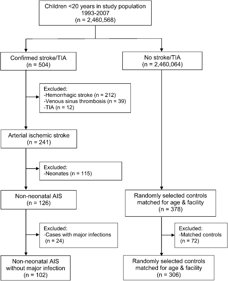
The methods of case and control ascertainment for this nested case-control study, part of the Kaiser Pediatric Stroke Study (KPSS), have been previously described7; for the current study, we performed additional chart abstraction on the same childhood AIS cases and controls drawn from the study cohort of 2.5 million children (>28 days and ≤19 years of age) enrolled in Kaiser Permanente Northern California (KPNC), 1993–2007 (figure 1). We included only strokes that occurred between 29 days and ≤19 years, excluding any strokes assumed to occur perinatally but diagnosed after the perinatal period. Because our focus was minor infections, the current analyses excluded all cases with a preceding (prior 4 weeks) or concurrent diagnosis of major infections associated with stroke (meningitis, sepsis, or endocarditis) and their matched controls.
Figure 1. Identification of study cases and controls.
Flow diagram demonstrates how study cases and controls were identified within the cohort of 2.5 million children enrolled in Kaiser Permanente Northern California from 1993 through 2007. AIS = arterial ischemic stroke.
Setting.
KPNC, the largest nonprofit managed care organization in the United States, encompasses 21 hospitals and 36 outpatient facilities, serves approximately 30% of the people in Northern California, and is largely representative of that population except at the outlying socioeconomic extremes.22
Case ascertainment and confirmation.
We have previously described the methodology used to identify and confirm AIS cases.7,23,24 We searched inpatient and outpatient electronic databases for diagnoses indicative of stroke including ischemic stroke (ICD-9 codes 433–436, 437.6, and 325), subarachnoid hemorrhage (ICD-9 codes 430 and 772.2), and intracerebral hemorrhage (ICD-9 code 431). We also performed text string searches of all head imaging reports (MRI and CT) using the following text strings: stroke, infarct, infarction, infarcted, thrombus, thromboembolic, thromboembolism, thrombotic, thrombosis, ischemia, ischemic, lacune, lacunar, vascular event, porencephaly, and porencephalic. A single pediatric stroke neurologist (H.J.F.) reviewed identified reports.24 Traditional (paper) and electronic medical records of potential stroke patients were reviewed by 2 neurologists to confirm AIS using pre-established clinical and radiographic criteria, with arbitration by a third. Criteria included (1) documented clinical presentation consistent with stroke, such as a sudden onset focal neurologic deficit; and (2) CT or MRI showing a focal ischemic infarct in a location and of a maturity consistent with the neurologic signs and symptoms. We excluded cases if the stroke occurred prior to the child's Kaiser enrollment or outside of the study period.24
Control selection.
Three population-based controls per case were randomly selected from the study cohort and matched on age, year of KPNC enrollment, and primary care facility. Each control was assigned the stroke date of the matched case, which was used as an index date for the purpose of collecting control data on infectious visits occurring prior to stroke.
Data abstraction.
A single pediatric RN medical records analyst abstracted data through review of electronic and traditional medical records for both cases and controls. Previously examined KPSS data included only a dichotomous classification of a visit for infection within the 28 days prior to stroke diagnosis.7 For the current study, the analyst abstracted detailed data regarding all documented medical visits specifically for infectious illnesses in the 2 years prior to the stroke/index date.
Classification of primary stroke etiology.
In our historical cohort, we were limited by the fact that many early cases did not receive what would today be considered a complete stroke evaluation; contemporary childhood stroke classification systems could not be applied. We therefore used a simple construct for etiology, based on data in the medical record indicating the presence of comorbidities that increase the risk of childhood stroke. A single pediatric stroke neurologist (H.J.F.) reviewed all available records to identify broad categories of potential etiology: cardiac disease, major infection such as meningitis and sepsis (all excluded from this study), hypercoaguable state, primary arteriopathy (with subtypes based on imaging reports since actual imaging studies were not available for review), sickle cell disease, and cancer. We defined as idiopathic strokes in children with no documented comorbidities suggestive of increased susceptibility.24 In the current study, we adjusted for the presence of these factors to control for potential confounding. When comorbidities were not documented in the records, we assumed that they were absent.
Analyses of timing of infection.
Data collection focused on the 2 years prior to stroke/index date; children whose strokes occurred before age 2 (and their controls) had less time to accumulate infections. Analyses of specific time periods included only those who had accrued the requisite amount of time. Children could have infections during multiple time periods; therefore, analyses of infections diagnosed during a specific time period were adjusted for infections during all other time periods, effectively holding them constant. Binary variables were created for infection occurring during the following distinct time periods (in days): 0–3, 4–7, 8–30, 31–60, 61–90, 91–120, 121–150, 151–180, 181–360, and 361–720.
Cumulative number of infectious exposures.
Because blood samples were not available in this retrospective study, we could not measure infectious burden as defined in adult studies using serologic assays for common pathogens.20,21 Instead, we used as a marker the total number of visits during the prior 2 years at which a specific diagnosis of 1 or more infections was made. Multiple visits for infection within 1 week were considered part of the same infectious process, and therefore counted as a single visit. We required that cases (and their matched controls) had a length of at least 2 years of prior observation to be included in this analysis and thus excluded children under 2 years of age.
Effect of vascular abnormalities.
In the 72 children with AIS who received vascular imaging (cerebral or cervical magnetic resonance angiography, CT angiography, or conventional angiography), we examined whether infection increases risk of arteriopathy, defined as a pathologic abnormality of a cerebral or cervical artery described on the formal imaging report. A single MD reviewer (H.J.F.) reviewed the reports, blinded to history of infection, to determine presence of arteriopathy.
Statistical analysis.
All analyses were conducted using conditional logistic regression to account for matching. Infection-related predictors were examined in models (as described above) adjusting for other time periods. Additional multivariable models were also adjusted for risk factors known to be associated with childhood stroke: sex; chronic conditions (e.g., autoimmune disease, cardiac disease, and hematologic disease); and preceding head or neck trauma (as previously defined).7 All analyses were done using Stata v12 (Stata Corp., College Station, TX).
Standard protocol approvals, registrations, and patient consents.
Study procedures were approved by the institutional review boards at KPNC and the University of California, San Francisco; consent was waived for this retrospective study.
RESULTS
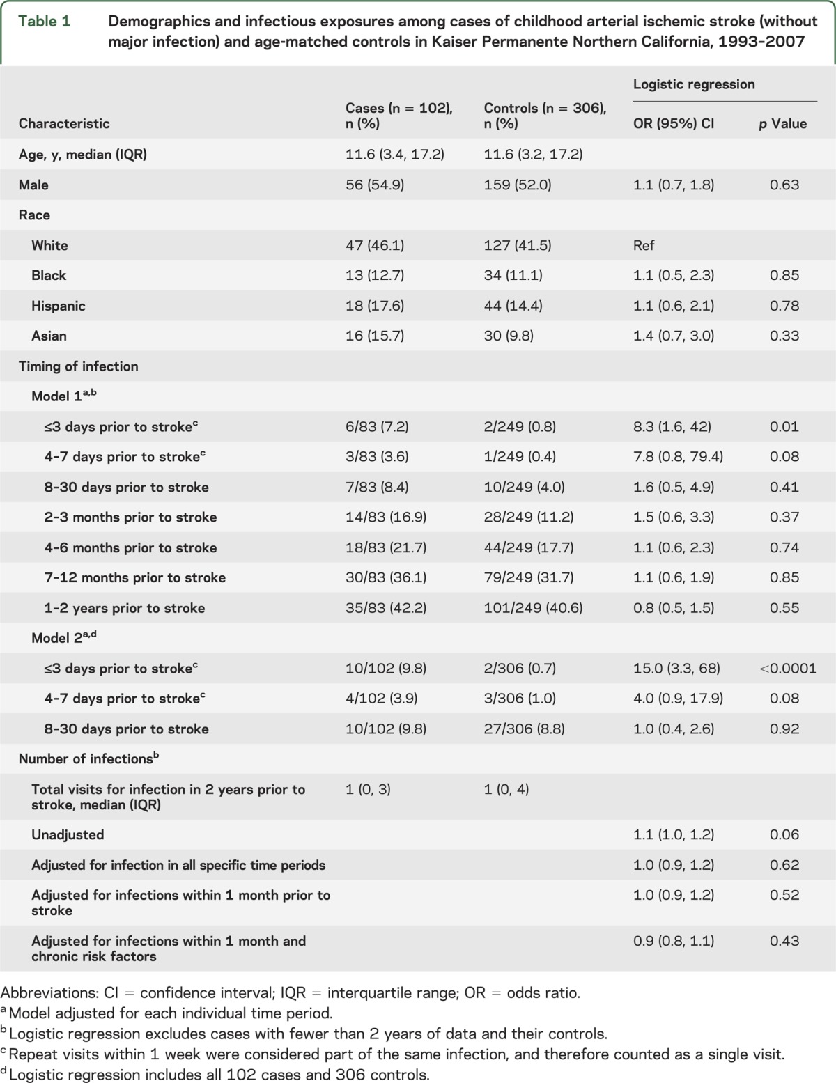
Our study included 102 children diagnosed with AIS, without a preceding or concurrent major infection, and 306 age-matched controls (figure 1). Primary comorbid factors that may have contributed to stroke etiology included cardiac disease (n = 12), hypercoaguable conditions (n = 10), primary arteriopathy (n = 45, including diagnoses of arterial dissection, n = 8, and moyamoya, n = 9), sickle cell disease (n = 2), and leukemia or lymphoma (n = 3). Six children had had a recent head or neck trauma without receiving a diagnosis of dissection. The strokes were idiopathic in the remaining 29 children. A total of 74 cases (73%) and 187 controls (61%) had at least 1 visit for infection during the 2-year observation period; allowing for multiple visits per child, the 261 study subjects accrued a total of 969 visits with an infection diagnosis. Analyses requiring the full 2 years of prior data included 81% of the study subjects: 83 cases and 249 controls (table 1; model 1). All children had at least 1 month of data prior to stroke/index date, and could be included in analyses of this time period (table 1; model 2).
Table 1.
Demographics and infectious exposures among cases of childhood arterial ischemic stroke (without major infection) and age-matched controls in Kaiser Permanente Northern California, 1993–2007
Timing of infection.
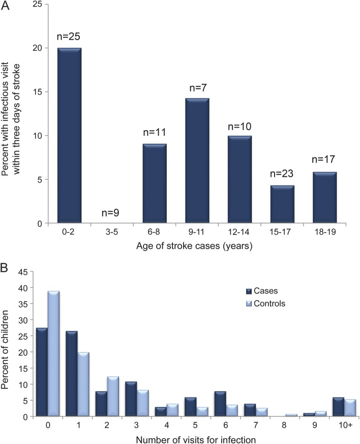
Rates of infection prior to stroke/index date were greater for cases than controls in each time period (table 1). Excluding children with fewer than 2 years of data and adjusting for all time periods (model 1), only a visit for infection within 3 days increased risk of stroke. In an analysis focused on the month prior to stroke and including all children (model 2), the risk again appeared to be concentrated in the most proximate time period. These recent infections occurred in children across the age spectrum (figure 2A). When we adjusted for other childhood stroke risk factors (table 2), a diagnosis of infection 3 days prior remained an independent risk factor, conferring a 12-fold increased risk of AIS.
Figure 2. Minor infections preceding childhood arterial ischemic stroke.
(A) Recent minor infection by age at stroke. Proportion of childhood arterial ischemic stroke cases (n = 102) with a visit for minor infection within 3 days prior to stroke, stratified by age group. (B) Number of infection visits in prior 2 years. Cumulative number of visits for minor infection in the 2-year period prior to stroke/index date in cases of childhood arterial ischemic stroke (n = 83) and age-matched controls (n = 249). Repeat visits within 1 week were considered part of the same infection, and therefore counted as a single visit.
Table 2.
Multivariable analysis of minor infections within specific time periods prior to stroke/index date in 102 cases of childhood arterial ischemic stroke and 306 age-matched controlsa
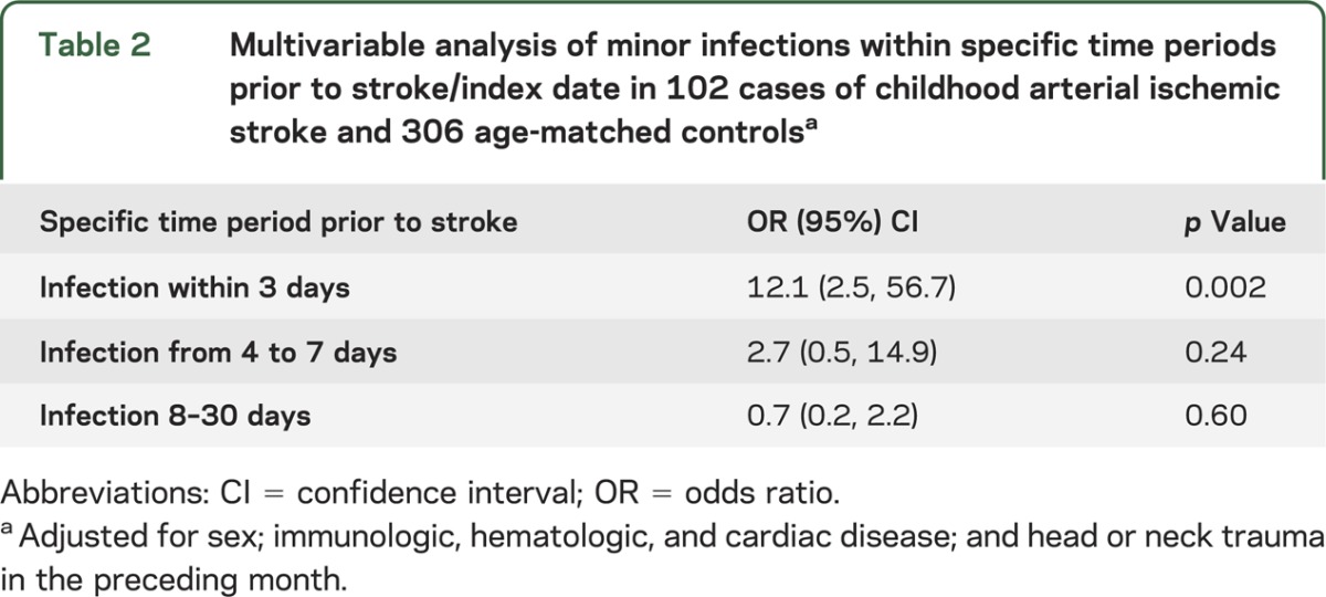
Our 102 cases included 3 children with a prior stroke that occurred either before the study period or before enrollment in KPNC. We conducted a sensitivity analysis of infection in the prior 3 days as a predictor of first stroke by excluding these 3 cases and their controls, and adjusting for the same variables as in the primary multivariable model (table 2): the OR was 10.7 (95% CI 2.2, 51, p = 0.003), compared to 12.1 in the primary analysis.
Cumulative number of infectious exposures.
We compared the total number of visits for infection in the 2 years preceding the stroke/index date between cases and controls with at least 2 years of prior observation time (figure 2B). When adjusted for the timing of these infections, the cumulative number of infectious exposures did not increase stroke risk (table 1).
Type of infection.
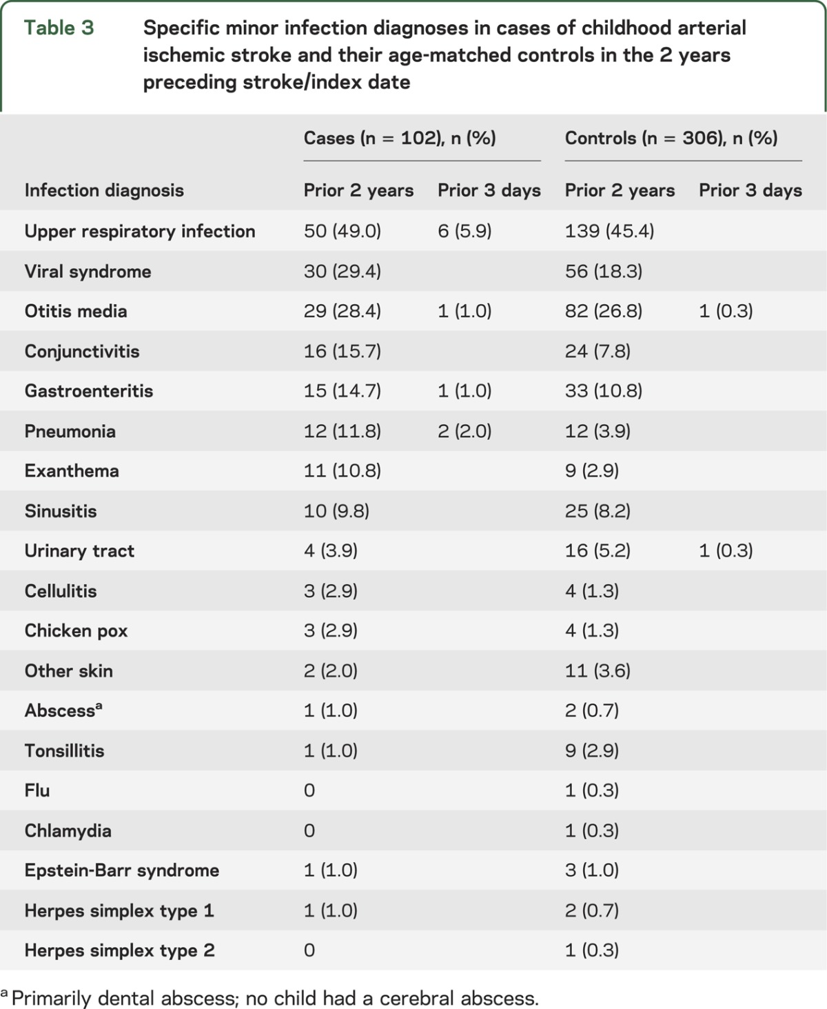
The most frequently diagnosed infections over the observation period were upper respiratory in both cases and controls (table 3). Respiratory infections accounted for 8 of the 10 case infections in the 3 days prior to stroke; only 2 cases (1 with upper respiratory infection, 1 with pneumonia) had been admitted to the hospital. Seven of the 8 cases with respiratory infections had vascular imaging, but none received a diagnosis of arterial dissection.
Table 3.
Specific minor infection diagnoses in cases of childhood arterial ischemic stroke and their age-matched controls in the 2 years preceding stroke/index date
Association of infection with arteriopathy.
Of 72 cases with vascular imaging data, 28 (39%) had an imaging report consistent with a cerebral or cervical arteriopathy; 17 had a specific diagnosis of moyamoya (n = 9) or arterial dissection (n = 8), while the others had arterial abnormalities without a specific diagnosis. We observed no difference in the rate or timing of infections prior to stroke between those with and without arteriopathy. Within 3 days prior, 11% of those with arteriopathy vs 14% of those without arteriopathy were diagnosed with a minor infection (p = 1.00); within 7 days prior, 11% vs 18% (p = 0.51); within 30 days prior, 14% vs 27% (p = 0.25). Likewise, the total sum of infectious visits over 2 years did not differ: median (interquartile range) visits were 1 (0, 4.5) for cases with arteriopathy; 1 (0, 4) for those with no arteriopathy; and 2 (0, 4) for those with no vascular imaging (p = 0.83, Kruskal-Wallis test).
DISCUSSION
In our prior study of this population-based cohort, a medical visit for minor infection in the preceding 4 weeks was a risk factor for childhood AIS, even after adjusting for chronic diseases that increase stroke risk and could lower the threshold for seeking medical attention.7 In the current report, we further explore infection as a risk factor for stroke, and find that (1) elevated stroke risk after infection may be transient, lasting only a few days; (2) the cumulative number of infections over time does not appear to play a role in stroke risk; and (3) respiratory infections are the most common type of infection immediately preceding childhood AIS.
Prior studies examining acute infection and adult AIS have typically assessed a single time window for exposure, ranging from 1 to 8 weeks before the stroke.6,9,11,25 Most of these studies used broad definitions of acute infection including bacterial and viral etiologies and any system involvement. Overall, the ORs for the association with AIS ranged from 2.9 (95% CI 1.6–5.3) for an infection in the prior 2 months (although 93% of the case infections occurred in the week prior to stroke)11 to 4.5 (95% CI 2.1–9.7) for an infection in the prior week.6 The OR was larger in younger patients, suggesting that infection is a more important risk factor in the young.9 Indeed, the largest OR was observed in young adults (<50 years old) with infection in the prior month: 14.5 (95% CI 1.9–112) after adjustment for traditional stroke risk factors.13 The association between infection and stroke did not depend on type of infection, suggesting that it is the infectious/inflammatory state, rather than a specific pathogen, that increases risk.
In our prior study of childhood AIS, the OR for the association with infection in the preceding 4 weeks, adjusted for chronic disease diagnoses, was 4.6, similar to reports from adult studies.7 However, in the current study, when we further assessed the role of timing of infection, we found that the period of highest risk is the 3 days after a visit for infection: OR 12.1 (95% CI, 2.5, 57). The risk rapidly diminishes after this period, with no increased risk of stroke beyond 1 week. One adult study examined timing of infection using within-person comparisons, and similarly found that risk of stroke was highest within the first 3 days after an infectious encounter, although their reported relative risk was lower (incidence ratio 3.2, 95% CI 2.8–3.6).10 This suggests that infection has an acute and powerful effect on stroke risk that is transient, wanes quickly over days, and may be more robust in children than adults. Our study cannot rule out, however, a more modest but persistent effect of infection on stroke risk: cases had more visits for infection than controls in all time periods up to 2 years, and the lack of significance could be due to inadequate power to detect a modest effect.
Overall, the spectrum of minor infections was remarkably similar between cases and controls. Among infections in the prior 3 days, the period of greatest risk, respiratory infections predominated among the stroke cases. This is consistent with studies of acute infection and stroke risk in adults, which similarly did not point to a particular organism, but consistently found respiratory infections the most common, comprising 41%–80% of all recent infections.6,8,11–13
Studies of atherosclerosis in adults have suggested a role for infectious burden leading to a chronic inflammatory condition that exacerbates atherosclerosis and increases stroke risk.3,26 In these studies, infectious burden is typically defined as the number of positive serologies to common pathogens, reflecting both clinical and subclinical infections. Without access to blood samples, we assessed the cumulative total medical visits for infection over the preceding 2 years, but did not detect a greater risk of childhood AIS with increased visits. Since we were unable to perform serologies, we cannot rule out an increased risk due to infectious burden as defined in adult studies. However, our results may suggest that repeated exposure to infection over time is less important for the pathogenesis of childhood than adult stroke.
Our findings may shed some light on the potential causal pathway between minor infection and childhood AIS. The transient nature of the risk seems more consistent with infection-related activation of the coagulation cascade, rather than arterial injury. (We observed similar rates of recent infection in stroke cases with and without arteriopathy, but we may have been underpowered to assess this.) Alternative explanations include exposure to cold remedies containing vasoactive medications and mechanical forces from coughing or sneezing leading to cervical arterial dissection; although we did not observe recent respiratory infections in the small number of dissection cases in this study, these mechanisms could also plausibly cause a short-lived increase in stroke risk. Infection in childhood is exceedingly common, while AIS is not; we hypothesize that infection may act as a trigger in a child who is otherwise predisposed to stroke.
Study limitations include its retrospective nature, which precluded measurement of precise timing of infection onset or exposure to over-the-counter medications. Defining infections by medical visits likely underestimates actual infectious exposure, although this should not differ between cases and controls after adjustment for chronic disease. Although the medical records analyst was unblinded to case/control status, we defined medical visit for infection as objectively and broadly as possible by specifying a list of key words to identify an infection-related visit. Serologic assays were infrequent; infections were identified by physician diagnosis alone. The Kaiser population lacks socioeconomic extremes, although infectious exposures may be higher in children of low socioeconomic status. The unavailability of vascular imaging for centralized review precluded meaningful analyses of the role infection might play in the presence of arteriopathy. However, advantages of our study are that it is population-based, and therefore not subject to referral bias, and that our measure of infection was resistant to recall bias.
An ongoing 40-center NIH-funded prospective study, Vascular effects of Infection in Pediatric Stroke, overcomes many of these limitations by collecting vascular imaging for consistent classification of arteriopathies and blood samples for objective measurement of infectious exposure.27 This large international effort will shed further light on the role of infection in childhood AIS.
Supplementary Material
ACKNOWLEDGMENT
Barbara B. Rowe, RN, performed the data collection for this study.
GLOSSARY
- AIS
arterial ischemic stroke
- CI
confidence interval
- ICD-9
International Classification of Diseases, 9th revision
- KPNC
Kaiser Permanente Northern California
- KPSS
Kaiser Pediatric Stroke Study
- OR
odds ratio
Footnotes
Editorial, page 872
AUTHOR CONTRIBUTIONS
Dr. Hills performed the statistical analyses, interpreted the results, and drafted the Introduction, Methods, and Results sections of the manuscript. Dr. Sidney assisted with the study design, supervised the acquisition of data, interpreted the results, and critically reviewed and revised the manuscript. Dr. Fullerton designed the study, obtained funding, interpreted the results, drafted the discussion, and critically reviewed and revised the full manuscript.
STUDY FUNDING
Funded by National Institute of Neurological Disorders and Stroke grant K02 NS053883 (H.J.F.). The funding agency played no role in the design or execution of the study.
DISCLOSURE
N. Hills received research support from the Thrasher Research Foundation and an NIH grant (R01 NS062820HJ). S. Sidney is supported by NIH grants (Stroke Prevention/Intervention Research Program U54). H. Fullerton is supported by NIH grants (R01 NS062820HJ; R01 HL096789-01RJ; U10 NS086494) and an American Heart Association Established Investigator Award. Go to Neurology.org for full disclosures.
REFERENCES
- 1.Ganesan V, Prengler M, McShane MA, Wade AM, Kirkham FJ. Investigation of risk factors in children with arterial ischemic stroke. Ann Neurol 2003;53:167–173 [DOI] [PubMed] [Google Scholar]
- 2.Bornstein NM, Bova IY, Korczyn AD. Infections as triggering factors for ischemic stroke. Neurology 1997;49:S45–S46 [DOI] [PubMed] [Google Scholar]
- 3.Elkind MS, Cole JW. Do common infections cause stroke? Semin Neurol 2006;26:88–99 [DOI] [PubMed] [Google Scholar]
- 4.Emsley HC, Tyrrell PJ. Inflammation and infection in clinical stroke. J Cereb Blood Flow Metab 2002;22:1399–1419 [DOI] [PubMed] [Google Scholar]
- 5.Grau AJ. Infection, inflammation, and cerebrovascular ischemia. Neurology 1997;49:S47–S51 [DOI] [PubMed] [Google Scholar]
- 6.Grau AJ, Buggle F, Heindl S, et al. Recent infection as a risk factor for cerebrovascular ischemia. Stroke 1995;26:373–379 [DOI] [PubMed] [Google Scholar]
- 7.Hills NK, Johnston SC, Sidney S, Zielinski BA, Fullerton HJ. Recent trauma and acute infection as risk factors for childhood arterial ischemic stroke. Ann Neurol 2012;72:850–858 [DOI] [PMC free article] [PubMed] [Google Scholar]
- 8.Roquer J, Cuadrado-Godia E, Giralt-Steinthauer E, et al. Previous infection and stroke: a prospective study. Cerebrovasc Dis 2012;33:310–315 [DOI] [PubMed] [Google Scholar]
- 9.Grau AJ, Buggle F, Becher H, et al. Recent bacterial and viral infection is a risk factor for cerebrovascular ischemia: clinical and biochemical studies. Neurology 1998;50:196–203 [DOI] [PubMed] [Google Scholar]
- 10.Smeeth L, Thomas SL, Hall AJ, Hubbard R, Farrington P, Vallance P. Risk of myocardial infarction and stroke after acute infection or vaccination. N Engl J Med 2004;351:2611–2618 [DOI] [PubMed] [Google Scholar]
- 11.Bova IY, Bornstein NM, Korczyn AD. Acute infection as a risk factor for ischemic stroke. Stroke 1996;27:2204–2206 [DOI] [PubMed] [Google Scholar]
- 12.Macko RF, Ameriso SF, Barndt R, Clough W, Weiner JM, Fisher M. Precipitants of brain infarction: roles of preceding infection/inflammation and recent psychological stress. Stroke 1996;27:1999–2004 [DOI] [PubMed] [Google Scholar]
- 13.Syrjanen J, Valtonen VV, Livanainen M, Kaste M, Huttunen JK. Preceding infection as an important risk factor for ischaemic brain infarction in young and middle aged patients. Br Med J 1988;296:1156–1160 [DOI] [PMC free article] [PubMed] [Google Scholar]
- 14.Smieja M, Gnarpe J, Lonn E, et al. Multiple infections and subsequent cardiovascular events in the Heart Outcomes Prevention Evaluation (HOPE) Study. Circulation 2003;107:251–257 [DOI] [PubMed] [Google Scholar]
- 15.Zhu J, Quyyumi AA, Norman JE, et al. Effects of total pathogen burden on coronary artery disease risk and C-reactive protein levels. Am J Cardiol 2000;85:140–146 [DOI] [PubMed] [Google Scholar]
- 16.Zhu J, Nieto FJ, Horne BD, Anderson JL, Muhlestein JB, Epstein SE. Prospective study of pathogen burden and risk of myocardial infarction or death. Circulation 2001;103:45–51 [DOI] [PubMed] [Google Scholar]
- 17.Auer JW, Berent R, Weber T, Eber B. Immunopathogenesis of atherosclerosis. Circulation 2002;105:E64–E64 [PubMed] [Google Scholar]
- 18.Pugh PJ, Malkin CJ, Channer KS. Cytomegalovirus seropositivity, infectious burden, and coronary artery disease. Am Heart J 2003;145:e11. [DOI] [PubMed] [Google Scholar]
- 19.Basinkevich AB, Shakhnovich RM, Martynova VR, et al. Role of Chlamydia, mycoplasma and cytomegalovirus infection in the development of coronary artery disease [in Russian]. Kardiologiia 2003;43:4–9 [PubMed] [Google Scholar]
- 20.Ngeh J, Gupta S, Goodbourn C, McElligott G. Mycoplasma pneumoniae in elderly patients with stroke: a case-control study on the seroprevalence of M pneumoniae in elderly patients with acute cerebrovascular disease: the M-PEPS Study. Cerebrovasc Dis 2004;17:314–319 [DOI] [PubMed] [Google Scholar]
- 21.Elkind MS, Moon YP, Liu K, et al. Infectious burden and carotid plaque thickness: the Northern Manhattan Study. Cerebrovasc Dis 2008;25:5 Abstract [DOI] [PMC free article] [PubMed] [Google Scholar]
- 22.Krieger N. Overcoming the absence of socioeconomic data in medical records: validation and application of a census-based methodology. Am J Public Health 1992;82:703–710 [DOI] [PMC free article] [PubMed] [Google Scholar]
- 23.Agrawal N, Johnston SC, Wu YW, Sidney S, Fullerton HJ. Imaging data reveal a higher pediatric stroke incidence than prior US estimates. Stroke 2009;40:3415–3421 [DOI] [PMC free article] [PubMed] [Google Scholar]
- 24.Fullerton HJ, Wu YW, Sidney S, Johnston SC. Risk of recurrent childhood arterial ischemic stroke in a population-based cohort: the importance of cerebrovascular imaging. Pediatrics 2007;119:495–501 [DOI] [PubMed] [Google Scholar]
- 25.Becher H, Grau A, Steindorf K, Buggle F, Hacke W. Previous infection and other risk factors for acute cerebrovascular ischaemia: attributable risks and the characterisation of high risk groups. J Epidemiol Biostat 2000;5:277–283 [PubMed] [Google Scholar]
- 26.Vercellotti GM. Overview of infections and cardiovascular diseases. J Allergy Clin Immunol 2001;108:S117–S120 [DOI] [PubMed] [Google Scholar]
- 27.Fullerton HJ, Elkind MS, Barkovich AJ, et al. The Vascular Effects of Infection in Pediatric Stroke (VIPS) Study. J Child Neurol 2011;26:1101–1110 [DOI] [PMC free article] [PubMed] [Google Scholar]
Associated Data
This section collects any data citations, data availability statements, or supplementary materials included in this article.


