Abstract
Conotruncal heart defects (CTDs) are among the most severe birth defects worldwide. Studies of CTDs indicate both lifestyle behaviors and genetic variation contribute to the risk of CTDs. Based on a hybrid design using data from 616 case-parental and 1645 control-parental triads recruited for the National Birth Defects Prevention Study between 1997 and 2008, we investigated whether the occurrence of CTDs is associated with interactions between 921 maternal and/or fetal single nucleotide polymorphisms (SNPs) and maternal obesity and tobacco use. The maternal genotypes of the variants in the glutamate-cysteine ligase, catalytic subunit (GCLC) gene and the fetal genotypes of the variants in the glutathione S-transferase alpha 3 (GSTA3) gene were associated with an elevated risk of CTDs among obese mothers. The risk of delivering infants with CTDs among obese mothers carrying AC genotype for a variant in the GCLC gene (rs6458939) was 2.00 times the risk among those carrying CC genotype (95% confidence interval: 1.41, 2.38). The maternal genotypes of several variants in the glutathione-S-transferase (GST) family of genes and the fetal genotypes of the variants in the GCLC gene interacted with tobacco exposures to increase the risk of CTDs. Our study suggests that the genetic basis underlying susceptibility of the developing heart to the adverse effects of maternal obesity and tobacco use involve both maternal and embryonic genetic variants. These results may provide insights into the underlying pathophysiology of CTDs, and ultimately lead to novel prevention strategies.
Introduction
Congenital heart defects (CHDs) are among the most common and severe birth defects worldwide, with reported estimated prevalence of 9.1 per 1,000 live births after 1995 [1]. Conotruncal heart defects (CTDs), a class of CHDs, affect the cardiac outflow tracts and great arteries which include truncus ateriosus, interrupted aortic arch type B, transposition of great arteries, double outlet right ventricle, conoventricular septal defect, tetralogy of Fallot, and pulmonary atresia with ventricular septal defect.
Nonsyndromic CTDs are due to a multifactorial etiology involving a complex interplay between genetic susceptibilities and environmental factors [2], [3]. One such interplay is between maternal folic acid supplement use and genetic variants in folate-related pathways. Based on the finding that maternal periconceptional folic acid intake decreases the occurrence of CTDs [4], [5], multiple studies have investigated associations between CTDs and polymorphisms in folate-related genes [6]–[8]. It has also been reported that genetic variants in folate-related pathways modify the association between birth defects and maternal intake of folic acid containing supplements [9].
Developmental toxicology studies using animal models have repeatedly demonstrated the unquestioned importance of genetic variation in determining risks to environmental factors [10]. Inbred strains of mice, representing different mouse genomes, vary in their susceptibility to teratogenic and xenobiotic agents. In reproductive age women, obesity and tobacco use have been associated with multiple adverse outcomes including intrauterine growth retardation [11], [12], prematurity [13], [14], and birth defects [15]–[17]. Obesity and cigarette smoking are also associated with alterations in folate and glutathione metabolism resulting in decreased folate [18], [19], increased homocysteine [20]–[24], and decreased glutathione [25]–[30] that may compromise the in-utero environment. Some studies have demonstrated that maternal genetic variants modulate the association between pregnancy smoking exposure and fetal growth restriction [31], [32]. It is possible that variants in genes that encode for critical enzymes in folate, homocysteine and glutathione pathways modify the adverse impact of obesity and tobacco on the developing heart.
In this study, we used a hybrid design which combines genetic and lifestyle data from case-parental and control-parental triads to investigate whether CTDs are associated with interactions between maternal and/or fetal single nucleotide polymorphisms (SNPs) and maternal obesity and tobacco use. In contrast to most published reports of Gene × Environment (G × E) interactions [4], [33], [34], we have evaluated maternal and fetal genetic effects simultaneously. A total of 1536 SNPs in 62 target genes were selected for this study from folate-related metabolic pathways.
Materials and Methods
Study Population
Families were recruited for the National Birth Defects Prevention Study (NBDPS) with estimated dates of delivery between October 1997 and August 2008 (www.nbdps.org). Detailed information about the NBDPS is outlined in Yoon et al [35]. Families were identified through population-based birth defects surveillance systems in 10 states: Arkansas, California, Iowa, Massachusetts, New Jersey (through 2002), New York, Texas, Georgia, North Carolina (beginning 2003), and Utah (beginning 2003). In this study, cases were singleton live-born infants with CTDs. NBDPS cardiac cases were reviewed by pediatric cardiologists using a classification strategy developed by investigators within the NBDPS. This strategy targeted etiologic investigations of CHDs that encourage explicit case definitions and aggregates of defects with a focus on simple, isolated phenotypes and associations [36]. Cases with recognized or strongly suspected monogenic or chromosomal conditions were excluded. Controls were singleton live-born infants without any major structural birth defects [37]. Both case and control mothers completed phone interviews. The study was approved by the University of Arkansas for Medical Sciences' Institutional Review Board and the NBDPS with protocol oversight by the Centers for Disease Control and Prevention Center for Birth Defects and Developmental Disabilities. All of the study subjects gave written informed consent. For minors, informed written consent was obtained from their legal guardian.
Maternal Interview
Participation in the NBDPS included a one-hour interview with mothers of cases and controls, conducted in English or Spanish, by interviewers using a computer-assisted telephone questionnaire [35]. In this study, we investigated how maternal obesity and tobacco use modify maternal and fetal genetic effects on the risk of CTDs. Obesity, using the Institute of Medicine definition [38], was defined as a body mass index (BMI) ≥30.0 and normal weight between 18.5 and 25.0. Smokers were defined as women who smoked cigarettes during the 3 months after conception and nonsmokers otherwise. Maternal use of folic acid supplements is a known risk factor for the occurrence of CTDs and warranted a separate manuscript [9].
DNA Collection
After completion of maternal interviews, a buccal cell collection kit was sent to participants to obtain cheek cell samples from case/control and parents. The collection kit included informed consent forms, instructions, $20 money order, materials for completing the specimen collection and prepaid US mail packets for specimen return [35].
Gene/SNP Selection
As previously described [39], a custom panel of 1536 SNPs in 62 genes involved in folate metabolism was developed jointly between our lab and Illumina. Candidate genes were required to encode an enzyme in one of the candidate metabolic pathways and be expressed in liver and/or heart tissue [40]. For each candidate gene, a maximally informative set of haplotype-tagging SNPs was selected using both linkage disequilibrium statistics and Illumina assay design scores. The custom genotyping panel was devised in 2005–2006. At that time there were two genes called RFC1 in the commonly used genetic databases. The genotype data presented here are for SNPs in the Replication Factor C (activator 1) 1 (RFC1) gene. This gene is an activator of DNA polymerase and is required for DNA synthesis and repair.
Genotyping and Quality Assessment
Genotyping was conducted on a total of 635 case and 1702 control families using 200 ng of WGA DNA on the Illumina Golden Gate platform [41]. Initial genotype calls were generated using Genome Studio's GenCall, Illumina's proprietary algorithm, with subsequent analysis performed using SNPMClust, a bivariate Gaussian model-based genotype clustering and calling algorithm developed in-house. A total of 297 individuals were removed due to study ineligibility (n = 33), high no-call rates (n = 63), or high rates of Mendelian inconsistency (n = 201). We found that the quality of genotype clustering varied substantially from SNP to SNP, which we attribute to the in silico design of the SNP panel based on data from phases I and II of the HapMap project, without the subsequent quality checks that would be applied to a standard commercial SNP panel. While the majority of SNPs exhibited well-segregated genotype clusters, a substantial percentage exhibited poor clustering and/or lower minor allele frequency (MAF) than expected. To ensure high-quality genotypes, we applied stringent quality control measures and excluded SNPs with obviously poor clustering behavior (60 SNPs), no-call rates >10% (328 SNPs), Mendelian error rates >5% (11 SNPs), MAF <5% (204 SNPs), or significant deviation from Hardy-Weinberg Equilibrium in at least one racial group (p<10−4, 12 SNPs). The final dataset included 4648 individuals, each with 921 SNPs.
Statistical Methods
Descriptive statistics were summarized for families. Comparisons of maternal characteristics were carried out between case and control families using the two-sample t-tests for continuous variables and chi-square tests for categorical variables. Because both case-parental and control-parental triads were enrolled and genotyped, a hybrid design provided optimal power by using a log-linear model [42]. This model has several advantages including incorporating case and control triads, providing population-based inferences related to maternal and fetal genetic effects, providing evidence related to fetal genetic effects that are robust to population admixture, and allowing inclusion of missing genotype data. To assess Gene × Environment (G × E) interactions, a log-linear model was fitted for each SNP as a function of mating types, an interaction between mating types and a maternal factor (E), case/control status (D), maternal genotype (D × M), fetal genotype (D × C), an interaction for maternal genotype (D × E × M), and an interaction for fetal genotype (D × E × C). The detailed information regarding the model is outlined in Hobbs et al [9]. Based on the log-linear model for counts and assuming a Poisson distribution, the relative risk (RR) (95% CI) of having one copy of the minor allele compared to no copy was estimated for each SNP. Because we assumed multiplicative risk of alleles, the estimated RR (95% CI) of having 2 copies of the minor allele is square of the estimated RR (95% CI) of having 1 copy of the minor allele. We also performed sensitivity analysis by restricting our models to the Caucasian families only. The Caucasian families were selected for the sensitivity analysis because among all the racial groups, we had the largest sample size for Caucasian families. For other racial groups, we did not have power to perform any subgroup analyses.
The Bayesian false-discovery probability (BFDP) [43]–[48] was used to evaluate the chance of false-positive associations using estimates of the RR and corresponding 95% CI obtained from log-linear models. In the results section, we reported associations where BFDP <0.80, which is a commonly used threshold suggested by Wakefield [43] for summary analyses. Patterns of linkage disequilibrium (LD) between significant SNPs in the same gene were constructed to assess the correlations among these SNPs using the parents of control families. Data were analyzed using statistical software LEM [49] for fitting log-linear models, R v3.0.1 (R Foundation for Statistical Computing, Vienna, Austria) for computing descriptive statistics, and BFDPs and HaploView 4.2 [50] for developing LD maps.
Results
Study Sample
A total of 616 case families and 1645 control families were included in our analysis. Due to different call rates among SNPs, case and control triads/dyads/monads numbers differ for any given SNP. Maternal demographics and lifestyle factors are summarized in Table 1 . The case mothers were slightly older than the control mothers [28.3±6.1 vs. 27.5±6.0, p = 0.002]. Case and control mothers did not differ on any of the other demographics and lifestyle characteristics (p>0.05).
Table 1. Maternal characteristics for 616 case families and 1,645 control families enrolled in National Birth Defects Prevention Study between 1997 and 2008.
| Case | Control | |
| (N = 616) | (N = 1,645) | |
| Age at delivery (years) | 28.3±6.1 | 27.5±6.0 |
| Race | ||
| African American | 49 (8%) | 143 (9%) |
| Caucasian | 401 (66%) | 1,136 (69%) |
| Hispanic | 123 (20%) | 285 (17%) |
| Others | 39 (6%) | 78 (5%) |
| Missing information | 4 | 3 |
| Education | ||
| <12 years | 83 (14%) | 217 (13%) |
| High school degree or equivalent | 167 (27%) | 413 (25%) |
| 1–3 years of college | 173 (28%) | 454 (28%) |
| At least 4 years of college or Bachelor degree | 190 (31%) | 559 (34%) |
| Missing information | 3 | 2 |
| Household income | ||
| Less than 10 Thousand | 94 (16%) | 236 (15%) |
| 10 to 30 Thousand | 150 (26%) | 408 (27%) |
| 30 to 50 Thousand Dollars | 118 (20%) | 348 (23%) |
| More than 50 Thousand | 217 (37%) | 538 (35%) |
| Missing information | 37 | 115 |
| Folic acid supplements | ||
| No | 299 (49%) | 738 (45%) |
| Yes1 | 314 (51%) | 907 (55%) |
| Missing information | 3 | 0 |
| BMI | ||
| Underweight (BMI <18.5) | 31 (5%) | 74 (5%) |
| Normal weight (18.5< = BMI <25) | 298 (50%) | 880 (55%) |
| Overweight (25< = BMI <30) | 141 (24%) | 360 (23%) |
| Obese (> = 30) | 121 (20%) | 281 (18%) |
| Missing information | 25 | 50 |
| Alcohol consumption | ||
| No | 460 (76%) | 1,251 (76%) |
| Yes2 | 149 (24%) | 390 (24%) |
| Missing information | 7 | 4 |
| Tobacco use | ||
| No | 498 (81%) | 1,356 (82%) |
| Yes3 | 114 (19%) | 288 (18%) |
| Missing information | 4 | 1 |
Summary statistics were expressed as mean ± standard deviation for continuous variables, and frequency (percentage) for categorical variables.
Folic acid supplements yes is defined as maternal use of folic acid supplements at least two months during the exposure window (i.e. one month prior to conception and two months after conception).
Alcohol consumption yes is defined as drinking during the three months after pregnancy.
Tobacco use yes is defined as cigarette smoking during the three months after pregnancy.
Genetic Variants
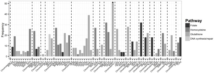
As described above, 921 candidate SNPs were included in the final analyses. The information about the number of SNPs in each gene, chromosome and pathway is displayed in Figure 1 . Hobbs et al. [9] identified the maternal genotypes of 17 SNPs and the fetal genotypes of 17 SNPs associated with CTD risks independently from interactions with maternal folic acid supplement use, obesity and tobacco use. Briefly, the maternal genotypes of 10 SNPs and the fetal genotypes of 2 SNPs in the glutamate-cysteine ligase, catalytic subunit (GCLC) gene were found to be significantly associated with the risk of CTDs through main genetic effects [9].
Figure 1. SNP, gene, chromosome and pathway information.
Information about the number of SNPs from each gene, chromosome and pathway (* there are a total of 142 candidate SNPs on MGMT, truncated at 50 for display purpose). The average number of SNPs per gene is 15±19. Among 60 genes, 10 (17%) genes are involved in folate pathway, 32 (53%) in glutathione pathway, and 17 (28%) in homocysteine pathway, and one gene RFC1 (2%) in DNA synthetic/repair pathway.
Gene × Environment Interactions
In Hobbs et al. [9], we identified SNPs that had a main effect on the occurrence of CTDs. To identify significant G × E interactions, we evaluated the combined effects of each maternal and infant SNP with maternal folic acid supplement use, obesity and tobacco use. Significant interactions are discussed below.
SNP × Folic Acid Supplement Use
In Hobbs et al. [9], we have studied the interactions between 921 SNPs and periconceptional folic acid supplement use, and found that the maternal genotypes of 19 SNPs and the fetal genotypes of 9 SNPs had BFDP <0.80 for testing the G × E interactions with maternal use of folic acid supplements. All these SNPs were not found to be significant based on the gene only models. Detailed information about the SNP × folic acid supplement use is outlined in Hobbs et al [9].
SNP × Obesity
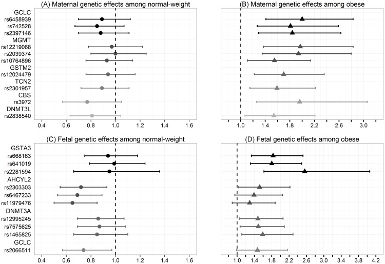
A main effect of the association between CTDs and obesity compared to normal weight resulted in an estimated RR = 1.27 (95% CI: 0.99, 1.63; p = 0.06). Because previous studies showed that obesity independent of pre-existing Type I or Type II diabetes was significantly associated with elevated risk of CTDs [15], [51], we computed a model for each SNP among normal-weight and obese women. The results are shown in Figure 2 and Table S1.
Figure 2. Interactions between gene and obesity.
The estimated relative risks and their 95% confidence intervals for each maternal and fetal SNPs that were identified to have a significant interaction with maternal obesity on the risk of CTDs. (A) Maternal genetic effects among normal-weight women; (B) Maternal genetic effects among obese women; (C) Fetal genetic effects among normal-weight women; (D) Fetal genetic effects among obese women.
Among 10 maternal SNPs with a significant gene × obesity interaction, three SNPs are in the GCLC gene, three SNPs are in the O-6-methylguanine-DNA methyltransferase (MGMT) gene, and the remaining four SNPs are in the glutathione S-transferase mu 2 (GSTM2), transcobalamin II (TCN2), cystathionine-beta-synthase (CBS), and DNA (cytosine-5-)-methyltransferase 3-like (DNMT3L) genes respectively. The most significant maternal gene × obesity interaction (i.e. most significant difference in the estimated RR for certain genotype among obese women compared to normal weight women) was embedded in the GCLC gene. The genotype of rs6458939 was not associated with the risk of CTDs among normal-weight women [estimated RR: 0.89, 95% CI: 0.70, 1.12]; however, the risk of delivering infants with CTDs among obese women carrying AC genotype for rs6458939 was estimated to be 2.00 (95% CI: 1.41, 2.83) times the risk among those carrying CC genotype. The RR (95% CI) for carrying two copies of the minor allele is square of the estimated RR (95% CI) for carrying one copy of the minor allele. Thus, only the RR (95% CI) is reported for carrying one copy of the minor allele compared to no copy of the minor allele. A similar pattern was observed for the maternal genotypes of two other SNPs in the GCLC gene. All three maternal SNPs in the GCLC gene were determined to be in high LD (D′≥0.98).
Among 10 fetal SNPs found to be associated with the risk of CTDs in combination with obesity, three SNPs reside in the glutathione S-transferase alpha 3 (GSTA3) gene, three SNPs are in the adenosylhomocysteinase-like 2 (AHCYL2) gene, three SNPs are in the DNA (cytosine-5-)-methyltransferase 3 alpha (DNMT3A) gene, and the other SNP resides in the GCLC gene. We observed the most significant difference in the fetal effect of the GSTA3 gene on the risk of CTDs between normal-weight and obese women. The risk of CTDs was elevated for infants with AG genotype for rs668163 compared to GG genotype among obese women (RR: 1.83, 95% CI: 1.33, 2.52), but was not significantly reduced among normal-weight women (RR: 0.94, 95% CI: 0.75, 1.18). Similar findings were observed for the fetal genotypes of two other SNPs in the GSTA3 gene. All three fetal SNPs in the GSTA3 gene were in high LD (D′≥0.96).
Similar results were observed when overweight women were compared to normal-weight women (data not shown) and based on Caucasian families only (data not shown). There was no main effect of any of the SNPs discussed above.
SNP × Tobacco Use
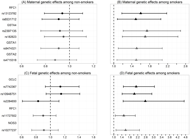
A main effect of the association between CTDs and maternal tobacco use compared to no tobacco use had an estimated RR = 1.08 (95% CI: 0.85, 1.37; p = 0.54). We investigated the interactions between SNPs and maternal tobacco use on the occurrence of CTDs by fitting the G × E log-linear model with maternal tobacco use for each SNP. The maternal and fetal SNPs that had a significant interaction with maternal tobacco use are presented in Figure 3 and Table S2.
Figure 3. Interactions between gene and maternal tobacco use.
The estimated relative risks and their 95% confidence intervals for each maternal and fetal SNPs that were identified to have a significant interaction with maternal tobacco use on the risk of CTDs. (A) Maternal genetic effects among non-smokers; (B) Maternal genetic effects among smokers; (C) Fetal genetic effects among non-smokers; (D) Fetal genetic effects among smokers.
Among 6 maternal SNPs found to have BFDP <0.80, two of them are located in the RFC1 gene, two SNPs are in the glutathione S-transferase alpha 4 (GSTA4) gene, and the other two SNPs are in the glutathione S-transferase alpha 1 (GSTA1) and glutathione S-transferase alpha 2 (GSTA2) genes respectively. The GSTA1, GSTA2 and GSTA4 genes are glutathione-S-transferase (GST) family of genes. The RRs of the maternal AC genotype for rs2397135 in the GSTA4 gene compared to CC genotype were estimated to be 0.92 (95% CI: 0.76, 1.11) among women without tobacco use, and 1.74 (95% CI: 1.21, 2.49) among women with tobacco use, interaction BFDP = 0.68<0.80. The two maternal SNPs in the RFC1 gene were in moderate LD (D′ = 0.82).
Among 5 fetal SNPs identified to have BFDP <0.80, three SNPs are in the GCLC gene, one SNP (rs11727502) is in the RFC1 gene, and the other SNP (rs10277237) is located in the nitric oxide synthase 3 (endothelial cell) (NOS3) gene. The RRs of the fetal AG genotype for rs7742367 in the GCLC gene compared to AA genotype were estimated to be 1.14 (95% CI: 0.83, 1.40) among women without tobacco use, and 2.12 (95% CI: 1.48, 3.04) among women with tobacco use, interaction BFDP = 0.71<0.80. The two fetal SNPs (rs7742367 and rs10948751) in the GCLC gene were in high LD (D′ = 0.99), and in moderate LD with the other SNP (rs2284650) respectively (D′ = 0.89 and 0.93).
Similar results were found based on Caucasian families only (data not shown). There was no SNP main effect.
Discussion
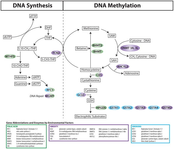
This study provides evidence for a moderating effect of multiple SNPs related to one-carbon metabolism and glutathione metabolism on the relationship between CTDs and maternal obesity and tobacco use. Significant genes and their pathway information are displayed in Figure 4 . Our findings support the hypothesis that CTDs are associated with G × E interactions between common genetic variants and lifestyle factors. Consistent with previous studies [52]–[54], our results provide supportive evidence that both maternal and fetal genotypes modify the susceptibility of the developing fetus to factors that may alter the in-utero environment.
Figure 4. Significant genes and their pathways.
Related pathways include the folate pathway, which is involved in DNA synthesis, and the methionine and glutathione pathways, which are necessary for DNA methylation and for conjugation of electrophilic substrates, respectively. Substrates and products of enzymatic reactions appear in boxes. Enzymes appear in color-coded bubbles according to their association with birth defects risk and maternal folic acid supplement use (Green), obesity (Purple) and tobacco use (Blue).
Maternal obesity independent of diabetes is a well-accepted risk factor for multiple nonsyndromic birth defects including CTDs [15], [51], [55]. Among obese women, the maternal genotypes of 10 SNPs within 6 genes (GCLC, MGMT, GSTM2, TCN2, CBS, and DNMT3L) and the fetal genotypes of 7 SNPs within 3 genes (GSTA3, AHCYL2, and DNMT3A) were associated with an increased risk of CTDs, whereas, among women of normal weight, none of the maternal genotypes had a significant impact on the RR, and the fetal genotypes of 4 SNPs in 2 genes (AHCYL2 and GCLC) decreased the risk of CTDs.
Among women who used tobacco in the periconceptional period, the maternal genotypes of 6 SNPs in 4 genes (RFC1, GSTA4, GSTA1, and GSTA2) were associated with an increased risk of CTDs. The GST genes function in the detoxification of several environmental exposures, including carcinogens, medications, and factors increasing oxidative stress. The fetal genotypes of 5 SNPs in 3 genes (GCLC, RFC1 and NOS3) increased the occurrence of CTDs among women who smoked. None of the maternal or fetal genotypes significantly impact the risk among women without tobacco use.
Our findings compel us to comment on some of the findings in the context of previous studies. Several maternal and fetal genotypes of SNPs in the glutathione transferase (GSTA4, GSTA1, GSTA2, GSTM2, and GSTA3) genes increased the impact of maternal obesity and tobacco use on the risk of CTDs. The Glutathione S-transferase complex of genes are involved in the detoxification of a large number of xenobiotics including chemical compounds in tobacco smoke [56]. Genetic variants in glutathione S-transferase genes may be a factor in determining individual susceptibility to embryotoxic effects of xenobiotics, including tobacco. Lending credence to the possibility that glutathione S-transferases in interaction with tobacco may impact the developing fetus, is a report that glutathione S-transferase genetic variants modified the impact of cigarette smoking on birth weight [32].
In earlier reports [57], [58], we demonstrated that women who had infants with CHDs had lower GSH levels. GSH level is essential for cell progression and serves several functions that are crucial during embryogenesis. GSH is important in the defense against oxidative stress, detoxification of xenobiotics, regulation of cell cycle progression and apoptosis, and modulation of immune function [56]. GCLC is the gene that encodes for the catalytic subunit glutamate-cysteine ligase, which is the enzyme that catalyzes the conversion of cysteine with glutamate, generating glutathione. Several maternal and fetal genotypes of SNPs in the GCLC gene increased the RR of CTDs among women who smoked and/or were obese.
Among women who did not use folic acid supplements, 8 SNPs in the hematopoietic prostaglandin D synthase gene (HPGDS) increased the risk of CTDs, but had no associated increased risk among folic acid supplement users [9]. HPGDS is a member of the sigma class of glutathione S-transferase gene family. The HPGDS gene catalyzes both isomerization of PGH (2) to PGD (2) and conjugation of glutathione to 1-chlore-2, 4-dinitrobenzene [59]–[61]. HPGDS is present in mast cells, macrophages and other cellular sources [62]. Little is known about the impact of prostaglandin synthase on the development of CHDs. Previous studies have shown that HPGDS plays a role in multiple physiological processes that are important during embryogenesis, including detoxification of xenobiotics [63], regulation of inflammatory pathways [59] and modification of reactive oxygen species [64]. Animal models and mechanistic studies are needed to validate and clarify the role of maternal HPGDS in the development of heart defects.
There are several strengths of this study including the use of log-linear models based on a hybrid design, its large sample size, population-based ascertainment of cases, and a rigorous analytic inquiry into potential G × E interactions on CTD risks. There are also limitations in our study. No adjustment was made for confounding effects from other exposures when fitting the model, which prohibited us from drawing further conclusions about the observed associations. Other limitations related to DNA and lifestyle data collection existed. The usage of buccal cell collection kits in collecting DNA samples created a disparity among the quality of the DNA samples, resulting in a reduction from 1534 to 921 candidate SNPs. Maternal obesity and tobacco use were self-reported, and not objectively measured.
It is now generally accepted that most CHDs are the result of a complex interplay between genetic and environmental factors. Our results suggest that, in the search for genetic variants related to CTDs, accounting for environmental and lifestyle factors may improve the ability to detect genetic effects. Because the typical effect size for maternal and fetal genetic variation acting on CHD phenotypes is small [65], [66], large sample sizes and validation within independent populations are necessary to draw firm conclusions about how certain polymorphisms modulate the experience of maternal lifestyles. The present findings will need to be replicated and further studies should incorporate functional genetics and targeted deep sequencing while enhancing information collected on environmental and lifestyle factors. One limitation of our current study is that to properly assess the accuracy of our findings, they will need to be replicated on a fully distinct and independent sample [67]. Nevertheless, our study is the largest family-based case-control study of CTDs ever conducted in the U.S., and to our knowledge, there is no comparable, independent sample which includes the maternal and fetal genotype data, and maternal lifestyle and demographic data, necessary to validate our results.
Supporting Information
Maternal and fetal SNPs with interactive effects with maternal obesity. For each significant SNP, the information about its pathway, chromosome, gene, allele, estimated relative risks and their 95% confidence intervals among normal weight and obese women, p-value and BFDP for the interaction term are presented.
(DOCX)
Maternal and fetal SNPs with interactive effects with maternal tobacco use. For each significant SNP, the information about its pathway, chromosome, gene, allele, estimated relative risks and their 95% confidence intervals among nonsmokers and smokers, p-value and BFDP for the interaction term are presented.
(DOCX)
Acknowledgments
The authors wish to thank the generous participation of the numerous families that made this research study possible. We also thank the Centers for Birth Defects Research and Prevention in California, Georgia, Iowa, Massachusetts, New Jersey, New York, North Carolina, Texas, and Utah for their contribution of data and manuscript review. The authors thank Ashley S. Block and Zuzana Gubrij for assistance in preparation of the manuscript.
The contents of this manuscript are solely the responsibility of the authors and do not necessarily represent the official views of the Centers for Disease Control and Prevention.
Data Availability
The authors confirm that all data underlying the findings are fully available without restriction. All relevant data are within the paper and its Supporting Information files.
Funding Statement
This work is supported by the National Institute of Child Health and Human Development (NICHD) under award number 5R01HD039054-12 and the National Center on Birth Defects and Developmental Disabilities (NCBDDD) under award number 5U01DD000491-05. The funders had no role in study design, data collection and analysis, decision to publish, or preparation of the manuscript.
References
- 1. van der Linde D, Konings EE, Slager MA, Witsenburg M, Helbing WA, et al. (2011) Birth prevalence of congenital heart disease worldwide: a systematic review and meta-analysis. J Am Coll Cardiol 58: 2241–2247. [DOI] [PubMed] [Google Scholar]
- 2. Pierpont ME, Basson CT, Benson DW Jr, Gelb BD, Giglia TM, et al. (2007) Genetic basis for congenital heart defects: current knowledge: a scientific statement from the American Heart Association Congenital Cardiac Defects Committee, Council on Cardiovascular Disease in the Young: endorsed by the American Academy of Pediatrics. Circulation 115: 3015–3038. [DOI] [PubMed] [Google Scholar]
- 3. Jenkins KJ, Correa A, Feinstein JA, Botto L, Britt AE, et al. (2007) Noninherited risk factors and congenital cardiovascular defects: current knowledge: a scientific statement from the American Heart Association Council on Cardiovascular Disease in the Young: endorsed by the American Academy of Pediatrics. Circulation 115: 2995–3014. [DOI] [PubMed] [Google Scholar]
- 4. Shaw GM, O'Malley CD, Wasserman CR, Tolarova MM, Lammer EJ (1995) Maternal periconceptional use of multivitamins and reduced risk for conotruncal heart defects and limb deficiencies among offspring. Am J Med Genet 59: 536–545. [DOI] [PubMed] [Google Scholar]
- 5. Botto LD, Khoury MJ, Mulinare J, Erickson JD (1996) Periconceptional multivitamin use and the occurrence of conotruncal heart defects: results from a population-based, case-control study. Pediatrics 98: 911–917. [PubMed] [Google Scholar]
- 6. Pediatric Cardiac Genomics C, Gelb B, Brueckner M, Chung W, Goldmuntz E, et al. (2013) The Congenital Heart Disease Genetic Network Study: rationale, design, and early results. Circ Res 112: 698–706. [DOI] [PMC free article] [PubMed] [Google Scholar]
- 7. Shaw GM, Lu W, Zhu H, Yang W, Briggs FB, et al. (2009) 118 SNPs of folate-related genes and risks of spina bifida and conotruncal heart defects. BMC Med Genet 10: 49. [DOI] [PMC free article] [PubMed] [Google Scholar]
- 8. Lupo PJ, Goldmuntz E, Mitchell LE (2010) Gene-gene interactions in the folate metabolic pathway and the risk of conotruncal heart defects. J Biomed Biotechnol 2010: 630940. [DOI] [PMC free article] [PubMed] [Google Scholar]
- 9. Hobbs CA, Cleves MA, Macleod SL, Erickson SW, Tang X, et al. (2014) Conotruncal heart defects and common variants in maternal and fetal genes in folate, homocysteine, and transsulfuration pathways. Birth Defects Res A Clin Mol Teratol 100: 116–126. [DOI] [PMC free article] [PubMed] [Google Scholar]
- 10. Wlodarczyk BJ, Palacios AM, Chapa CJ, Zhu H, George TM, et al. (2011) Genetic basis of susceptibility to teratogen induced birth defects. Am J Med Genet C Semin Med Genet 157: 215–226. [DOI] [PubMed] [Google Scholar]
- 11. Radulescu L, Munteanu O, Popa F, Cirstoiu M (2013) The implications and consequences of maternal obesity on fetal intrauterine growth restriction. J Med Life 6: 292–298. [PMC free article] [PubMed] [Google Scholar]
- 12. Delpisheh A, Brabin L, Drummond S, Brabin BJ (2008) Prenatal smoking exposure and asymmetric fetal growth restriction. Ann Hum Biol 35: 573–583. [DOI] [PubMed] [Google Scholar]
- 13. Cnattingius S, Villamor E, Johansson S, Edstedt Bonamy AK, Persson M, et al. (2013) Maternal obesity and risk of preterm delivery. JAMA 309: 2362–2370. [DOI] [PubMed] [Google Scholar]
- 14. Wang T, Zhang J, Lu X, Xi W, Li Z (2011) Maternal early pregnancy body mass index and risk of preterm birth. Arch Gynecol Obstet 284: 813–819. [DOI] [PubMed] [Google Scholar]
- 15. Gilboa SM, Correa A, Botto LD, Rasmussen SA, Waller DK, et al. (2010) Association between prepregnancy body mass index and congenital heart defects. Am J Obstet Gynecol 202: 51 e51–51 e10. [DOI] [PubMed] [Google Scholar]
- 16. Hackshaw A, Rodeck C, Boniface S (2011) Maternal smoking in pregnancy and birth defects: a systematic review based on 173 687 malformed cases and 11.7 million controls. Hum Reprod Update Sep-Oct 17: 589–604. [DOI] [PMC free article] [PubMed] [Google Scholar]
- 17. Malik S, Cleves MA, Honein MA, Romitti PA, Botto LD, et al. (2008) Maternal smoking and congenital heart defects. Pediatrics 121: e810–816. [DOI] [PubMed] [Google Scholar]
- 18. Sen S, Iyer C, Meydani SN (2014) Obesity during pregnancy alters maternal oxidant balance and micronutrient status. J Perinatol 34: 105–111. [DOI] [PubMed] [Google Scholar]
- 19. Stark KD, Pawlosky RJ, Sokol RJ, Hannigan JH, Salem N Jr (2007) Maternal smoking is associated with decreased 5-methyltetrahydrofolate in cord plasma. Am J Clin Nutr 85: 796–802. [DOI] [PubMed] [Google Scholar]
- 20. Davis W, van Rensburg SJ, Cronje FJ, Whati L, Fisher LR, et al. (2014) The fat mass and obesity-associated FTO rs9939609 polymorphism is associated with elevated homocysteine levels in patients with multiple sclerosis screened for vascular risk factors. Metab Brain Dis 29: 409–419. [DOI] [PubMed] [Google Scholar]
- 21. Linnebank M, Moskau S, Semmler A, Hoefgen B, Bopp G, et al. (2012) A possible genetic link between MTHFR genotype and smoking behavior. PLoS One 7: e53322. [DOI] [PMC free article] [PubMed] [Google Scholar]
- 22. Vaya A, Rivera L, Hernandez-Mijares A, de la Fuente M, Sola E, et al. (2012) Homocysteine levels in morbidly obese patients: its association with waist circumference and insulin resistance. Clin Hemorheol Microcirc 52: 49–56. [DOI] [PubMed] [Google Scholar]
- 23. Sanchez-Margalet V, Valle M, Ruz FJ, Gascon F, Mateo J, et al. (2002) Elevated plasma total homocysteine levels in hyperinsulinemic obese subjects. J Nutr Biochem 13: 75–79. [DOI] [PubMed] [Google Scholar]
- 24. Pfeiffer CM, Sternberg MR, Schleicher RL, Rybak ME (2013) Dietary supplement use and smoking are important correlates of biomarkers of water-soluble vitamin status after adjusting for sociodemographic and lifestyle variables in a representative sample of U.S. adults. J Nutr 143: 957S–965S. [DOI] [PMC free article] [PubMed] [Google Scholar]
- 25. Amirkhizi F, Siassi F, Djalali M, Shahraki SH (2014) Impaired enzymatic antioxidant defense in erythrocytes of women with general and abdominal obesity. Obes Res Clin Pract 8: e26–34. [DOI] [PubMed] [Google Scholar]
- 26. Chitty KM, Lagopoulos J, Hickie IB, Hermens DF (2014) The impact of alcohol and tobacco use on in vivo glutathione in youth with bipolar disorder: An exploratory study. J Psychiatr Res 55: 59–67. [DOI] [PubMed] [Google Scholar]
- 27. Fisher-Wellman KH, Weber TM, Cathey BL, Brophy PM, Gilliam LA, et al. (2014) Mitochondrial respiratory capacity and content are normal in young insulin-resistant obese humans. Diabetes 63: 132–141. [DOI] [PMC free article] [PubMed] [Google Scholar]
- 28. Igosheva N, Abramov AY, Poston L, Eckert JJ, Fleming TP, et al. (2010) Maternal diet-induced obesity alters mitochondrial activity and redox status in mouse oocytes and zygotes. PLoS One 5: e10074. [DOI] [PMC free article] [PubMed] [Google Scholar]
- 29. Fratta Pasini A, Albiero A, Stranieri C, Cominacini M, Pasini A, et al. (2012) Serum oxidative stress-induced repression of Nrf2 and GSH depletion: a mechanism potentially involved in endothelial dysfunction of young smokers. PLoS One 7: e30291. [DOI] [PMC free article] [PubMed] [Google Scholar]
- 30. Ermis B, Ors R, Yildirim A, Tastekin A, Kardas F, et al. (2004) Influence of smoking on maternal and neonatal serum malondialdehyde, superoxide dismutase, and glutathione peroxidase levels. Ann Clin Lab Sci 34: 405–409. [PubMed] [Google Scholar]
- 31. Delpisheh A, Brabin L, Topping J, Reyad M, Tang AW, et al. (2009) A case-control study of CYP1A1, GSTT1 and GSTM1 gene polymorphisms, pregnancy smoking and fetal growth restriction. Eur J Obstet Gynecol Reprod Biol 143: 38–42. [DOI] [PubMed] [Google Scholar]
- 32. Danileviciute A, Grazuleviciene R, Paulauskas A, Nadisauskiene R, Nieuwenhuijsen MJ (2012) Low level maternal smoking and infant birthweight reduction: genetic contributions of GSTT1 and GSTM1 polymorphisms. BMC Pregnancy Childbirth 12: 161. [DOI] [PMC free article] [PubMed] [Google Scholar]
- 33. Shaw GM, Iovannisci DM, Yang W, Finnell RH, Carmichael SL, et al. (2005) Risks of human conotruncal heart defects associated with 32 single nucleotide polymorphisms of selected cardiovascular disease-related genes. Am J Med Genet A 138: 21–26. [DOI] [PubMed] [Google Scholar]
- 34. Botto LD, Mulinare J, Erickson JD (2000) Occurrence of congenital heart defects in relation to maternal mulitivitamin use. Am J Epidemiol 151: 878–884. [DOI] [PubMed] [Google Scholar]
- 35. Yoon PW, Rasmussen SA, Lynberg MC, Moore CA, Anderka M, et al. (2001) The National Birth Defects Prevention Study. Public Health Rep 116 Suppl 1: 32–40. [DOI] [PMC free article] [PubMed] [Google Scholar]
- 36. Botto LD, Lin AE, Riehle-Colarusso T, Malik S, Correa A, et al. (2007) Seeking causes: Classifying and evaluating congenital heart defects in etiologic studies. Birth Defects Res A Clin Mol Teratol 79: 714–727. [DOI] [PubMed] [Google Scholar]
- 37. Cogswell ME, Bitsko RH, Anderka M, Caton AR, Feldkamp ML, et al. (2009) Control selection and participation in an ongoing, population-based, case-control study of birth defects: the National Birth Defects Prevention Study. Am J Epidemiol 170: 975–985. [DOI] [PubMed] [Google Scholar]
- 38.Institute of M (2009) Weight Gain During Pregnancy: Reexamining the Guidelines. [PubMed]
- 39. Chowdhury S, Hobbs CA, MacLeod SL, Cleves MA, Melnyk S, et al. (2012) Associations between maternal genotypes and metabolites implicated in congenital heart defects. Mol Genet Metab 107: 596–604. [DOI] [PMC free article] [PubMed] [Google Scholar]
- 40. Romero P, Wagg J, Green ML, Kaiser D, Krummenacker M, et al. (2005) Computational prediction of human metabolic pathways from the complete human genome. Genome Biol 6: R2. [DOI] [PMC free article] [PubMed] [Google Scholar]
- 41. Fan JB, Chee MS, Gunderson KL (2006) Highly parallel genomic assays. Nat Rev Genet 7: 632–644. [DOI] [PubMed] [Google Scholar]
- 42. Weinberg CR, Umbach DM (2005) A hybrid design for studying genetic influences on risk of diseases with onset early in life. Am J Hum Genet 77: 627–636. [DOI] [PMC free article] [PubMed] [Google Scholar]
- 43. Wakefield J (2007) A Bayesian measure of the probability of false discovery in genetic epidemiology studies. Am J Hum Genet 81: 208–227. [DOI] [PMC free article] [PubMed] [Google Scholar]
- 44. Liu Y, Shete S, Etzel CJ, Scheurer M, Alexiou G, et al. (2010) Polymorphisms of LIG4, BTBD2, HMGA2, and RTEL1 genes involved in the double-strand break repair pathway predict glioblastoma survival. J Clin Oncol 28: 2467–2474. [DOI] [PMC free article] [PubMed] [Google Scholar]
- 45. Park SL, Bastani D, Goldstein BY, Chang SC, Cozen W, et al. (2010) Associations between NBS1 polymorphisms, haplotypes and smoking-related cancers. Carcinogenesis 31: 1264–1271. [DOI] [PMC free article] [PubMed] [Google Scholar]
- 46. Oh SS, Chang SC, Cai L, Cordon-Cardo C, Ding BG, et al. (2010) Single nucleotide polymorphisms of 8 inflammation-related genes and their associations with smoking-related cancers. Int J Cancer 127: 2169–2182. [DOI] [PMC free article] [PubMed] [Google Scholar]
- 47. Spitz MR, Gorlov IP, Dong Q, Wu X, Chen W, et al. (2012) Multistage analysis of variants in the inflammation pathway and lung cancer risk in smokers. Cancer Epidemiol Biomarkers Prev 21: 1213–1221. [DOI] [PMC free article] [PubMed] [Google Scholar]
- 48. Zienolddiny S, Haugen A, Lie JA, Kjuus H, Anmarkrud KH, et al. (2013) Analysis of polymorphisms in the circadian-related genes and breast cancer risk in Norwegian nurses working night shifts. Breast Cancer Res 15: R53. [DOI] [PMC free article] [PubMed] [Google Scholar]
- 49.Vermunt JK (1997) LEM: A General Program for the Analysis of Categorical Data. Department of Methodology and Statistics, Tilburg University.
- 50. Barrett JC (2009) Haploview: Visualization and analysis of SNP genotype data. Cold Spring Harb Protoc 2009(10) pdb.ip71. doi:10.1101/pdb.ip71 [DOI] [PubMed] [Google Scholar]
- 51. Oddy WH, De Klerk NH, Miller M, Payne J, Bower C (2009) Association of maternal pre-pregnancy weight with birth defects: evidence from a case-control study in Western Australia. Aust N Z J Obstet Gynaecol 49: 11–15. [DOI] [PubMed] [Google Scholar]
- 52. Wang C, Xie L, Zhou K, Zhan Y, Li Y, et al. (2013) Increased risk for congenital heart defects in children carrying the ABCB1 Gene C3435T polymorphism and maternal periconceptional toxicants exposure. PLoS One 8: e68807. [DOI] [PMC free article] [PubMed] [Google Scholar]
- 53. Wang W, Wang Y, Gong F, Zhu W, Fu S (2013) MTHFR C677T polymorphism and risk of congenital heart defects: evidence from 29 case-control and TDT studies. PLoS One 8: e58041. [DOI] [PMC free article] [PubMed] [Google Scholar]
- 54. Finnell RH, Shaw GM, Lammer EJ, Brandl KL, Carmichael SL, et al. (2004) Gene-nutrient interactions: importance of folates and retinoids during early embryogenesis. Toxicol Appl Pharmacol 198: 75–85. [DOI] [PubMed] [Google Scholar]
- 55. Madsen NL, Schwartz SM, Lewin MB, Mueller BA (2013) Prepregnancy Body Mass Index and Congenital Heart Defects among Offspring: A Population-based Study. Congenit Heart Dis 8: 131–141. [DOI] [PubMed] [Google Scholar]
- 56. Lu SC (2013) Glutathione synthesis. Biochim Biophys Acta 1830: 3143–3153. [DOI] [PMC free article] [PubMed] [Google Scholar]
- 57. Hobbs CA, MacLeod SL, Jill James S, Cleves MA (2011) Congenital heart defects and maternal genetic, metabolic, and lifestyle factors. Birth Defects Res A Clin Mol Teratol 91: 195–203. [DOI] [PubMed] [Google Scholar]
- 58. Hobbs CA, Cleves MA, Zhao W, Melnyk S, James SJ (2005) Congenital heart defects and maternal biomarkers of oxidative stress. Am J Clin Nutr 82: 598–604. [DOI] [PubMed] [Google Scholar]
- 59. Pinzar E, Miyano M, Kanaoka Y, Urade Y, Hayaishi O (2000) Structural basis of hematopoietic prostaglandin D synthase activity elucidated by site-directed mutagenesis. J Biol Chem 275: 31239–31244. [DOI] [PubMed] [Google Scholar]
- 60. Inoue T, Irikura D, Okazaki N, Kinugasa S, Matsumura H, et al. (2003) Mechanism of metal activation of human hematopoietic prostaglandin D synthase. Nat Struct Biol 10: 291–296. [DOI] [PubMed] [Google Scholar]
- 61.Urade Y, Mohri I, Aritake K, Inoue T, Miyano M (2006) Biochemical and structural characteristics of hematopoietic prostaglandin D synthase: From evolutionary analysis to drug designing. In: Morikawa K, Tate S, editors. Functional and Structural Biology on the Lipo-network. Kerala, India: Trivandrum: Transworld Research Network. pp. 135–164. [Google Scholar]
- 62. Oguma T, Asano K, Ishizaka A (2008) Role of prostaglandin D(2) and its receptors in the pathophysiology of asthma. Allergol Int 57: 307–312. [DOI] [PubMed] [Google Scholar]
- 63. Vogel C (2000) Prostaglandin H synthases and their importance in chemical toxicity. Curr Drug Metab 1: 391–404. [DOI] [PubMed] [Google Scholar]
- 64. Lee CJ, Goncalves LL, Wells PG (2011) Embryopathic effects of thalidomide and its hydrolysis products in rabbit embryo culture: evidence for a prostaglandin H synthase (PHS)-dependent, reactive oxygen species (ROS)-mediated mechanism. FASEB J 25: 2468–2483. [DOI] [PubMed] [Google Scholar]
- 65. van Beynum IM, den HM, Blom HJ, Kapusta L (2007) The MTHFR 677C->T polymorphism and the risk of congenital heart defects: a literature review and meta-analysis. QJM 100: 743–753. [DOI] [PubMed] [Google Scholar]
- 66. Goldmuntz E, Woyciechowski S, Renstrom D, Lupo PJ, Mitchell LE (2008) Variants of folate metabolism genes and the risk of conotruncal cardiac defects. Circ Cardiovasc Genet 1: 126–132. [DOI] [PMC free article] [PubMed] [Google Scholar]
- 67.Yong E, editor (2013) Genetic Test for Autism Refuted.
Associated Data
This section collects any data citations, data availability statements, or supplementary materials included in this article.
Supplementary Materials
Maternal and fetal SNPs with interactive effects with maternal obesity. For each significant SNP, the information about its pathway, chromosome, gene, allele, estimated relative risks and their 95% confidence intervals among normal weight and obese women, p-value and BFDP for the interaction term are presented.
(DOCX)
Maternal and fetal SNPs with interactive effects with maternal tobacco use. For each significant SNP, the information about its pathway, chromosome, gene, allele, estimated relative risks and their 95% confidence intervals among nonsmokers and smokers, p-value and BFDP for the interaction term are presented.
(DOCX)
Data Availability Statement
The authors confirm that all data underlying the findings are fully available without restriction. All relevant data are within the paper and its Supporting Information files.


