Abstract
Salmonella enterica is a common cause of diarrhea. For eliciting disease, the pathogen has to colonize the gut lumen, a site colonized by the microbiota. This process/initial stage is incompletely understood. Recent work established that one particular strain, Salmonella enterica subspecies 1 serovar Typhimurium strain SL1344, employs the hyb H2-hydrogenase for consuming microbiota-derived H2 to support gut luminal pathogen growth: Protons from the H2-splitting reaction contribute to the proton gradient across the outer bacterial membrane which can be harvested for ATP production or for import of carbon sources. However, it remained unclear, if other Salmonella strains would use the same strategy. In particular, earlier work had left unanswered if strain ATCC14028 might use H2 for growth at systemic sites. To clarify the role of the hydrogenases, it seems important to establish if H2 is used at systemic sites or in the gut and if Salmonella strains may differ with respect to the host sites where they require H2 in vivo. In order to resolve this, we constructed a strain lacking all three H2-hydrogenases of ATCC14028 (14028hyd3) and performed competitive infection experiments. Upon intragastric inoculation, 14028hyd3 was present at 100-fold lower numbers than 14028WT in the stool and at systemic sites. In contrast, i.v. inoculation led to equivalent systemic loads of 14028hyd3 and the wild type strain. However, the pathogen population spreading to the gut lumen featured again up to 100-fold attenuation of 14028hyd3. Therefore, ATCC14028 requires H2-hydrogenases for growth in the gut lumen and not at systemic sites. This extends previous work on ATCC14028 and supports the notion that H2-utilization might be a general feature of S. Typhimurium gut colonization.
Introduction
The gut lumen is colonized by a dense microbial community called the microbiota. The microbiota performs numerous important functions which have been the topic of intense recent research (reviewed in [1]). One prominent function is the consumption of complex carbohydrates which the host is not able to digest. This is facilitated by primary fermenters which break down dietary and mucus-derived polymers and ferment the monomers into short chain fatty acids, lactate, CO2, formate and H2 [2]. These primary fermentation products are subsequently absorbed by the host, consumed by secondary fermenters or released into the atmosphere. Importantly, the metabolic activity of the microbiota limits gut luminal nutrient availability for incoming bacteria and thereby helps to prevent infection (“colonization resistance”; [2]–[4]). Enteric pathogens must have the ability to overcome colonization resistance in order to cause infection. However, these strategies are still not well understood.
Salmonella enterica is a Gram-negative bacterial species eliciting enteric infections in a wide range of hosts [5], [6]. In warm-blooded animals, most infections are caused by S. enterica subspecies 1, e.g. serovar Typhimurium. Using the S. Typhimurium strain SL1344, we have recently begun to investigate how the pathogen can establish in the host's gut in the face of an intact microbial community [7]–[9]. In this initial phase of colonization, the mucosa does not yet show any overt symptoms of disease and microbiota metabolism is thought to function normally. Here, SL1344 was found to capitalize on molecular hydrogen (H2), a central product of microbiota metabolism [8]. Specifically, H2 serves as an electron donor consumed by H2-hydrogenases, i.e. the hyb-hydrogenase. This is a well-characterized cytoplasmic membrane enzyme complex which abstracts the electrons from H2 and channels them into the ubiquinone pool [10]–[17]. During SL1344 growth in the mouse gut, about 90% of these electrons are transferred to fumarate, a step catalyzed by the fumarate reductase (frd; [8]). Overall, this anaerobic H2-consumption fuels SL1344 growth to such an extent that hydrogenase mutants are 100-fold attenuated in competitive gut colonization assays. This is true for the hyb mutant of SL1344 and for a SL1344 mutants lacking all three H2-hydrogenases. However, it had remained unclear, if this also holds for other Salmonella strains.
In many cases, mechanisms discovered in one strain are equally relevant for other strains of the serovar Typhimurium and often even for the entire S. enterica species. However, there is accumulating evidence that this is not always the case. Strain-specific differences in virulence, growth or other phenotypes can arise from sequence variations or differences in gene content (see below). While, S. enterica strains can differ by as much as 65 to 99% of their genetic content [18]–[22], many strains from the serovar Typhimurium are much more similar to each other [23], [24]. The S. Typhimurium strain ATCC14028 employed in this study differs from strain SL1344 by just 2.6% of its genome [24], [25]. These differences comprise the prohage SopEΦ (present in SL1344 [26], [27], not ATCC14028), the prophage Gifsy-3 (present in ATCC14028, not SL1344; [28]), different plasmid contents, a histidine auxotrophy (in SL1344, not ATCC14028) [29], as well as numerous sequence polymorphisms distributed throughout the genomes (e.g. one T→C change in a H2-hydrogenase operon, resulting in an R188→G188 amino acid exchange in HyaB2). In many cases (including the H2-hydrogenase operons), the functional consequences of the presence, the absence or the mutation of a particular gene have remained unclear. SopEΦ is a notable exception. This prophage encodes a gene cassette (“moron”) in its tail-fiber region which encodes SopE [30]–[32], a RhoGTPase activating effector protein which is injected into host cells via the SPI-1 type III secretion system [33], [34]. SopE dramatically enhances the capacity of S. Typhimurium strains to trigger membrane ruffling and elicit mucosal infection in cows and mice [33]–[36] Moreover, the absence of SopE (or SopEΦ) was found to explain why ATCC14028 (but not SL1344) utilizes the terminal electron acceptor tetrathionate for anaerobic respiration in the lumen of the inflamed gut [37]. This was of particular interest, as both strains encode for the genes required for anaerobic tetrathionate utilization. Thus, genetic comparison alone seems insufficient to predict the utilization of metabolic pathways in vivo, as genetic differences in unrelated genes (e.g. the virulence factor SopE) can substantially affect metabolic preferences in complex environments such as the mouse intestine. Therefore, experimental verification is indispensable to address the question whether a particular anaerobic pathway is used by a given Salmonella strain.
Indeed, earlier work on S. Typhimurium strain ATCC14028 suggested that differences in H2 metabolism might exist [14]. H2-hydrogenase mutants of this strain were found to be strongly attenuated at colonizing systemic sites. This was taken as evidence that ATCC14028 uses H2 to fuel growth, but it had remained unclear if this was attributable to H2-dependent growth in these organs or in the intestinal tract. In fact, this H2-fuelled growth of ATCC14028 at systemic sites seemed plausible, as microbiota-derived H2 is well known to diffuse even to distant sites in the body (an average of 40 µM of microbiota-derived H2 are found in the mouse liver/spleen [14]) and significant amounts of H2 are exhaled via the lungs [38], [39]. This left us with the possibility that different S. Typhimurium strains may use microbiota-derived H2 at different sites i.e. the gut lumen (strain SL1344) or at systemic organs (strain ATCC14028). However, it could not be excluded, that this was simply attributable to slight differences in the experimental design and the subsequent interpretation of the data. It is important to note that the ATCC14028 experiments had been performed in the typhoid fever model of Salmonella infection [14], [40]. In this type of experiment, the mice are inoculated via the oral route and the pathogen traverses the intestinal mucosa before disseminating to systemic sites. This left room for an alternative interpretation of the ATCC14028 data: the systemic colonization defect of ATCC14028 hydrogenase mutants might be attributable to a brief phase of gut luminal pathogen growth. A gut luminal growth defect of the ATCC14028 hydrogenase mutant could have skewed the ratio of wild type vs mutant bacteria before systemic colonization was initiated. However, gut luminal growth had not been monitored in the previous study, and it remained unresolved if H2-fuelled growth in the gut lumen may have contributed to the phenotype. Therefore, it remained to be established whether ATCC14028 uses microbiota-derived H2 for colonizing the gut lumen, or for growth at systemic sites.
Results and Discussion
H2-hydrogenases are required for efficient gut colonization by ATCC14028
ATCC14028 is known to encode three H2-hydrogenases which are largely identical to the operons in SL1344. In order to generate an isogenic H2-hydrogenase deficient mutant, we disrupted all three H2-hydrogenases (14028hyd3; Materials and Methods). For studying gut colonization in the face of an intact microbiota, we employed the LCM model. LCM mice are ex-germfree C57BL/6 mice which had been colonized by the 8 strains of the altered Schädler flora and which had incorporated several dozen of additional strains into their microbiota during subsequent housing [8], [9]. Importantly, the microbiota of LCM mice features most characteristics of a typical complex microbiota, including phylum-level composition, microbiota cell density and the ability to generate a steady state level of about 50 µM H2 in the cecum lumen [7]–[9], [14]. Importantly, these mice do feature an attenuated colonization resistance. This is quite different from mice with a complex, specified pathogen-free (SPF) microbiota (further termed SPF), which allow only low-level gut colonization by Salmonella spp. in most mice (approx. 102–106 cfu/g in 95% of the animals tested; [9], [41], [42]. Thus, efficient and reproducible gut colonization of SPF mice by S. Typhimurium is only achieved upon antibiotic treatment which transiently disrupts the microbiota and alleviates colonization resistance [41], [43]–[50]. In LCM mice, S. Typhimurium SL1344 can grow up in the gut lumen and reaches colonization densities of 108 cfu/g by day 1 p.i., reaches 109 cfu/g by day 3 and gut inflammation is triggered around day 3 p.i. [8]. Therefore, the LCM mice allow studying how S. Typhimurium establishes gut luminal colonization in the face of an intact microbiota.
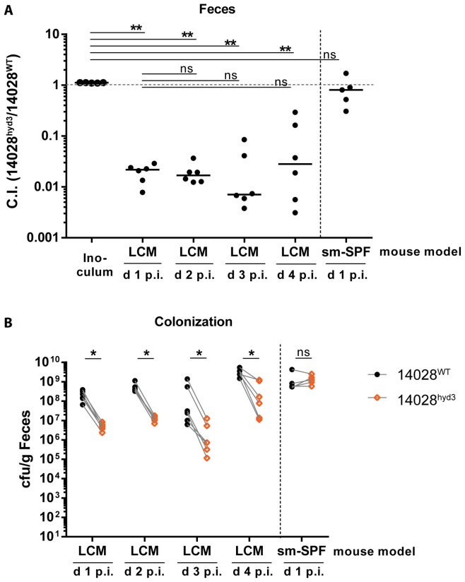
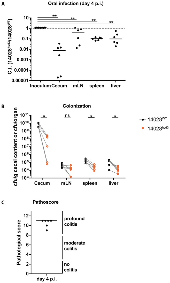
LCM-mice were infected with a 1∶1 mixture of wild type ATCC14028 (14028WT) and 14028hyd3 via the oral route (5×10⁷ cfu in total, by gavage). We analyzed the bacterial loads in the feces at days 1–4 p.i. (Fig. 1A, B), monitored pathogen loads in the cecum lumen, the mesenteric lymph nodes, the spleens and the livers, and analyzed the mucosal inflammation at day 4 p.i. (Fig. 2A–C). In the feces of the LCM-mice, 14028hyd3 featured a pronounced colonization defect already by day 1 p.i. (competitive index C.I. 0.02; Fig. 1A, B). During the subsequent three days, the total fecal pathogen loads rose from ≈108 cfu/g to about 109 cfu/g while the C.I. did not drop any further. Control infections were performed in streptomycin pretreated conventional mice (5×10⁷ cfu in total, by gavage; 1 day infection). In these animals, the microbiota is transiently disrupted by streptomycin and 14028hyd3 does not feature any gut luminal colonization defect (Fig. 1A, B). These data are strikingly similar to our earlier data obtained with H2-hydrogenase mutants of SL1344 [8] and indicated that ATCC14028 can subvert H2 for gut luminal colonization.
Figure 1. 14028hyd3 is impaired in early gut ecosystem invasion.
Gut colonization was monitored in two different mouse models: streptomycin-pretreated conventional mice (sm-SPF) and low complexity microbiota (LCM) mice. Mice were infected with a 1∶1 mixture (5×10⁷ cfu by gavage) of 14028hyd3 and the isogenic background strain (14028WT). Fecal loads of both strains were determined by selective plating. (A) Competitive infection indices were determined over 4 days. Ns = not significant (P≥0.05), ** P<0.01, Mann-Whitney U test. (B) Bacterial loads of both competing strains (14028WT and 14028hyd3) are depicted Ns = not significant (P≥0.05), * P<0.05; one-tailed Wilcoxon matched pairs signed rank test on paired data (dashed lines).
Figure 2. Oral infection experiments revealed that in ATCC14028, hydrogenases fuel pathogen growth in the intestine.
(A) LCM mice from Figure 1 were sacrificed at day 4 post infection and competitive indices in the cecum and at systemic sites were determined. ** P<0.01, Mann-Whitney U test. (B) Bacterial loads in the cecum and at systemic sites of both competing strains are plotted. Ns = not significant (P≥0.05), * P<0.05; one-tailed Wilcoxon matched pairs signed rank test on paired data (dashed lines). (C) Cecal tissue sections were HE-stained and scored for intestinal inflammation.
In the cecum lumen, 14028hyd3 had a similar colonization defect as in the feces (Fig. 2A, B) and all mice featured pronounced mucosal inflammation by day 4 p.i. (Fig. 2C). Furthermore, we detected a significant colonization defect of 14028hyd3 in the mLN, the spleens and the livers of the LCM mice (Fig. 2A, B). However the attenuation appeared to be slightly less pronounced than in the cecum lumen and in the feces. However, these data could not unequivocally settle whether H2-hydrogenase dependent growth might contribute to some extent to systemic colonization.
H2-hydrogenases do not contribute to systemic growth of ATCC14028
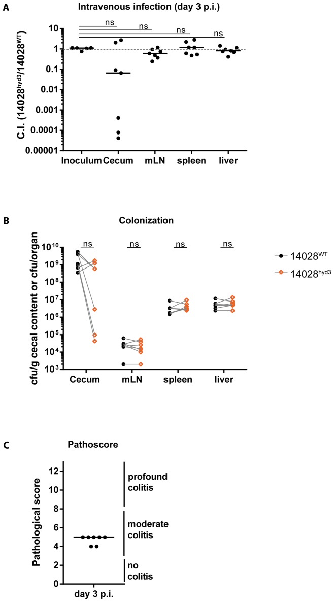
In a second approach, we specifically addressed whether H2-hydrogenases contribute to systemic colonization. To this end, we infected LCM-mice via the intravenous route with a 1∶1 mixture of 14028WT and 14028hyd3 (5×10³ cfu in total, i.v.). After three days, the animals were sacrificed and we analyzed the pathogen loads (and the C.I.) in the cecum lumen, the mLN, the spleens and the livers and assessed gut inflammation in the cecum tissue (Fig. 3A–C). The total pathogen loads in the mLN (≈104–105 cfu), the spleens (≈107 cfu) and the livers (≈107 cfu) were well in line with published data for i.v. infections in C57BL/6 mice [51]. Strikingly, 14028hyd3 did not feature any detectable colonization defect in the systemic organs after i.v. infection (p≥0.05; C.I.≈1; Fig. 3A). Colonization defects of 14028hyd3 were only detected in the cecum lumen in 5 out of 7 mice. This population must have arisen by pathogen dissemination from systemic sites to the gut lumen, e.g. by pathogen routing via the gall bladder, by phagocyte-mediated transport to the gut tissue [52]–[55] or by oral ingestion by licking the injection site at the tail. In any case, our data suggest that the growth defect of 14028hyd3 has most likely arisen after the pathogen had arrived in the gut lumen.
Figure 3. Intravenous infection experiments verified that in ATCC14028, hydrogenases are not required for growth at systemic sites.
(A) LCM mice were intravenously infected with a 1∶1 mixture of the 14028hyd3 and the isogenic background strain (14028WT) (5×10³ cfu). Animals were sacrificed at day 3 p.i. and competitive indices in the cecum and at systemic sites were determined. Ns = not significant (P≥0.05), Mann-Whitney U test. (B) Bacterial loads in the cecum and at systemic sites of both competing strains are plotted. Ns = not significant (P≥0.05); one-tailed Wilcoxon matched pairs signed rank test on paired data (dashed lines). (C) Cecal tissue sections were HE-stained and scored for intestinal inflammation.
These data established that ATCC14028 does not require H2-hydrogenases for growth at systemic sites if the gut is bypassed during the infection procedure. In the typhoid fever model [14] or oral infections of LCM mice, gut luminal growth of the bacteria seems to precede the spread to systemic sites. This gut luminal growth most likely explains why H2-hydrogenase mutants are found in lower numbers in the mLN, livers and spleens of the animals than the isogenic wild type strain.
It should be noted that the 14028hyd3 mutant used in our study lacked all the three uptake-type H2-hydrogenases. Thus, formally we cannot rule out that the requirement of a single hydrogenase is masked by the absence of the other two hydrogenases. For example, deletion of one hydrogenase might increase S. Tm fitness, while deletion of another hydrogenase might decrease S. Tm fitness. By analyzing both deletions in combination, the two opposed effects will be compensated. However, this seems unlikely, as none of the H2-hydrogenase mutants of SL1344 or ATCC14028 that have been analyzed in the past had featured higher virulence than the isogenic wild type strain [8], [14]. Nevertheless, mutants lacking just one of the H2-hydrogenases at a time would have to be studied in detail to address this in a systematic fashion. In addition, differential expression of the three H2-hydrogenases [15], [17], strain-specific differences in the expression patterns and microbiota/environment-specific cues (e.g. different H2 availability) might play a role. Indeed, the three different hydrogenases have different hydrogenase activities [16]. Moreover, in typhoid fever model infections of SPF mice, ATCC14028 may utilize several different H2-hydrogenases [14]. In contrast, SL1344 growth in the gut lumen of LCM mice relied exclusively on hyb, not the other H2-hydrogenases [8]. The environmental cues steering the differential hydrogenase expression in vivo remain to be established. Nevertheless, it seems quite safe to assume that the gut lumen is the site where H2-utilization by S. Typhimurium is most prominent. Still, H2 could represent an auxiliary reductant for Salmonella at systemic sites under otherwise poor nutrient conditions, or when the microbiota is especially active in fermentative metabolism (e. g. high H2 production).
In conclusion, our data establish that ATCC14028 is strikingly similar to SL1344 in requiring H2-hydrogenases for growth in the gut, not at systemic sites. This may suggest that the use of H2 for gut luminal colonization is a general feature of Salmonella Typhimurium strains.
Materials and Methods
Bacterial strains
All strains used in this study are derivates of the Salmonella enterica serovar Typhimurium ATCC14028 (IR715), in which a streptomycin resistance was added by P22 phage transduction of the aadA gene from S. Tm SL1344 [35]. Deletions in the hydrogenase genes were constructed by lambda/red homologous recombination [56] as described previously [8] (Table 1).
Table 1. Bacterial strains used in this study.
| Strain | Genotype | Reference |
| 14028WT | Streptomycin-resistant derivative of IR715 (constructed by P22-transduction of aadA gene from S. Tm SL1344 into the ATCC14028 derivative IR715) | [35] |
| 14028hyd3 | ΔSTM3147-3150, STM1786-87::aphT, STM1538-1539::cat | This study |
Mouse infection experiments
All mice used in this study are C57BL/6 background and bred at the Rodent Center HCI (RCHCI) (ETH Zurich, Switzerland). Low complex microbiota (LCM) mice are ex-germfree mice which were colonized with the Altered Schaedler flora-cocktail in 2007 [9] and ever since bred under strict hygienic isolation. Co-infection experiments were performed as described previously [41] in 8 to 10 week old mice. Pre-treatment with 20 mg streptomycin was only performed if indicated (Figure 1). For infection, both bacterial strain (14028WT to 14028hyd3) were grown for 12 h in 0.3 M NaCl supplemented LB medium, diluted 1∶20 and sub-cultured for 4 h in the same medium and mixed in a 1∶1 ratio. For oral infections, mice were infected with 5·107 cfu bacteria by gavage. For intravenous infections, 5·103 cfu bacteria were injected into the tail vein. Mice were sacrificed on day 1 p.i., day 3 p.i or day 4 p.i. by cervical dislocation. Freshly collected fecal pellets, cecum content and organs were homogenized in PBS (0.5% tergitol, 0.5% bovine serum albumin). Differential plating on MacConkey agar (Oxoid) supplemented with the appropriate antibiotics (50 µg/mL streptomycin, 50 µg/mL kanamycin and 30 µg/mL chloramphenicol) was performed to determine bacterial population sizes. The competitive index was calculated by division of the population size of 14028hyd3 by the population size of 14028WT. This ratio as corrected for the ratio of both strains in the inoculum. Parts of the cecal tissue were embedded in OCT (Sakura), cryosections were prepared and stained with hematoxiline/eosine. HE-stained sections were evaluated by scoring for submucosal edema, PMN infiltration, presence of goblet cells and epithelial damage with a maximum score of 13 [57].
Statistical analysis
The one-sided Wilcoxon matched-pairs signed rank test and the exact Mann-Whitney U test were performed using the software Graphpad Prism Version 6.0 for Windows (GraphPad Software, La Jolla California USA, www.graphpad.com). P values of less than 0.05 were considered as statistically significant. To compare C.I.s to C.I. of inoculi, ratios of 14028hyd3 and 14028WT were compared to the ratio of both strains in the inoculum using an exact Mann-Whitney U test.
Ethical statement
All animal experiments were reviewed and approved by the Kantonales Veterinäramt, Zürich (license 223/2010 & 222/2013) and are subject to the Swiss animal protection law (TschG).
Acknowledgments
We are grateful to the members of the RCHCI staff for excellent support of our animal work, to Boas Felmy for help with i.v. injections and to Rebekka Bauer for experimental assistance.
Data Availability
The authors confirm that all data underlying the findings are fully available without restriction. All relevant data are within the paper.
Funding Statement
This work was supported in part by the Swiss National Science Foundation (310030-132997/1) and the Sinergia project CRSII3_136286 to WDH) and the UBS Optimus Foundation. The funders had no role in study design, data collection and analysis, decision to publish, or preparation of the manuscript.
References
- 1. Sommer F, Backhed F (2013) The gut microbiota – masters of host development and physiology. Nat Rev Microbiol 11: 227–238. [DOI] [PubMed] [Google Scholar]
- 2. Stecher B, Hardt WD (2008) The role of microbiota in infectious disease. Trends Microbiol 16: 107–114. [DOI] [PubMed] [Google Scholar]
- 3. van der Waaij D, Berghuis-de Vries JM, Lekkerkerk L-v (1971) Colonization resistance of the digestive tract in conventional and antibiotic-treated mice. J Hyg (Lond) 69: 405–411. [DOI] [PMC free article] [PubMed] [Google Scholar]
- 4. Ng KM, Ferreyra JA, Higginbottom SK, Lynch JB, Kashyap PC, et al. (2013) Microbiota-liberated host sugars facilitate post-antibiotic expansion of enteric pathogens. Nature. [DOI] [PMC free article] [PubMed] [Google Scholar]
- 5. Santos RL, Zhang S, Tsolis RM, Kingsley RA, Adams LG, et al. (2001) Animal models of Salmonella infections: enteritis versus typhoid fever. Microbes Infect 3: 1335–1344. [DOI] [PubMed] [Google Scholar]
- 6. Wallis TS, Galyov EE (2000) Molecular basis of Salmonella-induced enteritis. Mol Microbiol 36: 997–1005. [DOI] [PubMed] [Google Scholar]
- 7. Endt K, Stecher B, Chaffron S, Slack E, Tchitchek N, et al. (2010) The microbiota mediates pathogen clearance from the gut lumen after non-typhoidal Salmonella diarrhea. PLoS Pathog 6: e1001097. [DOI] [PMC free article] [PubMed] [Google Scholar]
- 8. Maier L, Vyas R, Cordova CD, Lindsay H, Schmidt TS, et al. (2013) Microbiota-derived hydrogen fuels salmonella typhimurium invasion of the gut ecosystem. Cell Host Microbe 14: 641–651. [DOI] [PubMed] [Google Scholar]
- 9. Stecher B, Chaffron S, Kappeli R, Hapfelmeier S, Freedrich S, et al. (2010) Like will to like: abundances of closely related species can predict susceptibility to intestinal colonization by pathogenic and commensal bacteria. PLoS Pathog 6: e1000711. [DOI] [PMC free article] [PubMed] [Google Scholar]
- 10. Lamichhane-Khadka R, Benoit SL, Maier SE, Maier RJ (2013) A link between gut community metabolism and pathogenesis: molecular hydrogen-stimulated glucarate catabolism aids Salmonella virulence. Open Biol 3: 130146. [DOI] [PMC free article] [PubMed] [Google Scholar]
- 11. Lamichhane-Khadka R, Frye JG, Porwollik S, McClelland M, Maier RJ (2011) Hydrogen-stimulated carbon acquisition and conservation in Salmonella enterica serovar Typhimurium. J Bacteriol 193: 5824–5832. [DOI] [PMC free article] [PubMed] [Google Scholar]
- 12.Lamichhane-Khadka R, Kwiatkowski A, Maier RJ (2010) The Hyb hydrogenase permits hydrogen-dependent respiratory growth of Salmonella enterica serovar Typhimurium. MBio 1. [DOI] [PMC free article] [PubMed]
- 13. Maier RJ (2005) Use of molecular hydrogen as an energy substrate by human pathogenic bacteria. Biochem Soc Trans 33: 83–85. [DOI] [PubMed] [Google Scholar]
- 14. Maier RJ, Olczak A, Maier S, Soni S, Gunn J (2004) Respiratory hydrogen use by Salmonella enterica serovar Typhimurium is essential for virulence. Infect Immun 72: 6294–6299. [DOI] [PMC free article] [PubMed] [Google Scholar]
- 15. Zbell AL, Benoit SL, Maier RJ (2007) Differential expression of NiFe uptake-type hydrogenase genes in Salmonella enterica serovar Typhimurium. Microbiology 153: 3508–3516. [DOI] [PubMed] [Google Scholar]
- 16. Zbell AL, Maier RJ (2009) Role of the Hya hydrogenase in recycling of anaerobically produced H2 in Salmonella enterica serovar Typhimurium. Appl Environ Microbiol 75: 1456–1459. [DOI] [PMC free article] [PubMed] [Google Scholar]
- 17. Zbell AL, Maier SE, Maier RJ (2008) Salmonella enterica serovar Typhimurium NiFe uptake-type hydrogenases are differentially expressed in vivo. Infect Immun 76: 4445–4454. [DOI] [PMC free article] [PubMed] [Google Scholar]
- 18. Gordienko EN, Kazanov MD, Gelfand MS (2013) Evolution of pan-genomes of Escherichia coli, Shigella spp., and Salmonella enterica. J Bacteriol 195: 2786–2792. [DOI] [PMC free article] [PubMed] [Google Scholar]
- 19. Jacobsen A, Hendriksen RS, Aaresturp FM, Ussery DW, Friis C (2011) The Salmonella enterica pan-genome. Microb Ecol 62: 487–504. [DOI] [PMC free article] [PubMed] [Google Scholar]
- 20. McClelland M, Sanderson KE, Clifton SW, Latreille P, Porwollik S, et al. (2004) Comparison of genome degradation in Paratyphi A and Typhi, human-restricted serovars of Salmonella enterica that cause typhoid. Nat Genet 36: 1268–1274. [DOI] [PubMed] [Google Scholar]
- 21. Thomson NR, Clayton DJ, Windhorst D, Vernikos G, Davidson S, et al. (2008) Comparative genome analysis of Salmonella Enteritidis PT4 and Salmonella Gallinarum 287/91 provides insights into evolutionary and host adaptation pathways. Genome Res 18: 1624–1637. [DOI] [PMC free article] [PubMed] [Google Scholar]
- 22. Nuccio SP, Baumler AJ (2014) Comparative analysis of Salmonella genomes identifies a metabolic network for escalating growth in the inflamed gut. MBio 5: e00929–00914. [DOI] [PMC free article] [PubMed] [Google Scholar]
- 23. Achtman M, Hale J, Murphy RA, Boyd EF, Porwollik S (2013) Population structures in the SARA and SARB reference collections of Salmonella enterica according to MLST, MLEE and microarray hybridization. Infect Genet Evol 16: 314–325. [DOI] [PubMed] [Google Scholar]
- 24. Kroger C, Dillon SC, Cameron AD, Papenfort K, Sivasankaran SK, et al. (2012) The transcriptional landscape and small RNAs of Salmonella enterica serovar Typhimurium. Proc Natl Acad Sci U S A 109: E1277–1286. [DOI] [PMC free article] [PubMed] [Google Scholar]
- 25. Jarvik T, Smillie C, Groisman EA, Ochman H (2010) Short-term signatures of evolutionary change in the Salmonella enterica serovar typhimurium 14028 genome. J Bacteriol 192: 560–567. [DOI] [PMC free article] [PubMed] [Google Scholar]
- 26. Mirold S, Rabsch W, Rohde M, Stender S, Tschape H, et al. (1999) Isolation of a temperate bacteriophage encoding the type III effector protein SopE from an epidemic Salmonella typhimurium strain. Proc Natl Acad Sci U S A 96: 9845–9850. [DOI] [PMC free article] [PubMed] [Google Scholar]
- 27. Pelludat C, Mirold S, Hardt WD (2003) The SopEPhi phage integrates into the ssrA gene of Salmonella enterica serovar Typhimurium A36 and is closely related to the Fels-2 prophage. J Bacteriol 185: 5182–5191. [DOI] [PMC free article] [PubMed] [Google Scholar]
- 28. Figueroa-Bossi N, Uzzau S, Maloriol D, Bossi L (2001) Variable assortment of prophages provides a transferable repertoire of pathogenic determinants in Salmonella. Mol Microbiol 39: 260–271. [DOI] [PubMed] [Google Scholar]
- 29. Hoiseth SK, Stocker BA (1981) Aromatic-dependent Salmonella typhimurium are non-virulent and effective as live vaccines. Nature 291: 238–239. [DOI] [PubMed] [Google Scholar]
- 30.Brussow H, Canchaya C, Hardt WD (2004) Phages and the evolution of bacterial pathogens: from genomic rearrangements to lysogenic conversion. Microbiol Mol Biol Rev 68: 560–602, table of contents. [DOI] [PMC free article] [PubMed]
- 31. Hardt WD, Urlaub H, Galan JE (1998) A substrate of the centisome 63 type III protein secretion system of Salmonella typhimurium is encoded by a cryptic bacteriophage. Proc Natl Acad Sci U S A 95: 2574–2579. [DOI] [PMC free article] [PubMed] [Google Scholar]
- 32. Mirold S, Rabsch W, Tschape H, Hardt WD (2001) Transfer of the Salmonella type III effector sopE between unrelated phage families. J Mol Biol 312: 7–16. [DOI] [PubMed] [Google Scholar]
- 33. Hardt WD, Chen LM, Schuebel KE, Bustelo XR, Galan JE (1998) S. typhimurium encodes an activator of Rho GTPases that induces membrane ruffling and nuclear responses in host cells. Cell 93: 815–826. [DOI] [PubMed] [Google Scholar]
- 34. Wood MW, Rosqvist R, Mullan PB, Edwards MH, Galyov EE (1996) SopE, a secreted protein of Salmonella dublin, is translocated into the target eukaryotic cell via a sip-dependent mechanism and promotes bacterial entry. Mol Microbiol 22: 327–338. [DOI] [PubMed] [Google Scholar]
- 35. Hapfelmeier S, Ehrbar K, Stecher B, Barthel M, Kremer M, et al. (2004) Role of the Salmonella pathogenicity island 1 effector proteins SipA, SopB, SopE, and SopE2 in Salmonella enterica subspecies 1 serovar Typhimurium colitis in streptomycin-pretreated mice. Infect Immun 72: 795–809. [DOI] [PMC free article] [PubMed] [Google Scholar]
- 36. Zhang S, Santos RL, Tsolis RM, Mirold S, Hardt WD, et al. (2002) Phage mediated horizontal transfer of the sopE1 gene increases enteropathogenicity of Salmonella enterica serotype Typhimurium for calves. FEMS Microbiol Lett 217: 243–247. [DOI] [PubMed] [Google Scholar]
- 37.Lopez CA, Winter SE, Rivera-Chavez F, Xavier MN, Poon V, et al. (2012) Phage-mediated acquisition of a type III secreted effector protein boosts growth of salmonella by nitrate respiration. MBio 3. [DOI] [PMC free article] [PubMed]
- 38. Carbonero F, Benefiel AC, Gaskins HR (2012) Contributions of the microbial hydrogen economy to colonic homeostasis. Nat Rev Gastroenterol Hepatol 9: 504–518. [DOI] [PubMed] [Google Scholar]
- 39. Nakamura N, Lin HC, McSweeney CS, Mackie RI, Gaskins HR (2010) Mechanisms of microbial hydrogen disposal in the human colon and implications for health and disease. Annu Rev Food Sci Technol 1: 363–395. [DOI] [PubMed] [Google Scholar]
- 40. Tsolis RM, Xavier MN, Santos RL, Baumler AJ (2011) How to become a top model: impact of animal experimentation on human Salmonella disease research. Infect Immun 79: 1806–1814. [DOI] [PMC free article] [PubMed] [Google Scholar]
- 41. Barthel M, Hapfelmeier S, Quintanilla-Martinez L, Kremer M, Rohde M, et al. (2003) Pretreatment of mice with streptomycin provides a Salmonella enterica serovar Typhimurium colitis model that allows analysis of both pathogen and host. Infect Immun 71: 2839–2858. [DOI] [PMC free article] [PubMed] [Google Scholar]
- 42. Stecher B, Hardt WD (2011) Mechanisms controlling pathogen colonization of the gut. Curr Opin Microbiol 14: 82–91. [DOI] [PubMed] [Google Scholar]
- 43. Bohnhoff M, Miller CP (1962) Enhanced susceptibility to Salmonella infection in streptomycin-treated mice. J Infect Dis 111: 117–127. [DOI] [PubMed] [Google Scholar]
- 44. Bohnhoff M, Miller CP, Martin WR (1964) Resistance of the Mouse's Intestinal Tract to Experimental Salmonella Infection. Ii. Factors Responsible for Its Loss Following Streptomycin Treatment. J Exp Med 120: 817–828. [DOI] [PMC free article] [PubMed] [Google Scholar]
- 45. Brown KJ, Tannock GW, Eyres RA, Elliott RB, Lines DR (1979) Colonization by Salmonella typhimurium and Shigella flexneri III of the gastrointestinal tract of mice treated with beta-2-thienylalanine and streptomycin. Antonie Van Leeuwenhoek 45: 531–546. [DOI] [PubMed] [Google Scholar]
- 46. Kaiser P, Diard M, Stecher B, Hardt WD (2012) The streptomycin mouse model for Salmonella diarrhea: functional analysis of the microbiota, the pathogen's virulence factors, and the host's mucosal immune response. Immunol Rev 245: 56–83. [DOI] [PubMed] [Google Scholar]
- 47. Meynell GG (1955) Some factors affecting the resistance of mice to oral infection by Salmonella typhimurium. Proc R Soc Med. [Google Scholar]
- 48. Meynell GG, Subbaiah TV (1963) Antibacterial mechanisms of the mouse gut. I. Kinetics of infection by Salmonella typhi-murium in normal and streptomycin-treated mice studied with abortive transductants. Br J Exp Pathol 44: 197–208. [PMC free article] [PubMed] [Google Scholar]
- 49. Murray RA, Lee CA (2000) Invasion genes are not required for Salmonella enterica serovar typhimurium to breach the intestinal epithelium: evidence that salmonella pathogenicity island 1 has alternative functions during infection. Infect Immun 68: 5050–5055. [DOI] [PMC free article] [PubMed] [Google Scholar]
- 50. Que JU, Hentges DJ (1985) Effect of streptomycin administration on colonization resistance to Salmonella typhimurium in mice. Infect Immun 48: 169–174. [DOI] [PMC free article] [PubMed] [Google Scholar]
- 51. Pie S, Matsiota-Bernard P, Truffa-Bachi P, Nauciel C (1996) Gamma interferon and interleukin-10 gene expression in innately susceptible and resistant mice during the early phase of Salmonella typhimurium infection. Infect Immun 64: 849–854. [DOI] [PMC free article] [PubMed] [Google Scholar]
- 52. Crawford RW, Rosales-Reyes R, Ramirez-Aguilar Mde L, Chapa-Azuela O, Alpuche-Aranda C, et al. (2010) Gallstones play a significant role in Salmonella spp. gallbladder colonization and carriage. Proc Natl Acad Sci U S A 107: 4353–4358. [DOI] [PMC free article] [PubMed] [Google Scholar]
- 53. Kingsley RA, Msefula CL, Thomson NR, Kariuki S, Holt KE, et al. (2009) Epidemic multiple drug resistant Salmonella Typhimurium causing invasive disease in sub-Saharan Africa have a distinct genotype. Genome Res 19: 2279–2287. [DOI] [PMC free article] [PubMed] [Google Scholar]
- 54. Knodler LA, Vallance BA, Celli J, Winfree S, Hansen B, et al. (2010) Dissemination of invasive Salmonella via bacterial-induced extrusion of mucosal epithelia. Proc Natl Acad Sci U S A 107: 17733–17738. [DOI] [PMC free article] [PubMed] [Google Scholar]
- 55. Kaiser P, Regoes RR, Dolowschiak T, Wotzka SY, Lengefeld J, et al. (2014) Cecum lymph node dendritic cells harbor slow-growing bacteria phenotypically tolerant to antibiotic treatment. PLoS Biol 12: e1001793. [DOI] [PMC free article] [PubMed] [Google Scholar]
- 56. Datsenko KA, Wanner BL (2000) One-step inactivation of chromosomal genes in Escherichia coli K-12 using PCR products. Proc Natl Acad Sci U S A 97: 6640–6645. [DOI] [PMC free article] [PubMed] [Google Scholar]
- 57. Hapfelmeier S, Muller AJ, Stecher B, Kaiser P, Barthel M, et al. (2008) Microbe sampling by mucosal dendritic cells is a discrete, MyD88-independent step in DeltainvG S. Typhimurium colitis. J Exp Med 205: 437–450. [DOI] [PMC free article] [PubMed] [Google Scholar]
Associated Data
This section collects any data citations, data availability statements, or supplementary materials included in this article.
Data Availability Statement
The authors confirm that all data underlying the findings are fully available without restriction. All relevant data are within the paper.


