

Abstract
Cancer prevalence and mortality are high in developing nations, where resources for cancer control are inadequate. Nearly one-quarter of cancers in resource-limited nations are infection related, and molecular assays can capitalize on this relationship by detecting pertinent pathogen genomes and human gene variants to identify those at highest risk for progression to cancer, to classify lesions, to predict effective therapy, and to monitor tumor burden over time. Prime examples are human papillomavirus in cervical neoplasia, Helicobacter pylori and Epstein-Barr virus in gastric adenocarcinoma and lymphoma, and hepatitis B or C virus in hepatocellular cancer. Research is underway to engineer devices that overcome social, economic, and technical barriers limiting effective laboratory support. Additional challenges include an educated workforce, infrastructure for quality metrics and record keeping, and funds to sustain molecular test services. The combination of well-designed interfaces, novel and robust electrochemical technology, and telemedicine tools will promote adoption by frontline providers. Fast turnaround is crucial for surmounting loss to follow-up, although increased use of cell phones, even in rural areas, enhances options for patient education and engagement. Links to a broadband network facilitate consultation and centralized storage of medical data. Molecular technology shows promise to address gaps in health care through rapid, user-friendly, and cost-effective devices reflecting clinical priorities in resource-poor areas.
CME Accreditation Statement: This activity (“JMD 2014 CME Program in Molecular Diagnostics”) has been planned and implemented in accordance with the Essential Areas and policies of the Accreditation Council for Continuing Medical Education (ACCME) through the joint sponsorship of the American Society for Clinical Pathology (ASCP) and the American Society for Investigative Pathology (ASIP). ASCP is accredited by the ACCME to provide continuing medical education for physicians.
The ASCP designates this journal-based CME activity (“JMD 2014 CME Program in Molecular Diagnostics”) for a maximum of 48 AMA PRA Category 1 Credit(s)™. Physicians should only claim credit commensurate with the extent of their participation in the activity.
CME Disclosures: The authors of this article and the planning committee members and staff have no relevant financial relationships with commercial interests to disclose.
Molecular tests represent a powerful strategy for screening, early detection, tumor classification, and monitoring efficacy of intervention. Emerging technologies marry advanced biochemical methods with innovative fluidics and electronics to address the need for robust, automated test systems. These technologies are poised to facilitate a quantum leap forward in cancer diagnostics in low- and middle-income countries.
In parallel with improved communication technology are advances in biochemical sensors that make feasible measurements of multiple DNA, RNA, or microRNA targets. Heightened capital investment in devices, often referred to as Lab on a Chip, is streamlining specimen processing, biochemical analysis, and data manipulation.1 Before a new device can be implemented, performance data must demonstrate that the test system is analytically sound and clinically useful in the hands of the professionals who use it.2 The bar for success might be lessened by innovative internal quality control, automation from input to reporting, and other engineering feats that promote good outcomes when implemented by minimally trained workers or patients. Thus, modern technology shows promise to address gaps in health care through rapid, inexpensive, automated test systems that identify and monitor the types of neoplasia prevalent in resource-poor areas.
Cancer Burden in Resource-Limited Nations
Noncommunicable diseases are projected to become the major global health burden in the near term, with cancer accounting for approximately one-quarter of this burden,3 of which at least one-third is preventable. The annual global incidence of cancer is projected to increase from 12.7 to 22.2 million by 2030, with 13.1 million expected deaths.4,5 More than two-thirds of the burden will occur in low- and middle-income countries, wherein seven cancer types (lung, colon, breast, stomach, liver, cervical, and esophageal) account for nearly two-thirds of incident cases (GLOBOCAN 2012, http://globocan.iarc.fr, last accessed September 8, 2014). In the world's poorest countries, women are more than twice as likely to die of their breast cancer, and children are up to ninefold less likely to be cured of acute lymphoblastic leukemia.6,7
Cancer Linked to Infectious Disease
In developing nations, nearly one-quarter of cancers are infection related,3 and four infectious agents account for >80% of the burden: human papillomavirus (HPV), Helicobacter pylori, hepatitis B virus (HBV), and hepatitis C virus (HCV). Epstein-Barr virus (EBV) adds a significant burden in several areas of the world (Figure 1).
Figure 1.

Infection-related cancers comprise 23% of all cancers worldwide. Oncogenic pathogens (A) are linked to higher cancer burden in less developed regions of the world (B). Data are from de Martel et al.3 HTLV1, human T-cell lymphotropic virus 1.
Approximately 30% of infection-associated cancers occur in people <50 years. Compared to more developed regions, less developed nations have more cancers of the stomach, uterine cervix, and liver, all three of which are infection related (GLOBOCAN 2012, http://globocan.iarc.fr, last accessed September 8, 2014). Helicobacter pylori and EBV infection are linked to gastric adenocarcinoma and lymphoma, whereas HPV is uniformly found in cervical neoplasia. HBV or HCV infections often predate hepatocellular carcinoma.
Research into the link between infection and cancer sheds light on the mechanisms of oncogenesis. Interestingly, the aforementioned infection-related cancers that are prevalent in the developing world are the same three cancer types with the most complex genomic signatures as elucidated by mutation analysis.8 Mutations in infection-related cancer have been attributed to the following: i) pathogen-induced effects, such as heightened oxidation that damages DNA, ii) viral integration events disrupting human genes or their regulatory factors, iii) viral properties that prolong cell survival and resist apoptosis or immune destruction, and iv) suppression of DNA repair.
There is an unmet need for affordable devices to detect cancer-related infections. Such devices could capitalize on the link between infection and cancer to assist in screening and early diagnosis, tumor classification, pathogen-targeted therapy, and monitoring tumor burden over time.9
HPV
The HPV genome is present in virtually all cervical carcinomas. Because infection precedes malignancy, tests for the relevant high-risk strains of HPV add value in cervical neoplasia screening programs. Interestingly, the spectrum of HPV strains in high-grade cervical lesions differs somewhat by geographic region and by HIV status.10,11 These population-specific differences emphasize the need to validate genomic assays for each target population.
Rapid, low-cost devices are being developed to test for HPV DNA and RNA.12–14 The careHPV test system (Qiagen, Hilden, Germany) was devised specifically for resource-poor areas, and published data suggest that its performance is similar to the US Food and Drug Administration–approved HC2 test.14 The battery-powered bench-top instrument requires neither electricity nor running water to perform hybrid capture of RNA probes bound to high-risk HPV DNA genomes. The manufacturer states that the reagents are tolerant of temperature swings that may characterize a rural laboratory having spotty electricity for refrigeration. The careHPV test can be performed by minimally trained technologists at threefold less cost and sixfold less time than the HC2 assay, potentially permitting same-day intervention for HPV-positive patients.15 HPV tests may complement or replace the visual inspection strategy that is currently favored over cytology and over laboratory tests for cervical neoplasia detection in many low-resource settings.14,16
High-risk HPV testing of a self-collected specimen, with or without additional molecular tests of human genes, shows promise to improve cost-effectiveness of screening programs in low-resource settings.17–20 High levels of HPV 16 DNA in mouthwash or tonsil swab samples likewise show promise for identifying patients with infection-associated oropharyngeal carcinoma.21
Helicobacter pylori
Gastric adenocarcinoma is the leading global cause of infection-related cancer mortality and overall is the second leading cause of cancer death, projected to increase to eighth in all-cause mortality in the near term (GLOBOCAN 2012, http://globocan.iarc.fr, last accessed September 8, 2014). Helicobacter pylori infection is the strongest known risk factor for gastric carcinogenesis. Risk depends not only on bacterial genetics (eg, cagA and vacAs1m1 virulence factors) but also on host genotypes [cytokine variants (eg, IL1B 511T)] and coinfections, which lend themselves to molecular testing.22–25 Molecular test panels are being developed to examine germline and somatic variants that, along with molecular evidence of H. pylori, EBV, and other infections, could improve prediction of gastric cancer progression.24,26,27
Helicobacter pylori is also associated with gastric mucosa-associated lymphoid tissue lymphoma (MALT), and eradicating the bacteria via antibiotic therapy is curative in some patients with lymphoma.28 Antibiotic drug selection is improved by molecular testing of bacterial DNA.29 Clinical trials examining efficacy of antibiotics may benefit from molecular tests of bacterial DNA in biopsy, stool, or body fluids, including saliva.30–32 A creative strategy for specimen collection from the upper gastrointestinal tract involves swallowing a capsule attached to a fixed string that is then retrieved through the mouth for nucleic acid analysis.33
HBV and HCV
HBV and HCV infections are common and predispose to hepatocellular carcinoma.34 Transfusion-mediated infection remains a concerning means of spread for these and other pathogens in countries lacking a centralized system for blood collection and laboratory testing for transmissible agents. Perinatal infection accounts for approximately half of the burden among the 350 million people with chronic HBV infection.35 Vaccination is recommended for HBV-related cancer prevention, whereas treatment of HCV is associated with reduced cancer risk.36 Viral genomes can be detected, characterized, and monitored using molecular tests, such as real-time quantitative PCR (qPCR).37,38 Proof of principle for a point-of-care device for HBV DNA was recently reported.39 In this test system, an inexpensive microfluidic chip moves DNA and wash/detection buffers through channels containing immobilized probe. Interestingly, the reporter probe was designed to enzymatically convert sucrose into glucose so that probe hybridization signals were measurable on the type of personal glucometer that is commonly used at point of care.
EBV
EBV is deemed a group 1 carcinogen because of its putative role in pathogenesis of endemic Burkitt’s lymphoma (BL), extranodal natural killer/T-cell lymphoma (nasal type), angioimmunoblastic T-cell lymphoma, aggressive natural killer cell leukemia, immunodeficiency-related lymphoid neoplasia including AIDS lymphoma, nasopharyngeal carcinoma, gastric adenocarcinoma, and Hodgkin lymphoma. In each type of neoplasm, the viral genome is localized within malignant cells, providing a convincing link between infection and neoplasia that forms the basis for quantifying tumor burden by blood EBV qPCR.9 Affected patients with cancer tend to have high circulating EBV DNA loads that predate clinical diagnosis of malignancy. Serial viral load measurements reflect efficacy of therapy.9
BL is an example of a cancer that is prevalent in resource-limited areas of tropical Africa and for which molecular tests can support a diagnosis. MYC translocation, clonal IGH gene rearrangement, and EBV viral load tests have been applied to blood, aspirate, or biopsy specimens suspected to represent BL.40–43 Results of testing support a clinical diagnosis of cancer pending definitive cytologic or histological diagnosis that may be delayed because of limited access to histopathology services.
HHV8
Another member of the human herpesvirus family, human herpesvirus 8 (HHV8; alias Kaposi’s sarcoma–associated herpesvirus), is a group 1 carcinogen by virtue of its putative role in causing primary effusion lymphoma and Kaposi’s sarcoma. Endemic and HIV-associated Kaposi’s sarcoma are prevalent in sub-Saharan Africa. Differentiation of Kaposi’s sarcoma from clinical look-alikes, bacillary angiomatosis (caused by Bartonella species) and pyogenic granuloma, was recently achieved using a novel colorimetric nanoparticle aggregation method targeting the DNA of each pathogen.44 In another advance, solar energy was used as the heat source for a thermocycler that assisted, by a smartphone-based camera, to detect fluorescence, and an accompanying app to analyze data was capable of detecting HHV8 DNA in skin biopsy specimens of patients with Kaposi’s sarcoma.45
Tuberculosis
Tuberculosis is responsible for 1.7 million deaths per year. Risk of lung cancer (especially adenocarcinoma) is increased 11-fold among patients with tuberculosis.46 Proposed mechanisms of mycobacteria-related carcinogenesis include long-term immune stimulation (eg, tumor necrosis factor effect on anti-apoptotic NF-κB signaling), neoangiogenesis, and DNA damage from reactive oxygen species. Molecular tests can detect and speciate mycobacteria and predict drug resistance to assist clinicians in selecting rifampin, isoniazid, or second-line medications.47 Cepheid was among first device manufacturers to develop a PCR test system specifically designed to be rapid and user friendly in low-volume testing laboratories. The performance of Cepheid’s Xpert MTB/RIF test has been shown in tuberculosis-endemic regions to help guide appropriate therapy of affected patients and also to promote infection control in the larger community, leading to reduced transmission rates and fewer drug-resistant organisms.48–51
HIV and Malaria
Immunodeficiency (eg, HIV infection or malaria-related immune dysregulation) is a well-established predisposing factor for selected forms of cancer, some of which, in turn, harbor other viral genomes.52,53 HIV and malaria appear to act indirectly to diminish T-cell defenses against viruses and against tumor cells.54 Progressive HIV infection increases risk of cancer up to 200-fold, whereas recent malaria infection reportedly increases risk of BL by 21-fold.55 Patients with HIV or malaria remain at increased risk of cancer, even with appropriate access to medical care.
The HIV epidemic markedly accelerated development of molecular technologies and associated laboratory services. The need for rapid HIV testing continues to drive improvements to laboratory infrastructure that is then applied to diagnose other infectious diseases, cancer, or heritable disease, and to provide pharmacogenetic predictions.
Recently, microfluidic chips were reported to detect HIV and associated opportunistic infections (mycobacteria and pneumocystis) by PCR or by isothermal loop-mediated nucleic acid amplification.56–58 A promising handheld device can detect and speciate malaria by isothermal rRNA amplification of 2 μL of blood without the need for extraction.59 An optimized molecular method claims to be 2500-fold more sensitive than conventional microscopy for malaria parasites.60 Cost savings accrue with proper diagnosis and treatment versus unnecessary spending associated with misdiagnosis.61,62
Multigene Test Panels Targeting Human and Pathogen Nucleic Acid
The Cancer Genome Atlas project is a major step forward in cataloging mutation patterns and gene expression profiles in concert with traditional diagnostic histopathological analysis. Results to date confirm known cancer-related infections and provide new insights into the genetic underpinnings of neoplasia.63 Most tumors harbor many somatic changes that, along with transcriptome and methylome data, reveal potential druggable pathways that can be explored in clinical trials.
Multigene test panels can characterize signaling pathways driving tumor growth. A proof-of-principle study showed that genotyping is achievable on fresh, frozen, or fixed tissue in just 70 minutes using an instrument platform that performs automated extraction, followed by Invader chemistry,64 to query 13 mutations in KRAS, BRAF, and PIK3CA genes.65 Another study detected heritable variants in cytochrome p450 coding sequences to facilitate bedside clopidogrel dosing.66 Although these technologic advances are exciting, the particular somatic and heritable variants targeted may be off target for the needs of resource-poor facilities, where downstream interventions are limited. A more practical test panel in hospitals lacking on-site pathology services might examine fine-needle aspirate material from a mass lesion to help distinguish infection from tumor, pending send out for a pathologist's definitive diagnosis days to weeks later.67
Most of the technologies described herein are not yet implemented because of an insufficient evidence base and/or inadequacy for particular local needs.68 Investment is required to devise suitable genomic assays that answer pertinent biological and medical questions in clinical trials and ultimately in routine patient care. When applied in resource-limited areas, one must consider the unique logistic, cultural, and technologic features that promote success. As with all clinical research, multidisciplinary collaboration is required to optimize assay design and to ensure the study plan is ethical when offered to patients with few alternatives for care. Judicious use of precious resources is critical. Batch testing in a reference laboratory is generally less expensive and more effective than is point-of-care testing,69 although suitably designed test systems could alter this equation. Even central laboratories will adopt one-off test systems when rapid turnaround is required.
Cancer Screening
There are marked global disparities in access to cancer screening tests and programs. For cervical neoplasia, screening programs are well established in many resource-poor areas. Recent studies suggest that HPV DNA testing is cost effective as a primary screening method on self-collected specimens or on samplings gathered by health care personnel.70,71 Nasopharyngeal carcinoma screening by qPCR of EBV DNA in saliva or in nasopharyngeal brushings shows promise as a means to identify tumor in high-risk individuals.72,73 Screening for other cancers that are prevalent in the developing world (lung, stomach, breast, liver, colon, and esophagus) is in early stages of investigation to find genomic signatures distinguishing precursor lesions likely to act benign from those that are at risk of progressing and, thus, require intervention (GLOBOCON 2012, http://globocan.iarc.fr, last accessed September 8, 2014). A recent preventive health initiative in New Mexico showed feasibility of biometric screening and education in a community-based mobile unit.74 Endoscopic imaging technologies are advancing in parallel with molecular devices, and the two strategies may synergize to better visualize lesions to collect by biopsy or brushing.
Design of Molecular Devices for Resource-Limited Settings
Government-sponsored research has devised test systems for military and aerospace use, and some of these advances have been adapted for benefit of civilian health care facilities. For example, in the 1990s, the US government funded Idaho Technologies (now BioFire Defense, Salt Lake City, UT) to develop a briefcase-bound version of a thermocycler that was later redesigned for clinical laboratory use (Figure 2). A modern Biofire FilmArray System is being developed to test 27 nucleic acid targets in 1 hour with only 2 minutes of hands-on time.75 Even smaller portable devices were designed for deployment by first responders in emergency situations.76 These devices tend to operate without electricity and have flexible test options with barcode readers to ensure proper selection reagents for each protocol. Reagents are freeze dried, which makes them light weight to transport, and stable at room temperature for 6 months. Software includes analysis and archiving functions.77 Integrated test systems include preanalytic and postanalytic steps that are as important as the hybridization phase for ensuring good outcomes.78
Figure 2.
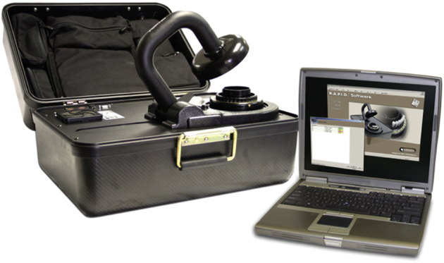
Devices initially developed for military or aerospace use can be re-appropriated for laboratory medicine. A portable thermocycler with laptop computer interface, initially developed for military field work, was later adapted for health care settings. The military version, dubbed the ruggedized advanced pathogen identification device (RAPID), was designed, in part, to examine environmental samples for biowarfare agents. The device requires limited space and technologist expertise, while still providing reliable real-time molecular results.
Image used with permission of BioFire Defense.
Although repurposing devices for resource-poor areas may be helpful, the optimal device is built from the ground up to meet the unique requirements of the locale and the intended use. Although most of the world's countries have at least one state-of-the-art medical complex, some tests must be performed in more remote facilities served by a meager workforce, perhaps with limited access to refrigeration, electricity, and potable water. Universally appealing device characteristics include low-cost, robust, and sustainable systems that are medically fit for purpose. Table 1 lists features promoting adoption of laboratory test devices, recognizing that various medical providers have differing priorities.
Table 1.
Desirable Characteristics of Laboratory Test Systems
| Medical performance |
| Reliable results (suitably sensitive, specific, linear, and reproducible) |
| Informative for actionable medical decision making |
| Same-day turnaround time to minimize loss to follow-up |
| Administrative qualities |
| Low cost to establish and to maintain the test system |
| Straightforward training of staff and supervisory personnel |
| User-friendly interface |
| Intact supply chain for supplies and reagents |
| Option to use expired reagents with documented explanation (eg, for training) |
| Simple equipment maintenance with reminders and record keeping |
| Available service and repairs |
| Safe for patients and for testing personnel |
| Accessible where medically necessary |
| Quality assurance |
| Document and report provider's ID, patient ID (option for barcoding), specimen type, date/time collected and reported, raw data, reportable result, interpretation, and comments |
| Automation to reduce risk of human error |
| Built-in quality checks with recorded results, such as endogenous control |
| Contamination alert (eg, statistically unlikely series of results) |
| Flags to alert for malfunction, aberrancies, and suggested corrective action |
| Barcoded supplies and reagent lot numbers recorded; alerts for expiration dates |
| Proficiency survey availability |
| Educational materials for clinicians and for testing personnel |
| Environmental factors |
| Minimal requirements for continuous electricity, Wi-Fi access, sterility, and clean water |
| Long shelf life under extreme conditions of transport and storage |
| Disposal of toxic chemicals, biohazards, cartridges, and sharps |
| Durable hardware, and in some cases waterproof or portable |
| Lockable in a physical way and from a patient privacy perspective |
Competition among vendors is a major driver of low cost and innovation, as is trade policy improving access to commercial products in the developing world. The recent US Supreme Court ruling that isolated DNA does not infringe on gene patents opens the door for molecular device design on the basis of medical and public health needs above costly legal quagmires.
Rapid, Inexpensive Molecular Technology
PCR is the mainstay of hybridization assays worldwide, but emerging molecular technologies may be less expensive and equally effective. A recent study of patients with hemorrhagic fever in Sudan showed that isothermal loop-mediated amplification (LAMP) was more rapid, less expensive, and performed as well as RT-PCR for detecting viral RNA in serum samples of patients with acute infection. The reaction is performed in a 63°C water bath rather than the more expensive thermocycler needed for PCR.
Another isothermal nucleic acid sequence-based amplification (NASBA) technology is being adapted to quantify HIV RNA and to predict drug resistance.79 NASBA is well suited for RNA profiling because it skips the need for reverse transcriptase used in RT-PCR. Expressed RNAs are naturally more abundant than DNA, although RNA is also more labile, thus generating the potential for specimen degradation unless the test is done promptly or preservatives are used.
Isothermal amplification eliminates the need for costly heating and cooling steps that characterize PCR. Instead, enzymes achieve strand separation, and these enzymes (eg, helicase) may be more tolerant of inhibitors than is the polymerase used in PCR, potentially reducing the need to purify analytes in crude specimens before hybridization.64,80,81 LAMP technology typically relies on six primers, two of which form stem loops that self-prime, and a strand-displacing polymerase then amplifies concatenated products. The assay yields an insoluble end product that can be quantified visually or in real time using a turbimetric detector, potentially saving cost by eliminating expensive fluorochromes. An option for fluorescent detection permits multiplexing of LAMP assays in one reaction vessel.
LAMP assays have been developed to detect tumor-specific RNA and also a range of microorganisms (bacteria, mycobacteria, viruses, parasites, amoeba, and protozoa) using handheld, battery-operated devices with disposable microfluidic cartridges.81–85 Open-source hardware, software, and protocols are intended to spur further innovation.82 Technical reviews of LAMP and other isothermal amplification technologies were recently published.58,64,80 Thus far, intellectual property concerns have hindered combining these promising chemistries with specimen collection tools, fluidics, and informatics modules required for a functional test system.
Next-generation sequencing is exceptionally powerful in its ability to identify genetic variants, although user-friendly sequencing devices remain to be commercialized. The technical costs of sequencing continue to plummet, but implementation in health care remains in its infancy due to the labor-intensive processes of preparing specimens and interpreting sequence variants. As with all cancer genetic tests, interpretation is done in the context of the clinical question being posed and the input material on which each test is performed (eg, tumor type, proportion of neoplastic cells, and fresh versus fixed tissue). Patient data are examined alongside multiple quality checks that reflect performance of the test system and adequacy of patient material.86 Research is ongoing to construct databases linking sequence variants to evidence of clinical actionability and improved patient outcome.
Chips, Assay Miniaturization, and Automation
Electronic sensors can detect DNA hybridization by its effect on current flowing through an electrode on a circuit board (Figure 3). This technology, combined with miniaturization (nanotechnology) and fluidics, could provide cost savings by using fewer and less expensive reagents, faster thermocycling, and less labor when compared with traditional plate-based hybridization.87 On-chip fluidics can accommodate multistep processing, including cell separation, cell lysis, and nucleic acid purification, before analysis. Automation promotes standardization and reproducibility. Several commercial test systems are capable of accommodating small input volume for multiwell analysis using disposable components.88,89 Interfaced barcode readers and laboratory information systems promote specimen identification and data analysis for interpretation and reporting.
Figure 3.

Electronic biosensors operate by electrochemical signal transduction on hybridization to complementary target sequence in the patient specimen. In this example, a hybridization-induced conformation change increases the distance between the electrode and the reporter molecule, which alters electron-transfer dynamics to generate a current in the electrode. Billions of sensors can be placed on a single semiconductor chip for simultaneous analysis of all relevant targets in human and pathogen genomes. System parts (chips, reagents, and detectors) are all inexpensive.
Preservation of Nucleic Acid before Analysis
Abundant data confirm that dried blood spots are amenable for genotyping.41,43,90–93 Dried specimens stored or transported at ambient temperature represent a cost-effective means of amassing specimens for batch testing. DNA, RNA, and microRNA targets are reportedly recoverable from filter paper on which blood or other body fluids have been dessicated. Alternative stabilizers have been described to promote specimen integrity between collection and analysis.94–97
Human Resources
The single most important aspect of high-quality laboratory service is competent personnel. This includes all personnel in the chain from test order through collection, transport, analysis, interpretation, reporting, and downstream action. A recent white paper from COLA highlights problems surrounding tests done in poorly regulated health care environments.98 Waived testing sites (ie, point-of-care sites) in the United States indicated that 35% of personnel did not properly record quality control data, and 31% did not maintain a log of tests performed. Poor preventive maintenance of equipment can result in a variety of problems, including contamination that may adversely affect sensitive molecular tests. The findings highlight the potential gap between a well-trained, well-supervised laboratory technologist and the reality when other (well-meaning) health care providers perform laboratory tests. It is, thus, recommended that testing be done in a centralized laboratory, except when there is medical need for rapid turnaround, to prevent loss to follow-up, or when transport would compromise specimen integrity.
When less-experienced personnel are expected to perform tests, it is important that test systems are engineered accordingly to address likely errors in technique, interpretation, or documentation. A supervisor should periodically oversee the work of testing personnel (trust but verify) and nurture those serving on the front lines who may otherwise feel isolated. Continuing medical education motivates workers to persist in and take pride in their specialized work. Poor-quality test results not only harm patients but ultimately undermine clinician's confidence in the utility of laboratory tools, making it even more difficult to establish and maintain laboratory services. Software is available to facilitate oversight of point-of-care testing.
It is difficult to recruit and retain pathologists and technologists in developing countries. For example, in Honduras there are only nine certified histotechnologists, none of whom are positioned in rural areas. Staffing is likely to get worse in the coming decades due to a workforce shortage of both pathologists and technologists. The looming crisis must be addressed through training programs in developed nations and in the developing world. A non-profit organization, Pathologists Overseas, has a 25-year track record of providing, advising, and promoting self-sustaining pathology and laboratory services.99 Many other noteworthy programs exist, such as those coordinated by the African Society for Laboratory Medicine, the Pan American Health Organization, the World Health Organization, the American Society for Clinical Pathology, the American Society of Cytopathology, the US and Canadian Academy of Pathology, The Diagnostic Microbiology Development Program, the Royal College of Pathologists, The International Network for Cancer Treatment and Research, and the College of American Pathologists Foundation. The programs are aimed at building local laboratory capacity.100–102 It is important to coordinate with in-country leaders, particularly with the Minister of Health, to ensure that effort is appropriate for local needs and that resources are strategically allocated.
To promote capacity building, ideally local practitioners are empowered for independence within the framework of multinational regional networks (eg, Central America).102–105 The American Association for Clinical Chemistry offers an online training program for point-of-care specialists. The Association for Molecular Pathology helps arrange regional chapters of molecular laboratory professionals. Much effort of the aforementioned organizations is focused on educating personnel working in central laboratories and academic centers, who, in turn, train others in the region. The College of American Pathologists offers proficiency surveys that form one component of quality assurance.
Telepathology
Telepathology encompasses the practice of pathology at a distance. Remote interpretation of qualitative or quantitative laboratory results is feasible, such as visualizing peaks on an electropherogram or analyzing BAM files generated by sequencing DNA of patients and associated controls. Guidance from the Canadian Association of Pathologists provides a framework for telepathology implementation and validation.106
Telepathology assumes that an infrastructure exists to perform the technical component of testing near the point of care, and that technical components should be overseen by a physician responsible for ensuring quality of transmitted data or images. Broadband connection is available to only one-third of the world's population, although efforts are underway to render access universal (eg, http://internet.org). A large bandwidth is required for image transmission.
Telepathology is a mechanism for second opinion consultation and for sharing case workload, reviewing unusual cases for continuing medical education, and promoting quality assurance. One concern about remote consultation is the lack of consultant familiarity with local medical practices and with regional diseases, such as pertussis (whooping cough), tropical enteropathy, or nutrient deficiencies. Video-conferencing is helpful for two-way learning, devising and updating work plans, and facilitating train the trainer programs.
Gopal et al107 recently described their experience establishing a histopathology laboratory in Malawi, including digital image transmission of scanned slides to a consulting pathologist at the University of North Carolina who reviews findings with the primary pathologist in Malawi. The establishment of new laboratory services is expensive and requires multidisciplinary coordination (eg, with clinicians and a wide range of staff). Medical equipment or supply donations can be arranged by agencies such as Project C.U.R.E. (Commission on Urgent Relief and Equipment; (http://www.projectcure.org), MedShare (http://www.MedShare.org), MedWish (http://www.MedWish.org), or Central American Medical Outreach (http://www.camo.org) (websites were last accessed on September 8, 2014).
Information Systems
Same-day turnaround is often crucial for surmounting loss to follow-up, although increasing availability of cell phones, even in rural areas, provides better opportunity for patient engagement. Text messaging is ubiquitous worldwide. Smartphones are much more expensive but offer added benefits of global positioning system geotagging and interfaced biometric sensors with optional data upload to a central databank.108,109 Patients and their providers can then view serial data and receive customized notices or educational materials. An application for testing personnel (eg, Radiometer America's Avoid Errors application) contains instructional videos on specimen collection, a competency test, and tips to interpret findings or to troubleshoot problems. Connectivity to fiberoptic or mobile broadband service not only offers a way to deliver health care but also to facilitate epidemiological monitoring of emerging disease, and to access consultants.75 Transcription errors are reduced when data are automatically transferred.
The Computing For Good (http://limswiki.org/index.php/C4G_BLIS, last accessed September 8, 2014) program at Georgia Institute of Technology recently teamed with the Centers for Disease Control and Prevention to field-test an open-source basic laboratory information system for computer data entry and retrieval in laboratory settings.
Conclusion
Advances in molecular technology show promise to improve cancer control in resource-limited nations through rapid, informative, reliable, user-friendly, and cost-effective laboratory tests. Much of the focus in developing nations is on infection-related cancers because of their prevalence and potential for cost-efficient prevention, as well as the unique opportunity to use the pathogen as a tumor marker promoting diagnosis, monitoring, and targeted therapy. Encouraging progress has been made in engineering devices to overcome the economic and social barriers that often limit effective laboratory support. Recent advances in technology are yielding small, but powerful, instruments that, along with accessible reagents, make feasible the types of molecular diagnostic systems that are suited to frontline providers.
Footnotes
Supported by the University of North Carolina (UNC) Department of Pathology and Laboratory Medicine, the University Cancer Research Fund, UNC Clinical Translational Science Award NIHUL1 TR001111, Innovative Technologies for Molecular Analysis of Cancer award NCIR21 CA155543, and Western Honduras Gastric Cancer Initiative NCI grants CA125588, CA197773, and HHSN261200800001.
Disclosures: None declared.
References
- 1.Chin C.D., Linder V., Sia S.K. Commercialization of microfluidic point-of-care diagnostic devices. Lab Chip. 2012;12:2118–2134. doi: 10.1039/c2lc21204h. [DOI] [PubMed] [Google Scholar]
- 2.Jennings L., Van Deerlin V.M., Gulley M.L. Recommended principles and practices for validating clinical molecular pathology tests. Arch Pathol Lab Med. 2009;133:743–755. doi: 10.5858/133.5.743. [DOI] [PubMed] [Google Scholar]
- 3.de Martel C., Ferlay J., Franceschi S., Vignat J., Bray F., Forman D., Plummer M. Global burden of cancers attributable to infections in 2008: a review and synthetic analysis. Lancet Oncol. 2012;13:607–615. doi: 10.1016/S1470-2045(12)70137-7. [DOI] [PubMed] [Google Scholar]
- 4.Murray C.J., Lopez A.D. Alternative projections of mortality and disability by cause 1990-2020: global Burden of Disease Study. Lancet. 1997;349:1498–1504. doi: 10.1016/S0140-6736(96)07492-2. [DOI] [PubMed] [Google Scholar]
- 5.Goss P.E., Lee B.L., Badovinac-Crnjevic T., Strasser-Weippl K., Chavarri-Guerra Y., St Louis J. Planning cancer control in Latin America and the Caribbean. Lancet Oncol. 2013;14:391–436. doi: 10.1016/S1470-2045(13)70048-2. [DOI] [PubMed] [Google Scholar]
- 6.Farmer P., Frenk J., Knaul F.M., Shulman L.N., Alleyne G., Armstrong L., Atun R., Blayney D., Chen L., Feachem R., Gospodarowicz M., Gralow J., Gupta S., Langer A., Lob-Levyt J., Neal C., Mbewu A., Mired D., Piot P., Reddy K.S., Sachs J.D., Sarhan M., Seffrin J.R. Expansion of cancer care and control in countries of low and middle income: a call to action. Lancet. 2010;376:1186–1193. doi: 10.1016/S0140-6736(10)61152-X. [DOI] [PubMed] [Google Scholar]
- 7.Magrath I., Steliarova-Foucher E., Epelman S., Ribeiro R.C., Harif M., Li C.K., Kebudi R., Macfarlane S.D., Howard S.C. Paediatric cancer in low-income and middle-income countries. Lancet Oncol. 2013;14:e104–e116. doi: 10.1016/S1470-2045(13)70008-1. [DOI] [PubMed] [Google Scholar]
- 8.Alexandrov L.B., Nik-Zainal S., Wedge D.C., Aparicio S.A., Behjati S., Biankin A.V. Signatures of mutational processes in human cancer. Nature. 2013;500:415–421. doi: 10.1038/nature12477. [DOI] [PMC free article] [PubMed] [Google Scholar]
- 9.Gulley M.L., Tang W. Laboratory assays for Epstein-Barr virus-related disease. J Mol Diagn. 2008;10:279–292. doi: 10.2353/jmoldx.2008.080023. [DOI] [PMC free article] [PubMed] [Google Scholar]
- 10.Quiroga-Garza G., Zhou H., Mody D.R., Schwartz M.R., Ge Y. Unexpected high prevalence of HPV 90 infection in an underserved population: is it really a low-risk genotype? Arch Pathol Lab Med. 2013;137:1569–1573. doi: 10.5858/arpa.2012-0640-OA. [DOI] [PubMed] [Google Scholar]
- 11.Ramogola-Masire D., McGrath C.M., Barnhart K.T., Friedman H.M., Zetola N.M. Subtype distribution of human papillomavirus in HIV-infected women with cervical intraepithelial neoplasia stages 2 and 3 in Botswana. Int J Gynecol Pathol. 2011;30:591–596. doi: 10.1097/PGP.0b013e31821bf2a6. [DOI] [PMC free article] [PubMed] [Google Scholar]
- 12.Gulliksen A., Keegan H., Martin C., O'Leary J., Solli L.A., Falang I.M., Gronn P., Karlgard A., Mielnik M.M., Johansen I.R., Tofteberg T.R., Baier T., Gransee R., Drese K., Hansen-Hagge T., Riegger L., Koltay P., Zengerle R., Karlsen F., Ausen D., Furuberg L. Towards a “sample-in, answer-out” point-of-care platform for nucleic acid extraction and amplification: using an HPV E6/E7 mRNA model system. J Oncol. 2012;2012:905024. doi: 10.1155/2012/905024. [DOI] [PMC free article] [PubMed] [Google Scholar]
- 13.Spizz G., Young L., Yasmin R., Chen Z., Lee T., Mahoney D., Zhang X., Mouchka G., Thomas B., Honey W., Roswech T., McGuire C., Montagna R., Zhou P. Rheonix CARD((R)) Technology: an innovative and fully automated molecular diagnostic device. Point Care. 2012;11:42–51. doi: 10.1097/POC.0b013e318222e184. [DOI] [PMC free article] [PubMed] [Google Scholar]
- 14.Zhao F.H., Jeronimo J., Qiao Y.L., Schweizer J., Chen W., Valdez M., Lu P., Zhang X., Kang L.N., Bansil P., Paul P., Mahoney C., Berard-Bergery M., Bai P., Peck R., Li J., Chen F., Stoler M.H., Castle P.E. An evaluation of novel, lower-cost molecular screening tests for human papillomavirus in rural China. Cancer Prev Res (Phila) 2013;6:938–948. doi: 10.1158/1940-6207.CAPR-13-0091. [DOI] [PubMed] [Google Scholar]
- 15.Ying H., Jing F., Fanghui Z., Youlin Q., Yali H. High-risk HPV nucleic acid detection kit-the careHPV test -a new detection method for screening. Sci Rep. 2014;4:4704. doi: 10.1038/srep04704. [DOI] [PMC free article] [PubMed] [Google Scholar]
- 16.Trope L.A., Chumworathayi B., Blumenthal P.D. Feasibility of community-based careHPV for cervical cancer prevention in rural Thailand. J Low Genit Tract Dis. 2013;17:315–319. doi: 10.1097/LGT.0b013e31826b7b70. [DOI] [PubMed] [Google Scholar]
- 17.Untiet S., Vassilakos P., McCarey C., Tebeu P.M., Kengne-Fosso G., Menoud P.A., Boulvain M., Navarria I., Petignat P. HPV self-sampling as primary screening test in sub-Saharan Africa: implication for a triaging strategy. Int J Cancer. 2014;135:1911–1917. doi: 10.1002/ijc.28834. [DOI] [PubMed] [Google Scholar]
- 18.Verhoef V.M., Bosgraaf R.P., van Kemenade F.J., Rozendaal L., Heideman D.A., Hesselink A.T., Bekkers R.L., Steenbergen R.D., Massuger L.F., Melchers W.J., Bulten J., Overbeek L.I., Berkhof J., Snijders P.J., Meijer C.J. Triage by methylation-marker testing versus cytology in women who test HPV-positive on self-collected cervicovaginal specimens (PROHTECT-3): a randomised controlled non-inferiority trial. Lancet Oncol. 2014;15:315–322. doi: 10.1016/S1470-2045(14)70019-1. [DOI] [PubMed] [Google Scholar]
- 19.Guan Y., Gravitt P.E., Howard R., Eby Y.J., Wang S., Li B., Feng C., Qiao Y.L., Castle P.E. Agreement for HPV genotyping detection between self-collected specimens on a FTA cartridge and clinician-collected specimens. J Virol Methods. 2013;189:167–171. doi: 10.1016/j.jviromet.2012.11.010. [DOI] [PMC free article] [PubMed] [Google Scholar]
- 20.Odesanmi T.Y., Wasti S.P., Odesanmi O.S., Adegbola O., Oguntuase O.O., Mahmood S. Comparative effectiveness and acceptability of home-based and clinic-based sampling methods for sexually transmissible infections screening in females aged 14-50 years: a systematic review and meta-analysis. Sex Health. 2013;10:559–569. doi: 10.1071/SH13029. [DOI] [PubMed] [Google Scholar]
- 21.Nordfors C., Vlastos A., Du J., Ahrlund-Richter A., Tertipis N., Grun N., Romanitan M., Haeggblom L., Roosaar A., Dahllof G., Dona M.G., Benevolo M., Ramqvist T., Munck-Wikland E., Dalianis T. Human papillomavirus prevalence is high in oral samples of patients with tonsillar and base of tongue cancer. Oral Oncol. 2014;50:491–497. doi: 10.1016/j.oraloncology.2014.02.012. [DOI] [PubMed] [Google Scholar]
- 22.Zabaleta J. Multifactorial etiology of gastric cancer. Methods Mol Biol. 2012;863:411–435. doi: 10.1007/978-1-61779-612-8_26. [DOI] [PMC free article] [PubMed] [Google Scholar]
- 23.He C., Tu H., Sun L., Xu Q., Li P., Gong Y., Dong N., Yuan Y. Helicobacter pylori-related host gene polymorphisms associated with susceptibility of gastric carcinogenesis: a two-stage case-control study in Chinese. Carcinogenesis. 2013;34:1450–1457. doi: 10.1093/carcin/bgt079. [DOI] [PubMed] [Google Scholar]
- 24.Essawi T., Hammoudeh W., Sabri I., Sweidan W., Farraj M.A. Determination of Helicobacter pylori virulence genes in gastric biopsies by PCR. ISRN Gastroenterol. 2013;2013:606258. doi: 10.1155/2013/606258. [DOI] [PMC free article] [PubMed] [Google Scholar]
- 25.Ek C., Whary M.T., Ihrig M., Bravo L.E., Correa P., Fox J.G. Serologic evidence that ascaris and toxoplasma infections impact inflammatory responses to Helicobacter pylori in Colombians. Helicobacter. 2012;17:107–115. doi: 10.1111/j.1523-5378.2011.00916.x. [DOI] [PMC free article] [PubMed] [Google Scholar]
- 26.Tan I.B., Ng I., Tai W.M., Tan P. Understanding the genetic basis of gastric cancer: recent advances. Expert Rev Gastroenterol Hepatol. 2012;6:335–341. doi: 10.1586/egh.12.7. [DOI] [PubMed] [Google Scholar]
- 27.Tang W., Morgan D.R., Meyers M.O., Dominguez R.L., Martinez E., Kakudo K., Kuan P.F., Banet N., Muallem H., Woodward K., Speck O., Gulley M.L. Epstein-barr virus infected gastric adenocarcinoma expresses latent and lytic viral transcripts and has a distinct human gene expression profile. Infect Agent Cancer. 2012;7:21. doi: 10.1186/1750-9378-7-21. [DOI] [PMC free article] [PubMed] [Google Scholar]
- 28.Morgner A., Schmelz R., Thiede C., Stolte M., Miehlke S. Therapy of gastric mucosa associated lymphoid tissue lymphoma. World J Gastroenterol. 2007;13:3554–3566. doi: 10.3748/wjg.v13.i26.3554. [DOI] [PMC free article] [PubMed] [Google Scholar]
- 29.Schmitt B.H., Regner M., Mangold K.A., Thomson R.B., Jr., Kaul K.L. PCR detection of clarithromycin-susceptible and -resistant Helicobacter pylori from formalin-fixed, paraffin-embedded gastric biopsies. Mod Pathol. 2013;26:1222–1227. doi: 10.1038/modpathol.2013.48. [DOI] [PubMed] [Google Scholar]
- 30.Singh V., Mishra S., Rao G.R., Jain A.K., Dixit V.K., Gulati A.K., Mahajan D., McClelland M., Nath G. Evaluation of nested PCR in detection of Helicobacter pylori targeting a highly conserved gene: HSP60. Helicobacter. 2008;13:30–34. doi: 10.1111/j.1523-5378.2008.00573.x. [DOI] [PMC free article] [PubMed] [Google Scholar]
- 31.Perkins T.T., Tay C.Y., Thirriot F., Marshall B. Choosing a benchtop sequencing machine to characterise Helicobacter pylori genomes. PLoS One. 2013;8:e67539. doi: 10.1371/journal.pone.0067539. [DOI] [PMC free article] [PubMed] [Google Scholar]
- 32.Falsafi T., Favaedi R., Mahjoub F., Najafi M. Application of stool-PCR test for diagnosis of Helicobacter pylori infection in children. World J Gastroenterol. 2009;15:484–488. doi: 10.3748/wjg.15.484. [DOI] [PMC free article] [PubMed] [Google Scholar]
- 33.Arboleda R.N., Schneider B.G., Bravo L.E., Romero-Gallo J., Peek R.M., Jr., Mera R.M., Yepez M.C., Campo C., Correa P. Use of the noninvasive entero-test in the detection of Helicobacter pylori in children in an endemic area in Colombia. J Pediatr Gastroenterol Nutr. 2013;57:192–196. doi: 10.1097/MPG.0b013e318293e1e1. [DOI] [PMC free article] [PubMed] [Google Scholar]
- 34.Nishida N., Kudo M. Recent advancements in comprehensive genetic analyses for human hepatocellular carcinoma. Oncology. 2013;84(Suppl 1):93–97. doi: 10.1159/000345897. [DOI] [PubMed] [Google Scholar]
- 35.Han G.R., Xu C.L., Zhao W., Yang Y.F. Management of chronic hepatitis B in pregnancy. World J Gastroenterol. 2012;18:4517–4521. doi: 10.3748/wjg.v18.i33.4517. [DOI] [PMC free article] [PubMed] [Google Scholar]
- 36.Morgan R.L., Baack B., Smith B.D., Yartel A., Pitasi M., Falck-Ytter Y. Eradication of hepatitis C virus infection and the development of hepatocellular carcinoma: a meta-analysis of observational studies. Ann Intern Med. 2013;158:329–337. doi: 10.7326/0003-4819-158-5-201303050-00005. [DOI] [PubMed] [Google Scholar]
- 37.Cabezas-Fernandez M., Cabeza-Barrera M. Introduction of an automated system for the diagnosis and quantification of hepatitis B and hepatitis C viruses. Open Virol J. 2012;6:122–134. doi: 10.2174/1874357901206010122. [DOI] [PMC free article] [PubMed] [Google Scholar]
- 38.Chevaliez S., Rodriguez C., Pawlotsky J.M. New virologic tools for management of chronic hepatitis B and C. Gastroenterology. 2012;142:1303–1313.e1. doi: 10.1053/j.gastro.2012.02.027. [DOI] [PMC free article] [PubMed] [Google Scholar]
- 39.Wang Q., Wang H., Yang X., Wang K., Liu F., Zhao Q., Liu P., Liu R. Multiplex detection of nucleic acids using a low cost microfluidic chip and a personal glucose meter at the point-of-care. Chem Commun (Camb) 2014;50:3824–3826. doi: 10.1039/c4cc00133h. [DOI] [PubMed] [Google Scholar]
- 40.Petrara M.R., Elefanti L., Quaggio M., Zanchetta M., Scaini M.C., Masalu N., De Rossi A., Menin C. Dried blood spot sampling for detection of monoclonal immunoglobulin gene rearrangement. Leuk Res. 2013;37:1265–1270. doi: 10.1016/j.leukres.2013.08.003. [DOI] [PubMed] [Google Scholar]
- 41.Snijdewind I.J., van Kampen J.J., Fraaij P.L., van der Ende M.E., Osterhaus A.D., Gruters R.A. Current and future applications of dried blood spots in viral disease management. Antiviral Res. 2012;93:309–321. doi: 10.1016/j.antiviral.2011.12.011. [DOI] [PubMed] [Google Scholar]
- 42.Tang W., Harmon P., Gulley M.L., Mwansambo C., Kazembe P.N., Martinson F., Wokocha C., Kenney S.C., Hoffman I., Sigel C., Maygarden S., Hoffman M., Shores C. Viral response to chemotherapy in endemic burkitt lymphoma. Clin Cancer Res. 2010;16:2055–2064. doi: 10.1158/1078-0432.CCR-09-2424. [DOI] [PMC free article] [PubMed] [Google Scholar]
- 43.Petrara M.R., Penazzato M., Massavon W., Nabachwa S., Nannyonga M., Mazza A., Gianesin K., Del Bianco P., Lundin R., Sumpter C., Zanchetta M., Giaquinto C., De Rossi A. Epstein-Barr virus load in children infected with human immunodeficiency virus type 1 in Uganda. J Infect Dis. 2014;210:392–399. doi: 10.1093/infdis/jiu099. [DOI] [PubMed] [Google Scholar]
- 44.Mancuso M., Jiang L., Cesarman E., Erickson D. Multiplexed colorimetric detection of Kaposi's sarcoma associated herpesvirus and Bartonella DNA using gold and silver nanoparticles. Nanoscale. 2013;5:1678–1686. doi: 10.1039/c3nr33492a. [DOI] [PMC free article] [PubMed] [Google Scholar]
- 45.Jiang L., Mancuso M., Lu Z., Akar G., Cesarman E., Erickson D. Solar thermal polymerase chain reaction for smartphone-assisted molecular diagnostics. Sci Rep. 2014;4:4137. doi: 10.1038/srep04137. [DOI] [PMC free article] [PubMed] [Google Scholar]
- 46.Yu Y.H., Liao C.C., Hsu W.H., Chen H.J., Liao W.C., Muo C.H., Sung F.C., Chen C.Y. Increased lung cancer risk among patients with pulmonary tuberculosis: a population cohort study. J Thorac Oncol. 2011;6:32–37. doi: 10.1097/JTO.0b013e3181fb4fcc. [DOI] [PubMed] [Google Scholar]
- 47.Dheda K., Ruhwald M., Theron G., Peter J., Yam W.C. Point-of-care diagnosis of tuberculosis: past, present and future. Respirology. 2013;18:217–232. doi: 10.1111/resp.12022. [DOI] [PubMed] [Google Scholar]
- 48.Scott L.E., McCarthy K., Gous N., Nduna M., Van Rie A., Sanne I., Venter W.F., Duse A., Stevens W. Comparison of Xpert MTB/RIF with other nucleic acid technologies for diagnosing pulmonary tuberculosis in a high HIV prevalence setting: a prospective study. PLoS Med. 2011;8:e1001061. doi: 10.1371/journal.pmed.1001061. [DOI] [PMC free article] [PubMed] [Google Scholar]
- 49.Dowdy D.W., Davis J.L., den Boon S., Walter N.D., Katamba A., Cattamanchi A. Population-level impact of same-day microscopy and Xpert MTB/RIF for tuberculosis diagnosis in Africa. PLoS One. 2013;8:e70485. doi: 10.1371/journal.pone.0070485. [DOI] [PMC free article] [PubMed] [Google Scholar]
- 50.Centers for Disease Control and Prevention (CDC) Availability of an assay for detecting Mycobacterium tuberculosis, including rifampin-resistant strains, and considerations for its use: United States, 2013. MMWR Morb Mortal Wkly Rep. 2013;62:821–827. [PMC free article] [PubMed] [Google Scholar]
- 51.Van Rie A., Page-Shipp L., Hanrahan C.F., Schnippel K., Dansey H., Bassett J., Clouse K., Scott L., Stevens W., Sanne I. Point-of-care Xpert(R) MTB/RIF for smear-negative tuberculosis suspects at a primary care clinic in South Africa. Int J Tuberc Lung Dis. 2013;17:368–372. doi: 10.5588/ijtld.12.0392. [DOI] [PMC free article] [PubMed] [Google Scholar]
- 52.Cutrell J., Bedimo R. Non-AIDS-defining cancers among HIV-infected patients. Curr HIV/AIDS Rep. 2013;10:207–216. doi: 10.1007/s11904-013-0166-8. [DOI] [PubMed] [Google Scholar]
- 53.Mulama D.H., Bailey J.A., Foley J., Chelimo K., Ouma C., Jura W.G., Otieno J., Vulule J., Moormann A.M. Sickle cell trait is not associated with endemic Burkitt lymphoma: an ethnicity and malaria endemicity-matched case-control study suggests factors controlling EBV may serve as a predictive biomarker for this pediatric cancer. Int J Cancer. 2014;134:645–653. doi: 10.1002/ijc.28378. [DOI] [PMC free article] [PubMed] [Google Scholar]
- 54.Ondondo B.O. Fallen angels or risen apes? A tale of the intricate complexities of imbalanced immune responses in the pathogenesis and progression of immune-mediated and viral cancers. Front Immunol. 2014;5:90. doi: 10.3389/fimmu.2014.00090. [DOI] [PMC free article] [PubMed] [Google Scholar]
- 55.Aka P., Vila M.C., Jariwala A., Nkrumah F., Emmanuel B., Yagi M., Palacpac N.M., Periago M.V., Neequaye J., Kiruthu C., Tougan T., Levine P.H., Biggar R.J., Pfeiffer R.M., Bhatia K., Horii T., Bethony J.M., Mbulaiteye S.M. Endemic Burkitt lymphoma is associated with strength and diversity of Plasmodium falciparum malaria stage-specific antigen antibody response. Blood. 2013;122:629–635. doi: 10.1182/blood-2012-12-475665. [DOI] [PMC free article] [PubMed] [Google Scholar]
- 56.Xu L., Kong J. A multiplexed nucleic acid microsystem for point-of-care detection of HIV co-infection with MTB and PCP. Talanta. 2013;117:532–535. doi: 10.1016/j.talanta.2013.08.056. [DOI] [PubMed] [Google Scholar]
- 57.Wang J.H., Cheng L., Wang C.H., Ling W.S., Wang S.W., Lee G.B. An integrated chip capable of performing sample pretreatment and nucleic acid amplification for HIV-1 detection. Biosens Bioelectron. 2013;41:484–491. doi: 10.1016/j.bios.2012.09.011. [DOI] [PubMed] [Google Scholar]
- 58.Haleyur Giri Setty M.K., Hewlett I.K. Point of Care Technologies for HIV. AIDS Res Treat. 2014;2014:497046. doi: 10.1155/2014/497046. [DOI] [PMC free article] [PubMed] [Google Scholar]
- 59.Li Y., Kumar N., Gopalakrishnan A., Ginocchio C., Manji R., Bythrow M., Lemieux B., Kong H. Detection and species identification of malaria parasites by isothermal tHDA amplification directly from human blood without sample preparation. J Mol Diagn. 2013;15:634–641. doi: 10.1016/j.jmoldx.2013.05.005. [DOI] [PMC free article] [PubMed] [Google Scholar]
- 60.Imwong M., Hanchana S., Malleret B., Rénia L., Day N.P., Dondorp A., Nosten F., Snounou G., White N.J. High-throughput ultrasensitive molecular techniques for quantifying low-density malaria parasitemias. J Clin Microbiol. 2014;52:3303–3309. doi: 10.1128/JCM.01057-14. [DOI] [PMC free article] [PubMed] [Google Scholar]
- 61.Goff D.A., Jankowski C., Tenover F.C. Using rapid diagnostic tests to optimize antimicrobial selection in antimicrobial stewardship programs. Pharmacotherapy. 2012;32:677–687. doi: 10.1002/j.1875-9114.2012.01137.x. [DOI] [PubMed] [Google Scholar]
- 62.Petti C.A., Polage C.R., Quinn T.C., Ronald A.R., Sande M.A. Laboratory medicine in Africa: a barrier to effective health care. Clin Infect Dis. 2006;42:377–382. doi: 10.1086/499363. [DOI] [PubMed] [Google Scholar]
- 63.Khoury J.D., Tannir N.M., Williams M.D., Chen Y., Yao H., Zhang J., Thompson E.J., Meric-Bernstam F., Medeiros L.J., Weinstein J.N., Su X. Landscape of DNA virus associations across human malignant cancers: analysis of 3,775 cases using RNA-Seq. J Virol. 2013;87:8916–8926. doi: 10.1128/JVI.00340-13. [DOI] [PMC free article] [PubMed] [Google Scholar]
- 64.Yan L., Zhou J., Zheng Y., Gamson A.S., Roembke B.T., Nakayama S., Sintim H.O. Isothermal amplified detection of DNA and RNA. Mol Biosyst. 2014;10:970–1003. doi: 10.1039/c3mb70304e. [DOI] [PubMed] [Google Scholar]
- 65.Kitano S., Myers J., Nakamura J., Yamane A., Yamashita M., Nakayama M., Tsukahara Y., Ushida H., Liu W., Ratain M.J., Amano M. A novel fully automated molecular diagnostic system (AMDS) for colorectal cancer mutation detection. PLoS One. 2013;8:e62989. doi: 10.1371/journal.pone.0062989. [DOI] [PMC free article] [PubMed] [Google Scholar]
- 66.Roberts J.D., Wells G.A., Le May M.R., Labinaz M., Glover C., Froeschl M., Dick A., Marquis J.F., O'Brien E., Goncalves S., Druce I., Stewart A., Gollob M.H., So D.Y. Point-of-care genetic testing for personalisation of antiplatelet treatment (RAPID GENE): a prospective, randomised, proof-of-concept trial. Lancet. 2012;379:1705–1711. doi: 10.1016/S0140-6736(12)60161-5. [DOI] [PubMed] [Google Scholar]
- 67.Bernacki K.D., Betz B.L., Weigelin H.C., Lao C.D., Redman B.G., Knoepp S.M., Roh M.H. Molecular diagnostics of melanoma fine-needle aspirates: a cytology-histology correlation study. Am J Clin Pathol. 2012;138:670–677. doi: 10.1309/AJCPEQJW3PLOOZTC. [DOI] [PubMed] [Google Scholar]
- 68.McCall B., Olsen R.J., Nelles N.J., Williams D.L., Jackson K., Richards-Kortum R., Graviss E.A., Tkaczyk T.S. Evaluation of a miniature microscope objective designed for fluorescence array microscopy detection of Mycobacterium tuberculosis. Arch Pathol Lab Med. 2014;138:379–389. doi: 10.5858/arpa.2013-0146-OA. [DOI] [PubMed] [Google Scholar]
- 69.Mosammaparast N., McAdam A.J., Nolte F.S. Molecular testing for infectious diseases should be done in the clinical microbiology laboratory. J Clin Microbiol. 2012;50:1836–1840. doi: 10.1128/JCM.00488-12. [DOI] [PMC free article] [PubMed] [Google Scholar]
- 70.Geraets D.T., van Baars R., Alonso I., Ordi J., Torne A., Melchers W.J., Meijer C.J., Quint W.G. Clinical evaluation of high-risk HPV detection on self-samples using the indicating FTA-elute solid-carrier cartridge. J Clin Virol. 2013;57:125–129. doi: 10.1016/j.jcv.2013.02.016. [DOI] [PubMed] [Google Scholar]
- 71.Quincy B.L., Turbow D.J., Dabinett L.N. Acceptability of self-collected human papillomavirus specimens as a primary screen for cervical cancer. J Obstet Gynaecol. 2012;32:87–91. doi: 10.3109/01443615.2011.625456. [DOI] [PubMed] [Google Scholar]
- 72.Ng R.H., Ngan R., Wei W.I., Gullane P.J., Phillips J. Trans-oral brush biopsies and quantitative PCR for EBV DNA detection and screening of nasopharyngeal carcinoma. Otolaryngol Head Neck Surg. 2014;150:602–609. doi: 10.1177/0194599813520136. [DOI] [PubMed] [Google Scholar]
- 73.Shan J., Pow E.H., Tsang P.C., Perera R.A., Kwong D.L. Comparison of two laboratory extraction techniques for the detection of Epstein-Barr virus in the saliva of nasopharyngeal carcinoma patients. J Investig Clin Dent. 2014;5:104–108. doi: 10.1111/jicd.12078. [DOI] [PubMed] [Google Scholar]
- 74.Brown-Connolly N.E., Concha J.B., English J. Mobile health is worth it! Economic benefit and impact on health of a population-based mobile screening program in New Mexico. Telemed J E Health. 2014;20:18–23. doi: 10.1089/tmj.2013.0080. [DOI] [PMC free article] [PubMed] [Google Scholar]
- 75.Seiner D.R., Colburn H.A., Baird C., Bartholomew R.A., Straub T., Victry K., Hutchison J.R., Valentine N., Bruckner-Lea C.J. Evaluation of the FilmArray(R) system for detection of Bacillus anthracis, Francisella tularensis and Yersinia pestis. J Appl Microbiol. 2013;114:992–1000. doi: 10.1111/jam.12107. [DOI] [PMC free article] [PubMed] [Google Scholar]
- 76.Fatah A.A., Arcilesi R., Peterson J., Lattin C.H., Wells C.Y., McClintock J.A. 3rd ed. National Institute of Standards and Technology; Gaithersburg, MD: 2007. Guide for the Selection of Chemical Detection Equipment for Emergency First Responders Guide 100–06, DHS Guide 100-06. [Google Scholar]
- 77.Chin C.D., Cheung Y.K., Laksanasopin T., Modena M.M., Chin S.Y., Sridhara A.A., Steinmiller D., Linder V., Mushingantahe J., Umviligihozo G., Karita E., Mwambarangwe L., Braunstein S.L., van de Wijgert J., Sahabo R., Justman J.E., El-Sadr W., Sia S.K. Mobile device for disease diagnosis and data tracking in resource-limited settings. Clin Chem. 2013;59:629–640. doi: 10.1373/clinchem.2012.199596. [DOI] [PubMed] [Google Scholar]
- 78.Zhang S., Lin Y., Wang J., Wang P., Chen J., Xue M., He S., Zhou W., Xu F., Liu P., Chen P., Ge S., Xia N. A convenient nucleic acid test on the basis of the capillary convective PCR for the on-site detection of enterovirus 71. J Mol Diagn. 2014;16:452–458. doi: 10.1016/j.jmoldx.2014.04.002. [DOI] [PubMed] [Google Scholar]
- 79.Morabito K., Kantor R., Tai W., Schreier L., Tripathi A. Detection of HIV-1 minority variants containing the K103N drug-resistance mutation using a simple method to amplify RNA targets (SMART) J Mol Diagn. 2013;15:401–412. doi: 10.1016/j.jmoldx.2013.02.005. [DOI] [PMC free article] [PubMed] [Google Scholar]
- 80.Craw P., Balachandran W. Isothermal nucleic acid amplification technologies for point-of-care diagnostics: a critical review. Lab Chip. 2012;12:2469–2486. doi: 10.1039/c2lc40100b. [DOI] [PubMed] [Google Scholar]
- 81.Perez L.E., Merrill G.A., DeLorenzo R.A., Schoenfeld T.W., Vats A., Moser M.J. Evaluation of the specificity and sensitivity of a potential rapid influenza screening system. Diagn Microbiol Infect Dis. 2013;75:77–80. doi: 10.1016/j.diagmicrobio.2012.09.005. [DOI] [PMC free article] [PubMed] [Google Scholar]
- 82.Myers F.B., Henrikson R.H., Bone J., Lee L.P. A handheld point-of-care genomic diagnostic system. PLoS One. 2013;8:e70266. doi: 10.1371/journal.pone.0070266. [DOI] [PMC free article] [PubMed] [Google Scholar]
- 83.Zhu Q., Gao Y., Yu B., Ren H., Qiu L., Han S., Jin W., Jin Q., Mu Y. Self-priming compartmentalization digital LAMP for point-of-care. Lab Chip. 2012;12:4755–4763. doi: 10.1039/c2lc40774d. [DOI] [PubMed] [Google Scholar]
- 84.Smolarz B., Krawczyk T., Westfal B., Maciejczyk R., Zadrozny M., Samulak D., Michalska M.M., Mojs E., Wilczak M., Romanowicz-Makowska H. Comparison of one-step nucleic acid amplification (OSNA) method and routine histological investigation for intraoperative detection of lymph node metastasis in Polish women with breast cancer. Pol J Pathol. 2013;64:104–108. doi: 10.5114/pjp.2013.36014. [DOI] [PubMed] [Google Scholar]
- 85.Muto Y., Matubara H., Tanizawa T., Nabeya Y., Kawahira H., Akai T., Hoshino I., Hayashi H. Rapid diagnosis of micrometastasis of gastric cancer using reverse transcription loop-mediated isothermal amplification. Oncol Rep. 2011;26:789–794. doi: 10.3892/or.2011.1389. [DOI] [PubMed] [Google Scholar]
- 86.Tang W., Hu Z., Muallem H., Gulley M.L. Quality assurance of RNA expression profiling in clinical laboratories. J Mol Diagn. 2012;14:1–11. doi: 10.1016/j.jmoldx.2011.09.003. [DOI] [PMC free article] [PubMed] [Google Scholar]
- 87.Lam B., Das J., Holmes R.D., Live L., Sage A., Sargent E.H., Kelley S.O. Solution-based circuits enable rapid and multiplexed pathogen detection. Nat Commun. 2013;4:2001. doi: 10.1038/ncomms3001. [DOI] [PubMed] [Google Scholar]
- 88.Guarnaccia M., Gentile G., Alessi E., Schneider C., Petralia S., Cavallaro S. Is this the real time for genomics? Genomics. 2014;103:177–182. doi: 10.1016/j.ygeno.2014.02.003. [DOI] [PubMed] [Google Scholar]
- 89.Patterson A.S., Hsieh K., Soh H.T., Plaxco K.W. Electrochemical real-time nucleic acid amplification: towards point-of-care quantification of pathogens. Trends Biotechnol. 2013;31:704–712. doi: 10.1016/j.tibtech.2013.09.005. [DOI] [PubMed] [Google Scholar]
- 90.Yapo V., d'Aquin Toni Td, Desmonde S., Amani-Bosse C., Oga M., Lenaud S., Menan H., Timité-Konan M., Leroy V., Rouzioux C. Evaluation of dried blood spot diagnosis using HIV1-DNA and HIV1-RNA Biocentric assays in infants in Abidjan, Côte d'Ivoire: the Pedi-Test DBS ANRS 12183 Study. J Virol Methods. 2013;193:439–445. doi: 10.1016/j.jviromet.2013.07.011. [DOI] [PubMed] [Google Scholar]
- 91.Mohamed S., Raimondo A., Penaranda G., Camus C., Ouzan D., Ravet S., Bourliere M., Khiri H., Dukan P., Olive D., Halfon P. Dried blood spot sampling for hepatitis B virus serology and molecular testing. PLoS One. 2013;8:e61077. doi: 10.1371/journal.pone.0061077. [DOI] [PMC free article] [PubMed] [Google Scholar]
- 92.Ivers L.C., Smith Fawzi M.C., Mann J., Jerome J.G., Raymonville M., Mukherjee J.S. Overseas processing of dried blood spots for timely diagnosis of HIV in Haitian infants. Rev Panam Salud Publica. 2008;24:331–335. doi: 10.1590/s1020-49892008001100005. [DOI] [PMC free article] [PubMed] [Google Scholar]
- 93.Smit P.W., Sollis K.A., Fiscus S., Ford N., Vitoria M., Essajee S., Barnett D., Cheng B., Crowe S.M., Denny T., Landay A., Stevens W., Habiyambere V., Perriens J.H., Peeling R.W. Systematic review of the use of dried blood spots for monitoring HIV viral load and for early infant diagnosis. PLoS One. 2014;9:e86461. doi: 10.1371/journal.pone.0086461. [DOI] [PMC free article] [PubMed] [Google Scholar]
- 94.Berry S.M., LaVanway A.J., Pezzi H.M., Guckenberger D.J., Anderson M.A., Loeb J.M., Beebe D.J. HIV viral RNA extraction in wax immiscible filtration assisted by surface tension (IFAST) devices. J Mol Diagn. 2014;16:297–304. doi: 10.1016/j.jmoldx.2014.01.004. [DOI] [PMC free article] [PubMed] [Google Scholar]
- 95.Hewitt S.M., Badve S.S., True L.D. Impact of preanalytic factors on the design and application of integral biomarkers for directing patient therapy. Clin Cancer Res. 2012;18:1524–1530. doi: 10.1158/1078-0432.CCR-11-2204. [DOI] [PMC free article] [PubMed] [Google Scholar]
- 96.Mathay C., Yan W., Chuaqui R., Skubitz A.P., Jeon J.P., Fall N., Betsou F., Barnes M. Short-term stability study of RNA at room temperature. Biopreserv Biobank. 2012;10:532–542. doi: 10.1089/bio.2012.0030. [DOI] [PubMed] [Google Scholar]
- 97.Howlett S.E., Castillo H.S., Gioeni L.J., Robertson J.M., Donfack J. Evaluation of DNAstable for DNA storage at ambient temperature. Forensic Sci Int Genet. 2014;8:170–178. doi: 10.1016/j.fsigen.2013.09.003. [DOI] [PubMed] [Google Scholar]
- 98.Commission on Office Laboratory Accreditation (COLA): Federal Government Questions Quality in Waived Testing. The Hard Facts and What Can Laboratories Do Now? A COLA White Paper. Available online at http://www.colainsider.com/cola-issues-waived-testing-white-paper-calls-for-more-education-at-waived-testing-sites (last updated April 22, 2013)
- 99.Hoenecke H., Lee V., Roy I. Pathologists overseas: coordinating volunteer pathology services for 19 years. Arch Pathol Lab Med. 2011;135:173–178. doi: 10.5858/2008-0450-SOR1.1. [DOI] [PubMed] [Google Scholar]
- 100.Cagle P.T. Pathologists as leaders, innovators, and devoted physicians: special section on pathology in resource-poor nations. Arch Pathol Lab Med. 2011;135:170. doi: 10.5858/135.2.170. [DOI] [PubMed] [Google Scholar]
- 101.Harder H.I. Pathology services in developing countries. Arch Pathol Lab Med. 2009;133:1911. doi: 10.5858/133.12.1911. [DOI] [PubMed] [Google Scholar]
- 102.Carlson J.W., Lyon E., Walton D., Foo W.C., Sievers A.C., Shulman L.N., Farmer P., Nose V., Milner D.A., Jr. Partners in pathology: a collaborative model to bring pathology to resource poor settings. Am J Surg Pathol. 2010;34:118–123. doi: 10.1097/PAS.0b013e3181c17fe6. [DOI] [PubMed] [Google Scholar]
- 103.Knaul F.M., Shulman L.N., Gralow J., Quick J., Bhadelia A., Frenk J. Improving pathology for better cancer care and control in countries of low and middle income. Lancet. 2012;379:2052. doi: 10.1016/S0140-6736(12)60894-0. [DOI] [PubMed] [Google Scholar]
- 104.Roberts D.J., Wilson M.L., Nelson A.M., Adesina A.M., Fleming K.A., Milner D., Guarner J., Rebbeck T.R., Castle P., Lucas S. The good news about cancer in developing countries–pathology answers the call. Lancet. 2012;379:712. doi: 10.1016/S0140-6736(12)60306-7. [DOI] [PubMed] [Google Scholar]
- 105.Rebbeck TR: Handbook for Cancer Research in Africa. African Organisation for Research and Training in Cancer. Brazzaville, Republic of Congo, World Health Organization, 2013
- 106.Tetu B., Evans A. Canadian licensure for the use of digital pathology for routine diagnoses: one more step toward a new era of pathology practice without borders. Arch Pathol Lab Med. 2014;138:302–304. doi: 10.5858/arpa.2013-0289-ED. [DOI] [PubMed] [Google Scholar]
- 107.Gopal S., Krysiak R., Liomba N.G., Horner M.J., Shores C.G., Alide N., Kamiza S., Kampani C., Chimzimu F., Fedoriw Y., Dittmer D.P., Hosseinipour M.C., Hoffman I.F. Early experience after developing a pathology laboratory in Malawi, with emphasis on cancer diagnoses. PLoS One. 2013;8:e70361. doi: 10.1371/journal.pone.0070361. [DOI] [PMC free article] [PubMed] [Google Scholar]
- 108.Gallegos D., Long K.D., Yu H., Clark P.P., Lin Y., George S., Nath P., Cunningham B.T. Label-free biodetection using a smartphone. Lab Chip. 2013;13:2124–2132. doi: 10.1039/c3lc40991k. [DOI] [PubMed] [Google Scholar]
- 109.Liu X., Lin T.Y., Lillehoj P.B. Smartphones for cell and biomolecular detection. Ann Biomed Eng. 2014 doi: 10.1007/s10439-014-1055-z. 10.1007/s10439-014-1055-z [Epub ahead of print] doi: [DOI] [PubMed] [Google Scholar]