

Abstract
Junctions that mediate excitation-contraction (e-c) coupling are formed between the sarcoplasmic reticulum (SR) and either the surface membrane or the transverse (T) tubules in normal skeletal muscle. Two structural components of the junctions, the feet of the SR and the tetrads of T tubules, have been identified respectively as ryanodine receptors (RyRs, or SR calcium-release channels), and as groups of four dihydropyridine receptors (DHPRs, or voltage sensors of e-c coupling). A targeted mutation (skrrm1) of the gene for skeletal muscle RyRs in mice results in the absence of e-c coupling in homozygous offspring of transgenic parents. The mutant gene is expected to produce no functional RyRs, and we have named the mutant mice "dyspedic" because they lack feet--the cytoplasmic domain of RyRs anchored in the SR membrane. We have examined the development of junctions in skeletal muscle fibers from normal and dyspedic embryos. Surprisingly, despite the absence of RyRs, junctions are formed in dyspedic myotubes, but the junctional gap between the SR and T tubule is narrow, presumably because the feet are missing. Tetrads are also absent from these junctions. The results confirm the identity of RyRs and feet and a major role for RyRs and tetrads in e-c coupling. Since junctions form in the absence of feet and tetrads, coupling of SR to surface membrane and T tubules appears to be mediated by additional proteins, distinct from either RyRs or DHPRs.
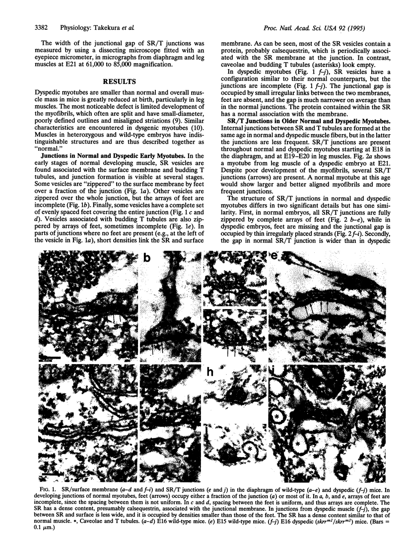
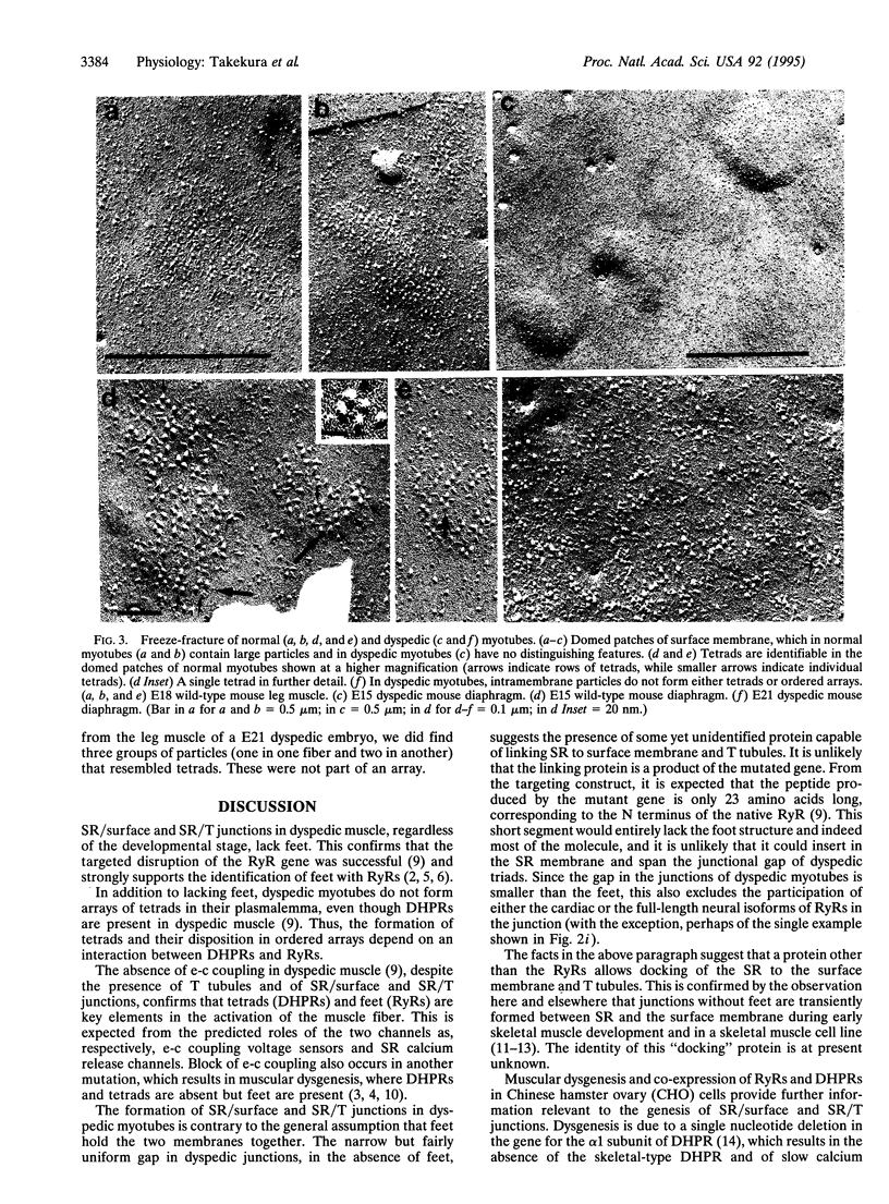
Full text
PDF


Images in this article

Selected References
These references are in PubMed. This may not be the complete list of references from this article.
- Adams B. A., Tanabe T., Mikami A., Numa S., Beam K. G. Intramembrane charge movement restored in dysgenic skeletal muscle by injection of dihydropyridine receptor cDNAs. Nature. 1990 Aug 9;346(6284):569–572. doi: 10.1038/346569a0. [DOI] [PubMed] [Google Scholar]
- Block B. A., Imagawa T., Campbell K. P., Franzini-Armstrong C. Structural evidence for direct interaction between the molecular components of the transverse tubule/sarcoplasmic reticulum junction in skeletal muscle. J Cell Biol. 1988 Dec;107(6 Pt 2):2587–2600. doi: 10.1083/jcb.107.6.2587. [DOI] [PMC free article] [PubMed] [Google Scholar]
- Chaudhari N., Beam K. G. mRNA for cardiac calcium channel is expressed during development of skeletal muscle. Dev Biol. 1993 Feb;155(2):507–515. doi: 10.1006/dbio.1993.1048. [DOI] [PubMed] [Google Scholar]
- Edge M. B. Development of apposed sarcoplasmic reticulum at the T system and sarcolemma and the change in orientation of triads in rat skeletal muscle. Dev Biol. 1970 Dec;23(4):634–650. doi: 10.1016/0012-1606(70)90144-2. [DOI] [PubMed] [Google Scholar]
- Flucher B. E., Phillips J. L., Powell J. A. Dihydropyridine receptor alpha subunits in normal and dysgenic muscle in vitro: expression of alpha 1 is required for proper targeting and distribution of alpha 2. J Cell Biol. 1991 Dec;115(5):1345–1356. doi: 10.1083/jcb.115.5.1345. [DOI] [PMC free article] [PubMed] [Google Scholar]
- Flucher B. E. Structural analysis of muscle development: transverse tubules, sarcoplasmic reticulum, and the triad. Dev Biol. 1992 Dec;154(2):245–260. doi: 10.1016/0012-1606(92)90065-o. [DOI] [PubMed] [Google Scholar]
- Franzini-Armstrong C., Jorgensen A. O. Structure and development of E-C coupling units in skeletal muscle. Annu Rev Physiol. 1994;56:509–534. doi: 10.1146/annurev.ph.56.030194.002453. [DOI] [PubMed] [Google Scholar]
- Franzini-Armstrong C., Pincon-Raymond M., Rieger F. Muscle fibers from dysgenic mouse in vivo lack a surface component of peripheral couplings. Dev Biol. 1991 Aug;146(2):364–376. doi: 10.1016/0012-1606(91)90238-x. [DOI] [PubMed] [Google Scholar]
- Inui M., Saito A., Fleischer S. Purification of the ryanodine receptor and identity with feet structures of junctional terminal cisternae of sarcoplasmic reticulum from fast skeletal muscle. J Biol Chem. 1987 Feb 5;262(4):1740–1747. [PubMed] [Google Scholar]
- Kim K. C., Caswell A. H., Talvenheimo J. A., Brandt N. R. Isolation of a terminal cisterna protein which may link the dihydropyridine receptor to the junctional foot protein in skeletal muscle. Biochemistry. 1990 Oct 2;29(39):9281–9289. doi: 10.1021/bi00491a025. [DOI] [PubMed] [Google Scholar]
- Lai F. A., Erickson H. P., Rousseau E., Liu Q. Y., Meissner G. Purification and reconstitution of the calcium release channel from skeletal muscle. Nature. 1988 Jan 28;331(6154):315–319. doi: 10.1038/331315a0. [DOI] [PubMed] [Google Scholar]
- Marks A. R., Taubman M. B., Saito A., Dai Y., Fleischer S. The ryanodine receptor/junctional channel complex is regulated by growth factors in a myogenic cell line. J Cell Biol. 1991 Jul;114(2):303–312. doi: 10.1083/jcb.114.2.303. [DOI] [PMC free article] [PubMed] [Google Scholar]
- Pinçon-Raymond M., Rieger F., Fosset M., Lazdunski M. Abnormal transverse tubule system and abnormal amount of receptors for Ca2+ channel inhibitors of the dihydropyridine family in skeletal muscle from mice with embryonic muscular dysgenesis. Dev Biol. 1985 Dec;112(2):458–466. doi: 10.1016/0012-1606(85)90418-x. [DOI] [PubMed] [Google Scholar]
- Takekura H., Bennett L., Tanabe T., Beam K. G., Franzini-Armstrong C. Restoration of junctional tetrads in dysgenic myotubes by dihydropyridine receptor cDNA. Biophys J. 1994 Aug;67(2):793–803. doi: 10.1016/S0006-3495(94)80539-9. [DOI] [PMC free article] [PubMed] [Google Scholar]
- Takekura H., Sun X., Franzini-Armstrong C. Development of the excitation-contraction coupling apparatus in skeletal muscle: peripheral and internal calcium release units are formed sequentially. J Muscle Res Cell Motil. 1994 Apr;15(2):102–118. doi: 10.1007/BF00130422. [DOI] [PubMed] [Google Scholar]
- Takeshima H., Iino M., Takekura H., Nishi M., Kuno J., Minowa O., Takano H., Noda T. Excitation-contraction uncoupling and muscular degeneration in mice lacking functional skeletal muscle ryanodine-receptor gene. Nature. 1994 Jun 16;369(6481):556–559. doi: 10.1038/369556a0. [DOI] [PubMed] [Google Scholar]
- Tanabe T., Beam K. G., Powell J. A., Numa S. Restoration of excitation-contraction coupling and slow calcium current in dysgenic muscle by dihydropyridine receptor complementary DNA. Nature. 1988 Nov 10;336(6195):134–139. doi: 10.1038/336134a0. [DOI] [PubMed] [Google Scholar]
- Yuan S. H., Arnold W., Jorgensen A. O. Biogenesis of transverse tubules and triads: immunolocalization of the 1,4-dihydropyridine receptor, TS28, and the ryanodine receptor in rabbit skeletal muscle developing in situ. J Cell Biol. 1991 Jan;112(2):289–301. doi: 10.1083/jcb.112.2.289. [DOI] [PMC free article] [PubMed] [Google Scholar]