Abstract
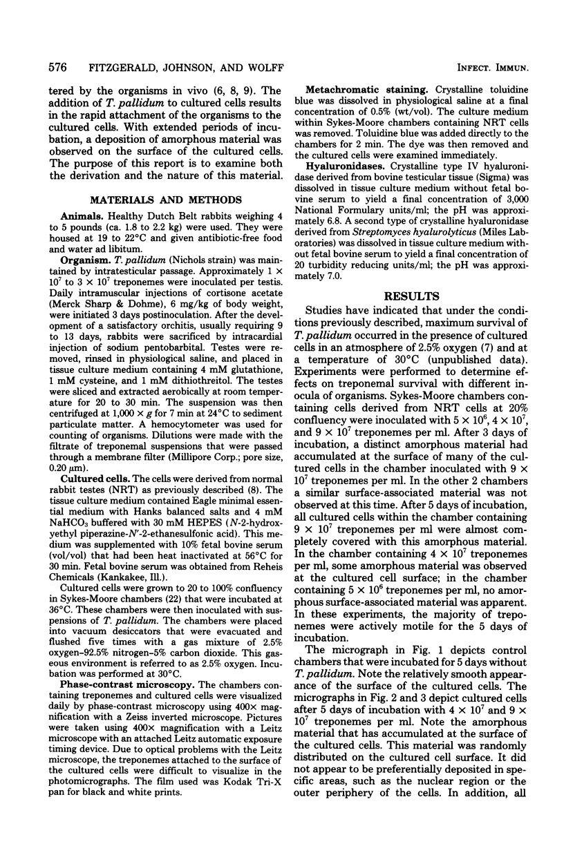
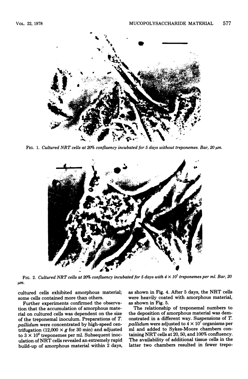
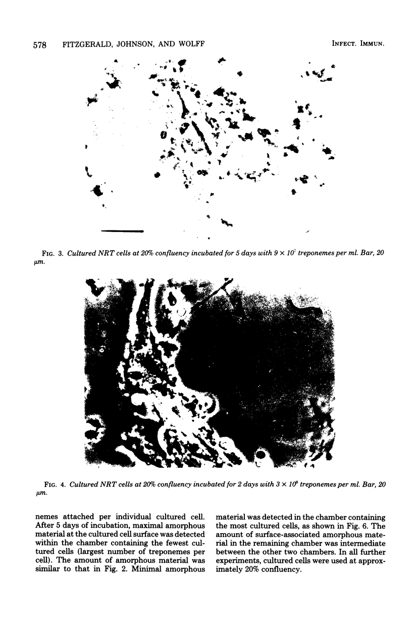
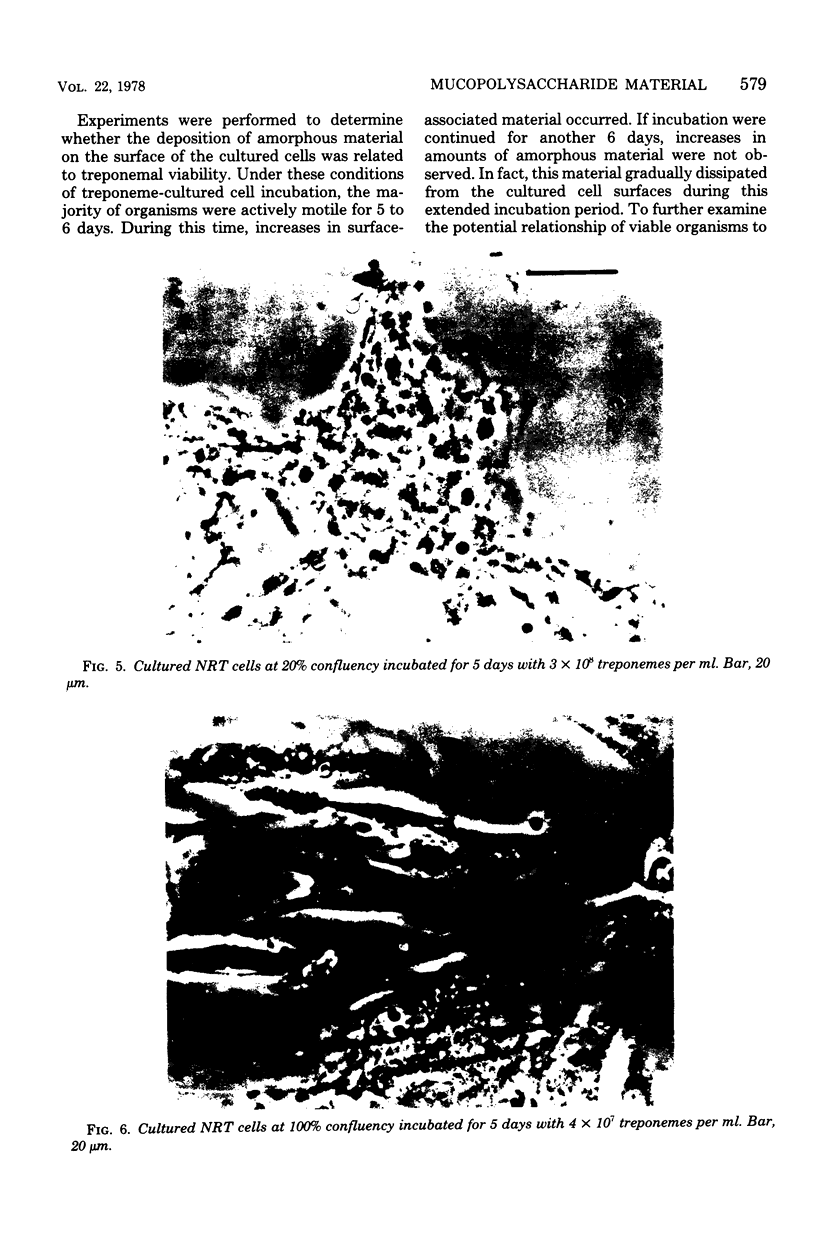
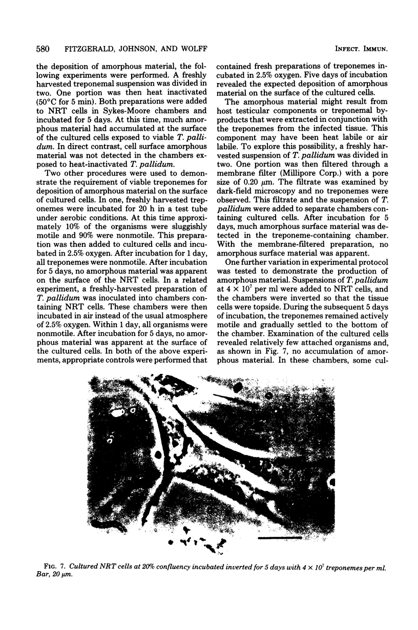
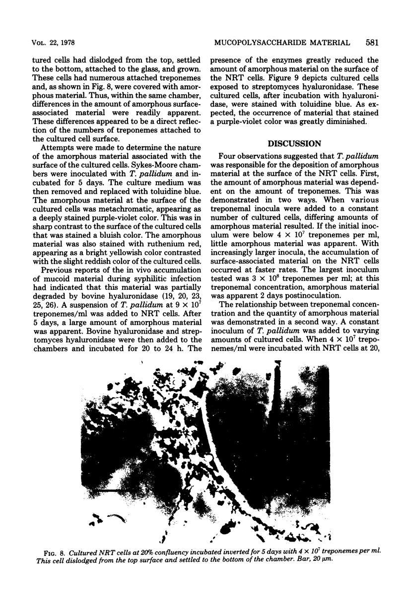
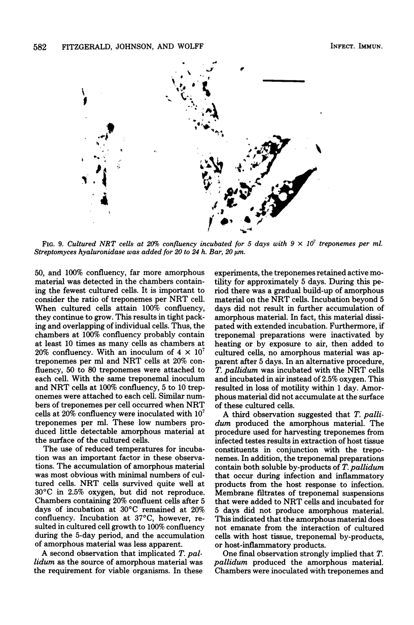
During incubation of Treponema pallidum (Nichols strain) with cultured mammlian cells derived from normal rabbit testes (NRT), an amorphous material accumulated at the surface of the cultured cells. This material was randomly distributed on all tissue cells within the culture chambers. The amount of amorphous material was dependent on the treponemal inocula. With 3 × 10⁸ organisms per ml, this material was readily apparent within 2 days; with 4 × 10⁷ organisms per ml, this material was detectable within 4 to 5 days; with lower inocula, the accumulation of amorphous material was far less apparent. Deposition of this surface-associated material required attachment of treponemes to the cultured cells, and the amount deposited was related to the number of treponemes attached per cell. This amorphous material was not detected when NRT cells were incubated with preparations of T. pallidum that were heat or air inactivated. In addition, the accumulaton of amorphous material was not due to a soluble component from host testicular tissue or to a soluble component developing during treponemal infection. This was demonstrated by the inability of membrane filtered preparations of T. pallidum to induce the deposition of amorphous material at the surface of the cultured cells. The nature of this material appeared to be acidic mucopolysaccharide as indicated by its metachromatic staining properties, its stainability with ruthenium red, and its partial degradation by bovine and streptomyces hyaluronidase. This amorphous material that accumulated in vitro at the surface of cultured cells may be similar to the mucoid material that accumulates in vivo during syphilitic infection.
Full text
PDF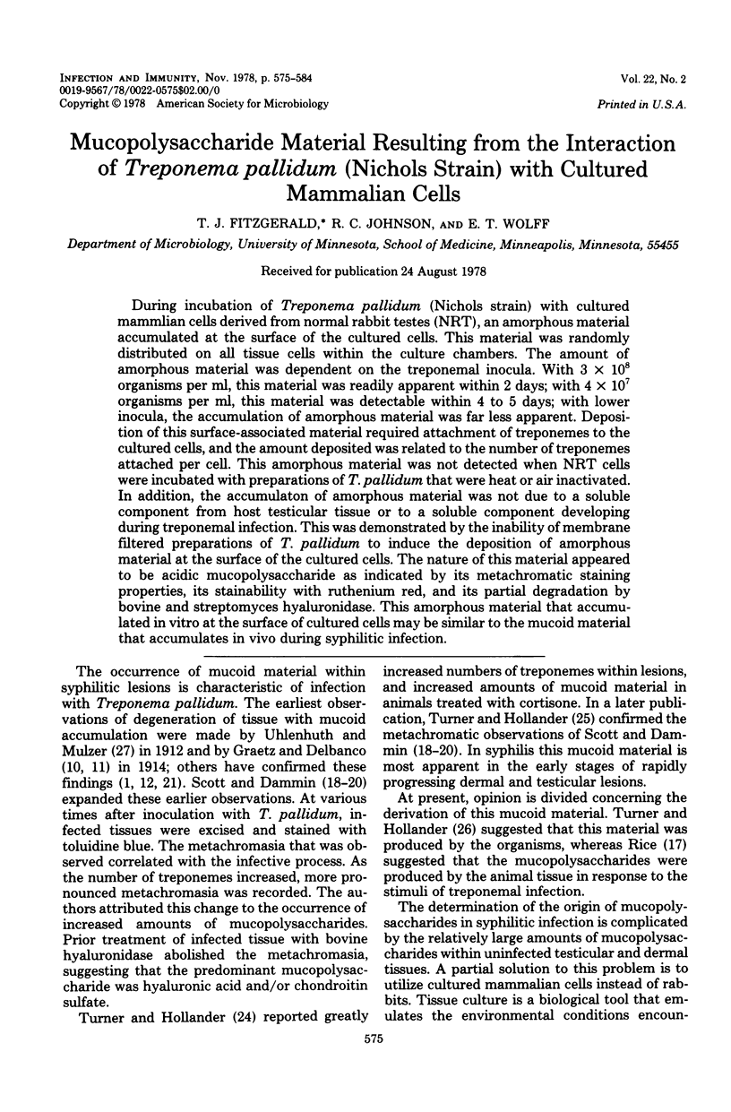
Images in this article
Selected References
These references are in PubMed. This may not be the complete list of references from this article.
- BELANGER L. F., HARTNETT A. Persistent toluidine blue metachromasia. J Histochem Cytochem. 1960 Jan;8:75–75. doi: 10.1177/8.1.75. [DOI] [PubMed] [Google Scholar]
- BELANGER L. F., MIGICOVSKY B. B. A comparison between different mucopolysacchwride stains as applied to chick epiphyseal cartilage. J Histochem Cytochem. 1961 Jan;9:73–78. doi: 10.1177/9.1.73. [DOI] [PubMed] [Google Scholar]
- BLUMENKRANTZ N. Microtest for mucopolysaccharides by means of toluidine blue: with special reference to hyaluronic acid. Clin Chem. 1957 Dec;3(6):696–702. [PubMed] [Google Scholar]
- Fitzgerald T. J., Johnson R. C., Miller J. N., Sykes J. A. Characterization of the attachment of Treponema pallidum (Nichols strain) to cultured mammalian cells and the potential relationship of attachment to pathogenicity. Infect Immun. 1977 Nov;18(2):467–478. doi: 10.1128/iai.18.2.467-478.1977. [DOI] [PMC free article] [PubMed] [Google Scholar]
- Fitzgerald T. J., Johnson R. C., Sykes J. A., Miller J. N. Interaction of Treponema pallidum (Nichols strain) with cultured mammalian cells: effects of oxygen, reducing agents, serum supplements, and different cell types. Infect Immun. 1977 Feb;15(2):444–452. doi: 10.1128/iai.15.2.444-452.1977. [DOI] [PMC free article] [PubMed] [Google Scholar]
- Fitzgerald T. J., Miller J. N., Sykes J. A. Treponema pallidum (Nichols strain) in tissue cultures: cellular attachment, entry, and survival. Infect Immun. 1975 May;11(5):1133–1140. doi: 10.1128/iai.11.5.1133-1140.1975. [DOI] [PMC free article] [PubMed] [Google Scholar]
- GROSSFELD H., MEYER K., GODMAN G., LINKER A. Mucopolysaccharides produced in tissue culture. J Biophys Biochem Cytol. 1957 May 25;3(3):391–396. doi: 10.1083/jcb.3.3.391. [DOI] [PMC free article] [PubMed] [Google Scholar]
- GROSSFELD H. Positive mucin clot test in supernates of cultures of avian embryonic brain. Proc Soc Exp Biol Med. 1957 Dec;96(3):844–846. doi: 10.3181/00379727-96-23627. [DOI] [PubMed] [Google Scholar]
- GROSSFELD H. Studies on production of hyaluronic acid in tissue culture; the presence of hyaluronidase in embryo extract. Exp Cell Res. 1958 Feb;14(1):213–216. doi: 10.1016/0014-4827(58)90231-3. [DOI] [PubMed] [Google Scholar]
- Luft J. H. Ruthenium red and violet. I. Chemistry, purification, methods of use for electron microscopy and mechanism of action. Anat Rec. 1971 Nov;171(3):347–368. doi: 10.1002/ar.1091710302. [DOI] [PubMed] [Google Scholar]
- RICE F. A. Chondroitin sulfate and hyaluronic acid in syphilomas of cortisone-treated rabbits. Science. 1956 Aug 10;124(3215):275–275. doi: 10.1126/science.124.3215.275. [DOI] [PubMed] [Google Scholar]
- SCOTT V., DAMMIN G. J. Hyaluronidase and experimental syphilis. III. Metachromasia in syphilitic orchitis and its relationship to hyaluronic acid. Am J Syph Gonorrhea Vener Dis. 1950 Nov;34(6):501–514. [PubMed] [Google Scholar]
- SCOTT V., DAMMIN G. J. Morphologic and histochemical sequences in syphilitic and in tuberculous orchitis in the rabbit. Am J Syph Gonorrhea Vener Dis. 1954 May;38(3):189–202. [PubMed] [Google Scholar]
- SYKES J. A., MOORE E. B. A simple tissue culture chamber. Tex Rep Biol Med. 1960;18:288–297. [PubMed] [Google Scholar]
- TURNER T. B., HOLLANDER D. H. Cortisone in experimental syphilis; a preliminary note. Bull Johns Hopkins Hosp. 1950 Nov;87(5):505–509. [PubMed] [Google Scholar]
- TURNER T. B., HOLLANDER D. H. Studies on the mechanism of action of cortisone in experimental syphilis. Am J Syph Gonorrhea Vener Dis. 1954 Sep;38(5):371–387. [PubMed] [Google Scholar]
- Zeigler J. A., Jones A. M., Jones R. H., Kubica K. M. Demonstration of extracellular material at the surface of pathogenic T. pallidum cells. Br J Vener Dis. 1976 Feb;52(1):1–8. doi: 10.1136/sti.52.1.1. [DOI] [PMC free article] [PubMed] [Google Scholar]


