Abstract
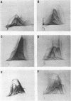
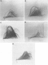
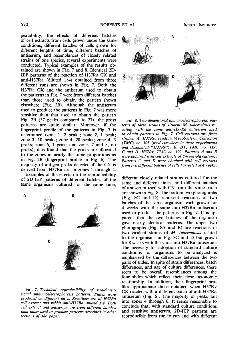
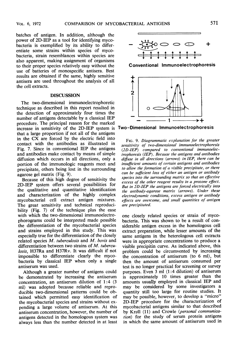
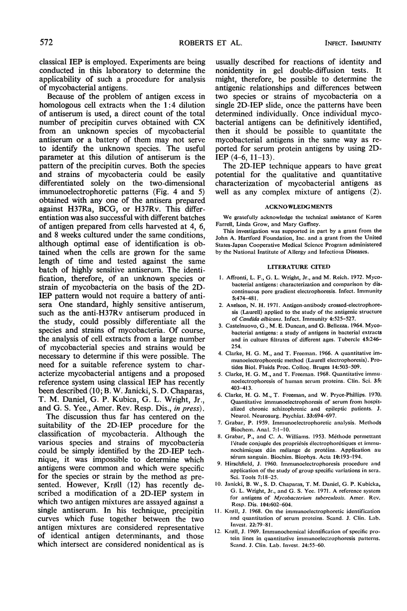
Two-dimensional immunoelectrophoresis (2D-IEP), in which a complex of antigens is subjected to electrophoresis first through an agarose matrix in one direction and secondly through an antiserum-agarose matrix at right angles to the first direction, was evaluated as a tool for analysis of mycobacterial antigens. Cell extracts from four species of mycobacteria, Mycobacterium tuberculosis (four strains), M. bovis strain BCG, M. scrofulaceum, and M. phlei, were assayed by 2D-IEP with four anti-mycobacterial antisera. Besides displaying the precipitin curves in a more easily interpreted format than did conventional immunoelectrophoresis (IEP), 2D-IEP offered greater sensitivity in terms of numbers of precipitin curves when like reactions were compared with IEP patterns. As many as 60 immunoprecipitates were observed on 2D-IEP slides compared to 18 on comparable IEP plates. Technical reproducibility of patterns from run to run was excellent. Other parameters, such as the influence of using different batches of antigen on the pattern, are discussed. Each of the cell extract antigens gave a unique pattern of precipitin peaks which could be easily differentiated from the patterns given by the other mycobacterial cell extracts when reacted with any of the antisera in 2D-IEP. Since both the species and strains of mycobacteria could be easily and reproducibly differentiated solely on the basis of two-dimensional immunoelectrophoretic patterns obtained with any of the antisera employed in this study, it may be possible, by using IEP, to differentiate and identify all species and strains of mycobacteria with one standard, highly sensitive antiserum, rather than a battery of antisera.
Full text
PDF

Images in this article
Selected References
These references are in PubMed. This may not be the complete list of references from this article.
- Affronti L. F., Wright G. L., Jr, Reich M. Characterization and comparison of mycobacterial antigens by discontinuous pore gradient gel electrophoresis. Infect Immun. 1972 Apr;5(4):474–481. doi: 10.1128/iai.5.4.474-481.1972. [DOI] [PMC free article] [PubMed] [Google Scholar]
- Axelsen N. H. Antigen-antibody crossed electrophoresis (Laurell) applied to the study of the antigenic structure of Candida albicans. Infect Immun. 1971 Nov;4(5):525–527. doi: 10.1128/iai.4.5.525-527.1971. [DOI] [PMC free article] [PubMed] [Google Scholar]
- CASTELNUOVO G., DUNCAN M. E., BELLEZZA G. MYCOBACTERIAL ANTIGENS: A STUDY OF ANTIGENS IN BACTERIAL EXTRACTS AND IN CULTURE FILTRATES OF DIFFERENT AGES. Tubercle. 1964 Sep;45:246–254. doi: 10.1016/s0041-3879(64)80015-5. [DOI] [PubMed] [Google Scholar]
- Clarke H. G., Freeman T., Pryse-Phillips W. Quantitative immunoelectrophoresis of serum from hospitalized chronic schizophrenic and epileptic patients. J Neurol Neurosurg Psychiatry. 1970 Oct;33(5):694–697. doi: 10.1136/jnnp.33.5.694. [DOI] [PMC free article] [PubMed] [Google Scholar]
- Clarke H. G., Freeman T. Quantitative immunoelectrophoresis of human serum proteins. Clin Sci. 1968 Oct;35(2):403–413. [PubMed] [Google Scholar]
- GRABAR P., WILLIAMS C. A. Méthode permettant l'étude conjuguée des proprietés électrophorétiques et immunochimiques d'un mélange de protéines; application au sérum sanguin. Biochim Biophys Acta. 1953 Jan;10(1):193–194. doi: 10.1016/0006-3002(53)90233-9. [DOI] [PubMed] [Google Scholar]
- Janicki B. W., Chaparas S. D., Daniel T. M., Kubica G. P., Wright G. L., Yee G. S. A reference system for antigens of Mycobacterium tuberculosis. Am Rev Respir Dis. 1971 Oct;104(4):602–604. doi: 10.1164/arrd.1971.104.4.602. [DOI] [PubMed] [Google Scholar]
- Kroll J. On the immunoelectrophoretical identification and quantitation of serum proteins. Scand J Clin Lab Invest. 1968;22(1):79–81. doi: 10.3109/00365516809160742. [DOI] [PubMed] [Google Scholar]
- Kröll J. Immunochemical identification of specific precipitin lines in quantitative immunoelectrophoresis patterns. Scand J Clin Lab Invest. 1969 Aug;24(1):55–60. doi: 10.3109/00365516909080132. [DOI] [PubMed] [Google Scholar]
- LAURELL C. B. ANTIGEN-ANTIBODY CROSSED ELECTROPHORESIS. Anal Biochem. 1965 Feb;10:358–361. doi: 10.1016/0003-2697(65)90278-2. [DOI] [PubMed] [Google Scholar]
- LOWRY O. H., ROSEBROUGH N. J., FARR A. L., RANDALL R. J. Protein measurement with the Folin phenol reagent. J Biol Chem. 1951 Nov;193(1):265–275. [PubMed] [Google Scholar]
- PARLETT R. C., YOUMANS G. P. Antigenic relationships between ninety-eight strains of mycobacteria using gel-diffusion precipitation techniques. Am Rev Tuberc. 1958 Mar;77(3):450–461. doi: 10.1164/artpd.1958.77.3.450. [DOI] [PubMed] [Google Scholar]
- Pickett M. J., Pedersen M. M., Goldstein D. I., Froman S. The antigens of Mycobacterium tuberculosis. Protein fractions from cytoplasm and culture filtrate. Am Rev Respir Dis. 1968 Mar;97(3):415–422. doi: 10.1164/arrd.1968.97.3.415. [DOI] [PubMed] [Google Scholar]
- RESSLER N. Electrophoresis of serum protein antigens in an antibody-containing buffer. Clin Chim Acta. 1960 May;5:359–365. doi: 10.1016/0009-8981(60)90140-6. [DOI] [PubMed] [Google Scholar]


