Abstract
Heart failure with preserved ejection fraction (HFpEF) has recently emerged as a major cause of cardiovascular morbidity and mortality. Contrary to initial beliefs, HFpEF is now known to be as common as heart failure with reduced ejection fraction (HFrEF) and carries an unacceptably high mortality rate. With a prevalence that has been steadily rising over the past two decades, it is very likely that HFpEF will represent the dominant heart failure phenotype over the coming few years. The scarcity of trials in this semi-discrete form of heart failure and lack of unified enrolment criteria in the studies conducted to date might have contributed to the current absence of specific therapies. Understanding the epidemiological, pathophysiological and molecular differences (and similarities) between these two forms of heart failure is cornerstone to the development of targeted therapies. Carefully designed studies that adhere to unified diagnostic criteria with the recruitment of appropriate controls and adoption of practical end-points are urgently needed to help identify effective treatment strategies.
Keywords: heart failure, heart failure with preserved ejection fraction, diastolic heart failure
Introduction
The rate of relaxation of the heart is quite as important as the systolic contraction. If an old man's heart relaxes slowly, his capacity for physical exertion is thus limited (Yandell Henderson, 1923).
Heart failure is a complex clinical syndrome characterized by a constellation of signs and symptoms involving various organ systems. Structural and/or functional cardiac abnormalities are cornerstone to the pathophysiology of heart failure; however, extracardiac dysfunction plays an equally important role in its development and progression. It is now well-recognized that two semi-discrete forms of heart failure exist; heart failure with reduced ejection fraction (HFrEF) and heart failure with preserved ejection fraction (HFpEF) (previously referred to as systolic heart failure and diastolic heart failure respectively). With diastole occupying approximately 65% of the cardiac cycle and despite Henderson's remarkably accurate observation 90 years ago, it is surprising that the latter form has only gained attention in the last 20 years.
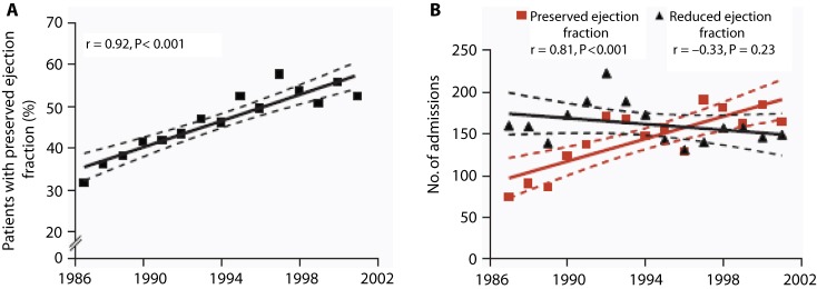
Over the past decennium, multiple features and epidemiological trends of HFpEF were uncovered prompting progressively increasing interest and research in this field. Contrary to initial beliefs, current data support that HFpEF is as common as HFrEF, representing approximately 50% of the world's heart failure population, and carries an almost similar dismal prognosis [1–3]. In contrast to HFrEF, the prevalence of HFpEF is steadily rising at an alarming rate with little improvement in outcome [ Fig. 1] [4]. Therapies of proven benefit in HFrEF have repeatedly been shown to add little if any benefit in HFpEF [5,6]. With the current increases in global life expectancy and significant advances in diagnosis, treatment and secondary prevention of almost all cardiovascular diseases, it is expected that the prevalence of heart failure will continue to rise reaching epidemic levels in both developed and developing countries [7–9]. HFpEF will likely represent the dominant heart failure phenotype in this epidemic over the next decade.
Figure 1. Secular trends in the prevalence of HFpEF. Data collected from the Mayo Clinic for 4596 patients discharged with a diagnosis of heart failure over a 15-year period (1987–2001). Panel A shows a steady increase in the percentage of patients with heart failure who had preserved ejection fraction (>50%) over the study period. Panel B shows the number of admissions for heart failure in patients with preserved ejection fraction (solid red line) and with reduced ejection fraction (solid black line). The dashed lines represent the 95% confidence intervals. From Theophilus E, et al. [4] with permission.
This review aims to address the current knowledge in this field including current understanding of the pathophysiology, diagnostic and therapeutic strategies as well as potential future directions and uncertainties that need to be addressed.
Terminology
Nomenclature for this syndrome has changed more than once over the past two decades reflecting initial uncertainties, progressively accumulating knowledge and, more recently, better understanding of the condition. Recent appreciation that diastolic dysfunction is not the only underlying abnormality in this syndrome has prompted a transition from a term that implies a single operating pathophysiology – diastolic heart failure (or primary diastolic heart failure) [10] – to a more observational term – heart failure with preserved ejection fraction (HFpEF). Moreover, diastolic dysfunction is not unique to HFpEF; it is also encountered in almost all patients with HFrEF [11,12]. A less popular term – heart failure with preserved systolic function - has also been abandoned after various studies showed that systolic function (assessed by parameters thought to be more sensitive than ejection fraction) is impaired in this patient population [13–18].
Perhaps the best feature of the current nomenclature – HFpEF – is being simply descriptive of the phenotype thus offering considerable flexibility and ability to accommodate evolving knowledge about the complex pathophysiology of the disease. The term however has its own limitations as the word “preserved” implies knowledge of pre-existing ejection fraction which is almost always not the case. In addition, the precise range of a “normal” ejection fraction (EF) is difficult to define [19,20].
Epidemiology
Given the data showing a significant increase in the number of heart failure related hospitalizations, it can be regarded as an emerging epidemic [7,21,22]. Heart failure continues to be the most common cause of hospitalization in patients aged 65 and above, accounting for more than one million hospitalizations in the United States alone. Recently, consistent figures regarding the prevalence of HFpEF became available despite different definitions, diagnostic modalities and criteria used in various studies.
Data from the Mayo Clinic registry revealed that 47% of 4596 consecutive patients admitted over a period of 15 years with a diagnosis of heart failure had a normal left ventricular ejection fraction (defined as LVEF ≥ 50%). HFpEF patients were older, more likely to be women, with a higher prevalence of hypertension, obesity, atrial fibrillation and anemia compared to patients with HFrEF. Both coronary artery disease and history of previous myocardial infarction were less frequently found in patients with HfpEF compared to patients with HFrEF [4]. Table 1 outlines the epidemiological differences between HFpEF and HFrEF including associated comorbidities. Other epidemiological studies conducted over the past 20 years have shown comparable results with the prevalence of HFpEF ranging from 40%–71% (mean 56%). Results of individual studies should be interpreted with caution as the diagnostic criteria for diastolic heart failure (the widely used term then) varied substantially, with a considerable number not requiring any objective evidence of LV diastolic dysfunction for establishing the diagnosis. The prevalence of HFpEF has consistently been shown to be higher in community-based compared to hospital-based studies; [3,4] an observation which can be in part explained by the fact that patients with a normal EF are less likely to be hospitalized. Underdiagnosis can also contribute to this finding owing to the limited sensitivity of symptoms and signs of heart failure. Furthermore, comorbidities such as obesity and pulmonary disease frequently encountered in hospitalized patients, particularly the elderly, can indeed make the diagnosis more challenging. The prevalence of HFpEF has been rising steadily over the past two decades at a rate of ≈1% per year. This is in contrast to the prevalence of HFrEF which has remained unchanged over the same period [23]. This alarming trend can be explained by a number of factors including an ageing population, increased prevalence of comorbidities (hypertension and obesity), growing awareness of HFpEF among physicians and other healthcare professionals, improved sensitivity of diagnostic tools (including tissue-Doppler imaging, deformation analysis parameters and natriuretic peptides) and the development of clear diagnostic algorithms in recently published guidelines.
Table 1. Comparison between epidemiological characteristics of HFpEF and HFrEF.
| Characteristics | HFpEF | HFrEF |
| Age | Older | Youger |
| Gender | Females > males | Males > females |
| Hypertension | +++ | ++ |
| Diabetes | +++ | ++ |
| CAD or previous MI | +/++ | +++ |
| Renal failure | ++ | + |
| Obesity | ++ | + |
| Atrial fibrillation | ++ | + |
| Chronic lung disease | ++ | − |
CAD, coronary artery disease; MI, myocardial infarction.
HFpEF has classically been considered as a relatively benign condition compared to HFrEF primarily based on a commonly held belief that mortality is inversely proportional to LV systolic function – namely LVEF – in the broad spectrum of heart failure patients [24,25]. However, over the past decade, we have come to realize that this was not entirely correct; the prognosis of HFpEF reported by two large community-based studies was as ominous as that of HFrEF with 5-year mortality ranging from 54% to 65% [1,26]. Both community-based studies suffered from defects inherent to their retrospective nature and the absence of baseline EF data in a large number of patients. On the other hand, two recent meta-analyses [27,28] found that HFpEF mortality is significantly less than that of HFrEF. In turn, these meta-analyses also suffered from a number of limitations including those of the original studies, selection bias, exclusion of a large number of patients due to incomplete data (or not meeting other inclusion criteria) and marked heterogeneity of patient populations included. Collectively these limitations might have led to an “artificial” population that does not accurately reflect the real-world HFpEF population (e.g. relatively young population with male predominance in the meta-analysis by Somaratne JB, et al. and exclusion of the I-PRESERVE patient population in the meta-analysis by Doughty RN, et al.) [27,28]. Further studies are required to determine the true prognosis of this condition in comparison to HFrEF, however, the mortality of HFpEF remains unacceptably high [29]. Furthermore, outpatient clinic consultations, rate of rehospitalization, length of hospital stay and subsequent healthcare costs are similar to those encountered in patients with HFrEF [1,26,30–33].
Pathophysiology
LV diastolic dysfunction – namely impaired myocardial relaxation and increased stiffness – is the hallmark of HFpEF [34–41], however, it is not the only underlying abnormality. Other factors – both cardiac and extracardiac – including increased arterial stiffness, altered ventricular-arterial coupling [42,43], endothelial dysfunction, reduced vasodilator reserve [44,45] and chronotropic incompetence [46,47] have been recently implicated in the pathophysiology of this complex syndrome. Furthermore, coexisting abnormalities in LV regional systolic function assessed by various parameters have been documented in HFpEF [13,15,17,18,48–50].
LV Diastolic Dysfunction
LV diastolic function is dependent on four major factors; myocardial relaxation, left ventricular stiffness/compliance, left atrial function and heart rate.
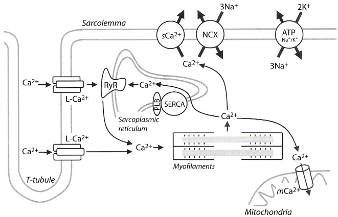
Myocardial relaxation is an active ATP-dependent non-uniform process that results from Ca2+ extrusion from the cytosol in order to achieve its diastolic levels essentially through phospholamban-modulated uptake of Ca2+ via the sarcoplasmic endoplasmic reticulum Ca2+ (SERCA-2a) and Ca2+ extrusion via the Na+/Ca2+ exchanger [ Fig. 2] [51]. The end result is cross-bridge detachment i.e. breaking the link between the actin molecule and myosin head. In HFpEF, SERCA-2a activity declines owing to reduced gene and protein expression and by reduced phosphorylation of its inhibitory modulating protein – phospholamban – resulting in reduced sarcoplasmic reticulum (SR) Ca2+ content [52–55]. This has the dual effect of incomplete removal of Ca2+ from the cytosol and diminished SR Ca2+ content available for the ensuing systole. Furthermore, hyperphosphorylation of the ryanodine receptor (RyR) has been observed in animal models and failing human hearts. This may result in diastolic Ca2+ leak (calcium sparks) into the cytosol, which in turn causes incomplete/delayed cross-bridge detachment and subsequently delayed relaxation, but to what extent this really occurs in heart failure is still controversial [56,57]. T-tubule disorganization has also been demonstrated in various human studies and animal models [58–60]. Recent evidence suggests that t-tubules might have a significant role in Ca2+ extrusion from the cell, however, the exact role of t-tubule dysfunction in HFpEF requires further studies [61–63].
Figure 2. Excitation–contraction and inactivation–relaxation coupling in cardiomyocytes. Cardiomyocyte depolarization promotes Ca2+ entry through the sarcolemmal L-type Ca2+ channels (L–Ca2+), leading to Ca2+ release for the sarcoplasmic reticulum (SR) through ryanodine receptors (RyR), thereby inducing contraction. During relaxation the four pathways involved in calcium removal from the cytosol are phospholamban (PLB)-modulated uptake of Ca2+ into the sarcoplasmic reticulum by a Ca2+-ATPase (SERCA), Ca2+ extrusion via the sodium-calcium exchanger (NCX), mitochondrial Ca2+ uniport and sarcolemmal Ca2+-ATPase, with the latter two being responsible for only about 1% of the total. From Roncon-Albuquerque R Jr et al. [48] with permission.
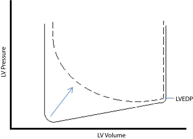
Collectively, these microstructural changes result in a depressed rate of LV pressure decay during the isovolumic relaxation phase leading to a prolonged time constant of LV relaxation (tau). The LV end-diastolic pressure (LVEDP), however, remains normal or is slightly elevated [Fig. 3]. Additionally, cross-bridge detachment can be disturbed by perturbations in loading conditions with enhanced preload delaying the onset and rate of relaxation [64]. On the other hand, effects of afterload on timing and rate of relaxation are complex and dependent on timing of load with afterload increases occurring late in systole having a more pronounced effect on the rate of relaxation [65,66].
Figure 3. Left ventricular pressure-volume loop in ventricles with delayed relaxation. Only the bottom half of the pressure-volume loop is shown. The rate of LV pressure decline is much slower in patients with a delayed relaxation pattern of diastolic filling (dashed line) compared to normal subjects (solid line). Left ventricular end-diastolic pressure (LVEDP) remains normal or slightly elevated.
LV stiffness is the main – but not the only – determinant of passive filling of the ventricle which in turn governs the late diastolic pressure-volume relationship of the chamber [64]. Stiffness is a physical property of the myocardium that is determined by its viscoelastic forces which are now thought to reside mostly in the macromolecule titin [67]. Titin functions as a bidirectional spring that enhances early diastolic LV recoil and late diastolic resistance to stretch [68,69]. It exists in two isoforms – N2B and N2BA – which differ substantially in length and stiffness, with the N2B isoform being smaller and stiffer. Increased expression of the N2B isoform has been demonstrated in hypertensive rats along with increased diastolic muscle stiffness [70], whereas N2BA is the predominant form in end-stage human ischemic/dilated cardiomyopathy [71]. Recent data suggest that aberrant mRNA splicing secondary to mutations in components of the cardiac splicing machinery is responsible for the expression of different titin isoforms in hereditary cardiomyopathies [72,73]. However, many questions about the relationship between titin and diastolic function remain unanswered including other mechanisms (and triggers) of isoform switching, relevance to human disease and the importance of other modifications of titin besides switching.
Changes in the structure of the myocardial extracellular matrix (ECM) – namely fibrillar proteins, proteoglycans and basement membrane proteins – also affect the viscoelastic properties of the myocardium. The most important components within the ECM that contribute to the development of diastolic dysfunction are fibrillar collagen, degree of cross-linking and ratio of collagen type I to III (with type I being more abundant in stiffer ventricles) [74]. Collagen synthesis is altered by both transcriptional and post-transcriptional regulation, neurohormonal activation (renin-angiotensin-aldosterone system), growth factors and cross-linking. Collagen degradation is under control of proteolytic enzymes, including matrix metalloproteinases (MMPs) and tissue inhibitors of matric metalloproteinases (TIMPs). Hypertensive patients and patients with aortic stenosis have decreased matrix degradation because of downregulation of MMPs and upregulation of TIMPs [75,76]. Plasma levels of TIMP-1 have recently been proposed as a potential biomarker of development of HFpEF in hypertensive patients [77].
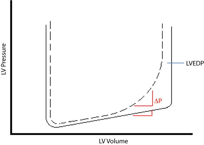
Increased myocardial stiffness – which describes changes in LV pressure in relation to changes in volume (dP/dV) – leads to an increase in the slope of the curvilinear diastolic pressure volume relationship expressed as the constant of LV passive stiffness (b) which ultimately raises the LVEDP. It is worth mentioning that the relatively small LV cavity (compared to HFrEF) which is a characteristic feature of HFpEF shifts the whole curve to the left thus leading to a more pronounced effect on the LVEDP with minor changes in the intravascular volume [ Fig. 4].
Figure 4. Left ventricular pressure-volume loop in ventricles with increased stiffness. Only the bottom half of the pressure-volume loop is shown. The slope of the late diastolic pressure volume relation in patients with stiff ventricles (dashed line) is increased leading to an exaggerated response in intraventricular pressure () to any given change in volume compared to normal subjects (solid line). Consequently, left ventricular end-diastolic pressure (LVEDP) is markedly elevated.
The left atrium (LA) acts as a reservoir of blood (mediated by both active and passive relaxation), as a passive conduit during early left ventricular filling and as an active pump at end-diastole. Atrial function therefore depends on its compliance, preload, afterload and intrinsic contractility [78,79]. In young and healthy normal individuals, the atrial contribution is less than 20% of the total stroke volume, whereas in older normal individuals, the atrial “kick” accounts for a greater proportion of the total LV filling. The pump phase of atrial systole is responsible for the fast ventricular filling at the end of diastole. In normal subjects and in patients with mild degrees of diastolic dysfunction, the pump function is closely related to the precontraction volume in accordance with the Starling mechanism. However, by end-stage ventricular diastolic dysfunction – when the limits of atrial preload are reached – the atrial pump function is blunted. This is further promoted by intrinsic depression of atrial contractility, afterload mismatch (due to high ventricular filling resistance) and the development of atrial arrhythmias or atrioventricular conduction disturbance. Episodes of atrial fibrillation (AF), even if very brief, may cause serious impairment of atrial contractility probably due to AF-induced Ca2+ overload and decreased release of Ca2+ from the sarcoplasmic reticulum. Additionally, the high cytosolic Ca2+ concentration and the “rigid” conformation of the extracellular matrix result in enhanced atrial stiffness [80]. Consequently, in the late stages of diastolic dysfunction, the atrium loses much of its reservoir and contractile capacities and predominantly behaves as a mere conduit [81]. LA stiffness has been found to correlate with pulmonary arterial pressures and has been recently proposed as an accurate measure in differentiating HFpEF from asymptomatic diastolic dysfunction [82].
Heart rate bears directly on cardiac output in diastolic dysfunction. As heart rate increases, the diastolic filling period preferentially decreases with respect to the systolic ejection period. As ventricular filling is functionally delayed, adequacy of inflow deteriorates and cardiac output paradoxically falls. However, in advanced degrees of diastolic dysfunction where most of ventricular filling occurs during early diastole, relatively rapid heart rates may help preserve the cardiac output [83].
Venrticular-arterial coupling and endothelial dysfunction
LV diastolic dysfunction is a common finding in the general population (present in up to 25%) and is particularly common in the elderly [84]. On the other hand, the prevalence of heart failure is much less common [85]. Several studies have therefore tried to identify other mechanisms – beyond diastolic dysfunction – that may contribute to the pathophysiology of HFpEF and in part explain why a limited number of individuals with diastolic dysfunction develop heart failure while others remain asymptomatic.
Arterial stiffness increases in the elderly, hypertension and diabetes mellitus. More recently, arterial stiffness was shown to be abnormally elevated in HFpEF with a strong impact on exercise capacity in this patient population [86,87]. Kawaguchi et al. [42] showed that both arterial elastance and LV end-systolic elastance are elevated in HFpEF. Borlaug et al. [88] used an integrated measure to evaluate global arterial stiffness – the effective arterial elastance calculated as the ratio of LV end-systolic pressure/stroke volume – and found it to be significantly elevated in HFpEF patients. Combined ventricular and arterial stiffening leads to a system that is vulnerable to minor changes in preload or afterload with exaggerated swings in blood pressure [88]. These effects are further amplified during exercise where increased preload, further elevations in filling pressures and impaired reduction of afterload (due to a blunted vasodilator response to exercise) collectively contribute to exercise intolerance [44,45,47]. Exercise-induced elevation of pulmonary artery pressure and the ensuing elevation of right-sided pressures may contribute to poor functional capacity secondary to ventricular interdependence and increased pericardial constraint [43,89,90]. Lam et al. [91] have recently shown that pulmonary hypertension may be present in up to 83% of HFpEF patients and found strong association between the severity of pulmonary hypertension and mortality. The same study also showed that pulmonary venous hypertension does not fully account for the severity of pulmonary hypertension observed in HFpEF and suggested that an element of “reactive” pulmonary arterial hypertension co-exists and may contribute to the pathophysiology of the disease [91].
Global cardiovascular reserve dysfunction
Similar to patients with HFrEF, most patients with HFpEF are asymptomatic at rest and become symptomatic only with exertion. In addition to unmasking symptoms, exercise amplifies subtle pathophysiological abnormalities that may be undetectable at rest and has greatly enhanced our understanding of this syndrome. Exercise entails increased cardiac output through integrated enhancements in venous return, lusitropy, contractility, heart rate and peripheral vasodilatation. Abnormalities in each of these components have been identified in HFpEF with blunted exercise-induced increases in preload, blunted increases in ejection fraction, contractility and regional systolic function, impaired chronotropic reserve (i.e. chronotropic incompetence) and attenuated exercise-mediated vasodilatation being documented in several studies [15,16,18,41,42,44–47,50,64,92–95]. Recent studies have also reported increased prevalence of diastolic dyssynchrony in patients with HFpEF [96–98]. The exact role of diastolic dyssynchrony in the pathophysiology of the disease and its relevance to exercise intolerance is not fully understood and warrants further studies.
Systolic function in HF pEF
HFpEF has classically been perceived as a distinct entity from HFrEF based on normal non-invasive and invasive measures of global LV systolic function (EF, stroke volume, peak dp/dt and end-systolic pressure-volume relationship) [14]. However, while some of these indices are relatively accurate measures of global contractility, they merely reflect the function of the heart as a “hemodynamic pump”. Prudent evaluation of cardiac function should include, in addition, properties of a “muscle” with sophisticated architecture that permits it to eject (and receive) blood using an integrated mechanism of longitudinal, radial and circumferential deformation in addition to twisting (torsion) [99]. Recent echo-Doppler analysis revealed that substantial systolic dysfunction of the ventricular muscular pump may be already present when mean overall hemodynamic pump performance including the EF is still well preserved. The long-axis function of the left ventricle (mitral annular plane systolic excursion) is particularly affected at an early stage given the longitudinal arrangement of the subendocardial fibers rendering them vulnerable to ischemia induced by high left ventricular end-diastolic pressures [11]. Multiple studies have elegantly demonstrated impaired torsion, longitudinal and radial systolic function in patients with HFpEF both at rest and during exercise [13,15–18,49,50,95,100,101].
Systole and diastole are closely linked events with systolic performance being dependent on diastole (preload) and vice-versa (diastole being sensitive to afterload changes as described earlier). It is therefore too simplistic to separate heart failure into two distinct entities. It remains to be fully understood why some patients preferentially progress towards a phenotype with eccentric LV remodeling and predominantly impaired systolic function while others progress towards another with concentric LV remodeling and predominantly impaired diastolic function. The exact role of certain “disease modifiers” including gender, age, blood pressure and diabetes in determining a specific phenotype needs further studies.
Diagnosis
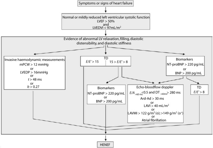
Four sets of guidelines have been published so far for the diagnosis of HFpEF [10,102–104]. All require the presence of symptoms and/or signs of heart failure, normal ejection fraction and evidence of LV diastolic dysfunction. The latest set of guidelines was published in 2007 by the Heart Failure and Echocardiography Associations of the European Society of Cardiology (ESC) and was the first to include tissue Doppler imaging (TDI) and brain natriuretic peptides (BNP) in the diagnostic criteria [104]. Fig. 5 illustrates the diagnostic algorithm recommended therein. Three criteria need to be fulfilled for the diagnosis of HFpEF to be established: 1) symptoms and/or signs of heart failure; 2) LVEF ≥ 50% in the absence of significant LV dilatation [left ventricular end-diastolic volume (LVEDVI) < 97 ml/m2]; and 3) evidence of diastolic dysfunction. The latter can be obtained via three routes: 1) invasive measurement of LVEDP, pulmonary capillary wedge pressure (PCWP), time constant of LV relaxation (tau) or LV stiffness modulus (b); 2) unequivocal tissue-Doppler findings; or 3) a combination of elevated natriuretic peptides and echocardiographic indices of LV diastolic dysfunction.
Figure 5. HFpEF Diagnostic Algorithm. LVEDVI, left ventricular end-diastolic volume index; mPCW, mean pulmonary capillary wedge pressure; LVEDP, left ventricular end-diastolic pressure; t, time constant of left ventricular relaxation; b, constant of left ventricular chamber stiffness; TD, tissue Doppler; E, early mitral valve flow velocity; E0, early TD lengthening velocity; NT-proBNP, N-terminal-pro brain natriuretic peptide; BNP, brain natriuretic peptide; E/A, ratio of early (E) to late (A) mitral valve flow velocity; DT, deceleration time; LVMI, left ventricular mass index; LAVI, left atrial volume index; Ard, duration of reverse pulmonary vein atrial systole flow; Ad, duration of mitral valve atrial wave flow. From Paulus et al. [96] with permission from Oxford press.
Invasive measurements: A time constant of LV relaxation (tau) > 48 ms or a diastolic LV stiffness modulus (b) > 0.27 is considered sufficient evidence of LV diastolic dysfunction. These two parameters require sophisticated calculations and special equipment which are usually not performed and/or available in routine clinical practice. Other accepted invasive measures include an elevated LVEDP (> 16 mmHg) or elevated mean PCWP (> 12 mmHg) which can be easily acquired during left or right heart catheterization respectively. Patients with symptoms and signs of heart failure with PCWPs of 13–15 mmHg and elevated pulmonary artery pressures may pose a diagnostic challenge as they do not fulfill the ESC definition of pulmonary hypertension due to left heart disease where a cut-off value of 15 mmHg to differentiate between pre- and post-capillary pulmonary hypertension was used [105]. Accordingly, using the current definitions, heart failure symptoms and signs may be attributed to either HFpEF or pulmonary arterial hypertension in this subgroup of patients; an overlap that can be an important source of confusion.
Echocardiographic measurements: Echocardiography has evolved as a reliable and practical tool for evaluating diastolic function. Recent appreciation that conventional Doppler evaluation of mitral and pulmonary venous flow patterns have significant limitations [106] [ Table 2] has prompted the introduction of a more sensitive parameter – peak early diastolic transmitral velocity to peak early diastolic mitral annulus velocity (E/E′) ratio – that has been shown to reliably reflect elevated LV filling pressures (when > 15) [107].
Table 2. Limitations of conventional Doppler indices in the assessment of diastolic function.
| Parameter | Limitations |
| Transmitral PW inflow pattern | - Preload dependent - Influenced by PW sample position - Difficult to analyze in AF and high heart rates - Influenced by age |
| Isovolumic relaxation time | - Technical difficulty in obtaining simultaneous tracings of transmitral inflow and LV outflow - Preload dependent - Low reproducibility |
| Pulmonary vein flow (Ard–Ad) | - Technically difficult to obtain in some patients - Cannot be used in atrial fibrillation - Dependent on heart rate |
| Mitral inflow propagation velocities | - Dependent on preload and LV cavity size - Low reproducibility |
Ard, atrial reversal of pulmonary venous flow duration; Ad, late diastolic transmitral flow duration; PW, pulsed wave
Accordingly, the latest guidelines consider an E/E′ ratio of more than 15 as sufficient independent evidence of diastolic dysfunction. An E/E′ratio between 8 and 15 is however associated with a very wide range of LVEDP [107] and hence, further parameters are required to establish the diagnosis in these patients. These parameters include one of the following: 1) ratio of peak early (E) to peak late (A) diastolic transmitral velocity < 0.5 and deceleration time (DT) > 280 ms; 2) difference between durations of atrial reversal of pulmonary venous flow (Ard) and late diastolic transmitral flow (Ad) > 30 ms; 3) left atrial volume index (LAVI) > 40 ml/m2; 4) left ventricular mass index (LVMI) > 122 g/m2 in females or 149 g/m2 in males; or 5) electrocardiographically documented atrial fibrillation. Elevated BNP levels (NT-proBNP > 220 pg/ml or BNP > 200 pg/ml) is also accepted as supportive evidence in these patients [104]. TDI also serves as a valuable tool in differentiating HFpEF from constrictive pericarditis with E’ being typically low in HFpEF [108].
Studies aiming to validate various criteria used in the latest Heart Failure and Echocardiography Associations of the European Society of Cardiology recommendations have yielded valuable information. Kasner et al. [109] demonstrated that the lateral annular E/E′ ratio > 8 closely correlated with increased LV stiffness modulus derived from pressure volume loops obtained by conductance catheter in 43 patients with HFpEF with 83% sensitivity, 92% specificity and an area under the receiver-operator characteristic (ROC) curve of 0.907. Contrary to the current recommendations, these findings suggest that E/E′ ratios > 8 may be able to provide sufficient independent evidence of LV diastolic dysfunction [110]. The left atrial volume indexed to body surface area (LAVI) is considered a simple yet excellent biomarker of the chronicity of diastolic dysfunction and of cardiovascular disease risk [111]. Severe left atrial dilatation (LAVI > 40 ml/m2) has recently been shown to be highly sensitive and specific in detecting an E/E′ ratio of > 15 [112] and is therefore considered in the current guidelines as sufficient evidence of diastolic dysfunction when E/E′ is non-conclusive (i.e. 15 > E/E′> 8) or when plasma BNP levels are elevated. In the same study, LVMI > 149 g/m2 in men or > 122 g/m2 in women provided a high specificity albeit with low sensitivity for E/E′> 15 [112].
Natriuretic peptides
In patients HFpEF, BNP and the N-terminal of its precursor peptide – NT-proBNP – correlate with various diastolic function indices and LVEDP [113,114]. Their values also correlate with the severity of the underlying degree of diastolic dysfunction [115,116]. The majority of data on specific cut-off values for natriuretic peptides are however derived from patients with HFrEF. It is well-known that the concentration of natriuretic peptides rises in a number of conditions other than LV systolic and/or diastolic dysfunction including age, female gender, sepsis, hepatic or renal impairment [117–121]. BNP and NT-proBNP concentrations are also elevated in conditions causing right atrial distension including pulmonary hypertension (of various etiologies) and pulmonary embolism [122,123]. In contrast, obesity lowers BNP levels and lower cut-off values need to be used when the body mass index (BMI) exceeds 35 kg/m2 [124,125]. The cut-off levels recommended by the guidelines (BNP ≥ 200 pg/ml or NT-proBNP ≥220 pg/ml) might be too low and thus too unspecific in two important subgroups of HFpEF patient – the elderly and females [126].
The current guidelines acknowledge these limitations and hence recommend the use of natriuretic peptides as supporting evidence for LV diastolic dysfunction in patients with non-conclusive E/E′ ratios. On the other hand, the guidelines recommend a cut-off value of 100 pg/ml (BNP) and 120 pg/ml (NT-proBNP) to exclude HFpEF in patients presenting with shortness of breath without signs of fluid overload with a negative predictive value of 96% and 93% respectively [104].
Diastolic stress test
As mentioned earlier, patients with HFpEF may be totally asymptomatic at rest with no apparent volume overload and develop symptoms only with exercise. A recent study found that some patients with normal physical exams, natriuretic peptides, echocardiography and normal resting hemodynamics may still develop pathological elevations of LV filling pressures characteristic of HFpEF during exercise [41]. The E/E′ ratio during exercise was recently found to correlate with invasively measured LVEDP and functional capacity in patients with both normal and elevated resting LVEDP [127]. Stress echocardiography may therefore serve as a useful tool in identifying patients with early-stage HFpEF and normal resting echocardiograms [83,127–129]. However, further validation of these findings and determination of accurate cut-off values are needed before incorporating non-invasive diastolic stress testing in the routine evaluation of patients in whom there is strong clinical suspicion of HFpEF but do not meet the established criteria [130].
Treatment
Evidence-based therapy for HFpEF remains largely unavailable. The few large randomized trials in this subset of heart failure patients have repeatedly reported neutral results—which is at least partially related to methodological issues in identifying and recruiting patients with HFpEF. Paulus and Van Ballegoij have recently published an extensive review of all HFpEF trials performed so far that provides valuable insight into the limitations of data available in this field [5]. Current recommendations are essentially based on potential advantages of managing underlying comorbidities and/or assumed counteraction of known pathophysiologic mechanisms that lead to diastolic dysfunction. However, regimens are largely limited to groups of drugs known to be of benefit in patients with HFrEF.
Management of patients with heart failure and preserved ejection fraction has two main goals:
A. To treat the presenting manifestations of heart failure, relieve venous congestion and eliminate precipitating factors.
B. To reverse the factors responsible for diastolic heart failure.
General measures used in the management of patients with HFpEF are no different from those pursued in patients with HFrEF including daily monitoring of weight, attention to diet and lifestyle, patient education and close medical follow-up. Aggressive control of hypertension, myocardial ischemia, tachycardia and other potential precipitants of heart failure decompensation is essential [8]. There is accumulating evidence that the morbidity and mortality burden of comorbidities such as chronic obstructive lung disease, diabetes, obesity and chronic kidney disease is almost similar to that of heart failure in this patient population with more than half the number of hospital admissions and deaths occurring secondary to causes other than heart failure. Hospitalization and mortality due to non-cardiac causes also seems to be higher in HFpEF patients compared to HFrEF [2,131–133]. Therefore, the importance of aggressive management of such comorbidities – both cardiac and non-cardiac – cannot be overemphasized [134,135]. There is some evidence that exercise training is effective in decreasing symptoms and improving quality of life [136]. The severity of diastolic dysfunction can serve as a useful guide to treatment. Patients with milder degrees of diastolic dysfunction (grade I and II) may benefit from beta blockers or heart rate slowing calcium channel blockers to improve diastolic filling. On the other hand, patients with advanced degrees of diastolic dysfunction (grade III and IV) have stiffer ventricles with limited end-diastolic passive filling and a fixed stroke volume. Diastolic filling is near-complete by mid-diastole; slowing the heart rate might be deleterious due to diminished cardiac output [83]. Therapeutic options of proven benefit in HFpEF include:
Diuretics: The ACC/AHA 2009 Guidelines for the Diagnosis and Management of Heart Failure in Adults recommend diuretic therapy to control pulmonary congestion and peripheral edema (Class I, LOE: C). The use of diuretics seems prudent in this patient population to improve symptoms as well as induce regression of LV hypertrophy in patients with hypertension. Overdiuresis and volume depletion should be avoided for their well-known detrimental cardiorenal effects especially in this patient population who are sensitive to volume changes and require higher than normal filling pressures [8].
Beta Blockers: The use of beta blockers in HFpEF has many theoretical advantages; prolonging diastolic filling time; lowering blood pressure; decrease myocardial oxygen demands; and reducing arterial stiffness with agents possessing vasodilator properties (carvedilol and nebivolol).
The guidelines recommend their use to control ventricular rate in patients with atrial fibrillation (Class I, LOE: C). However, their use in patients with sinus rhythm and controlled hypertension receives a class IIb recommendation given the scarcity of data in this setting with a limited number of small studies showing symptomatic and/or diastolic function parameters improvement with nebivolol and carvedilol [137–140]. Contrary to HFrEF where beta-blockers have shown a remarkable survival benefit, they have failed to show any effect on mortality in HFpEF patients to date. Apart from the SENIORS trial – which tested the effect of nebivolol in elderly people with both HFrEF and HFpEF (defined in the study as EF < 35% and EF > 35% respectively), no large randomized trial has tested beta blockers in this patient population so far.
ACEIs and ARBs: The guidelines strongly recommend blood pressure control in HFpEF patients (Class I, LOE: A). Given the high prevalence of diabetes and LV hypertrophy, there is a compelling indication for the use of ACEIs or ARBs in many patients [8].
The use of these agents beyond this indication is not as clear because of failure to show a survival benefit when compared to placebo in three large trials (perindopril in the Perindopril for Elderly People With Chronic Heart Failure [PEP-CHF] trial [141], candesartan in the Effects of Candesartan in Patients with Chronic Heart Failure and Preserved Left-ventricular Ejection Fraction [CHARM-Preserved] trial [132] and irbesartan in the Irbesartan in Heart Failure with Preserved Ejection Fraction Study [I-PRESERVE] trial [142]). The negative results should however be interpreted with caution as patient selection might have affected the results. Inclusion criteria in all three trials did not meet the current HFpEF diagnostic criteria and none used tissue-Doppler parameters. Results of the PEP-CHF trial were confounded by a high prevalence of patients with eccentric LV remodeling secondary to coronary artery disease (39%) and premature withdrawal of many patients after one year – a possibility raised by an interim analysis at one year of follow-up showing a significant reduction in heart failure hospitalizations in patients receiving daily perindopril compared to placebo [141]. Objective evidence of diastolic dysfunction was not required in the CHARM-Preserved trial [132] and was absent in 33% of patients in the echocardiographic substudy (Candesartan in Heart Failure Reduction in Mortality Echocardiography Substudy [CHARMES] trial) [106]. Again the clinical characteristics of recruited patients (56% suffered from coronary artery disease) suggested a high prevalence of eccentric LV remodeling. The mean LV mass in the CHARMES trial was 111 ±35 g/m2 which is within the normal reference range for men (49 to 115 g/m2) who comprised 66% of the study population [106]. The I-PRESERVE trial – the largest reported trial for HFpEF to date – also failed to show any mortality benefit, reduction in rate of hospitalization or improvement of the Minnesota Living with Heart Failure scale with irbesartan versus placebo [142]. Echocardiographic evidence of diastolic dysfunction was not required for inclusion. Several other factors might have confounded the results of the I-PRESERVE trial including high rate of drug discontinuation (40%), concomitant use of ACEIs in the control arm (29%) and possible inclusion of patients without heart failure with symptoms being attributable to other factors (e.g. obesity) given that many patients had normal NT-proBNP levels (25th percentile for NT-proBNP was 139 and 131 pg/ml in the irbesartan and the placebo groups respectively) [143]. Because of these uncertainties, the guidelines give a weak recommendation (Class IIb, LOE: C) for the use of ACEIs or ARBs in HFpEF beyond control of blood pressure mentioning that they “might be effective to minimize symptoms of heart failure” [8].
Digoxin: The Digitalis Investigation Group (DIG) investigated the use of digoxin in both HFrEF (main trial) and HFpEF settings (ancillary trial) [133,144]. In both trials, digoxin had no effect on the mortality of patients in normal sinus rhythm but significantly reduced the rate of hospitalization for worsening heart failure in patients with HFrEF [144]. In patients with HFpEF, digoxin resulted in a significant reduction in hospitalizations for worsening heart failure at 2 years (protocol prespecified period), however, this effect disappeared in the long-term (mean = 37 months) [133]. Digoxin might therefore be considered to minimize the symptoms of heart failure in patients with HFpEF (Class IIb recommendation, LOE: C) who remain symptomatic despite control of blood pressure and adequate diuresis [8].
Atrial fibrillation: Ventricular rate should be controlled in the presence of atrial fibrillation. Beta blockers, non-dihydropyridine calcium channel blockers and digoxin allow improved diastolic filling by slowing the heart rate (Class I, LOE C). If atrial fibrillation is poorly tolerated, restoration and maintenance of sinus rhythm seems of logical benefit but data supporting this approach is sparse (Class IIb, LOE: C) [8].
Coronary artery disease: Revascularization might be useful in patients with coronary artery disease with objective signs of ischemia or persistent symptoms in whom myocardial ischemia is judged to be adversely affecting ventricular function (Class IIa, LOE: C) [8].
Future directions and emerging therapies
Future trials should aim at recruiting more homogenous patient populations with strict adherence to uniform diagnostic criteria including objective evidence of LV diastolic dysfunction. Alternative diagnoses that causes diastolic dysfunction and mimic HFpEF e.g. fixed left ventricular outflow tract obstruction, hypertrophic cardiomyopathy, restrictive cardiomyopathies and constrictive pericarditis should be carefully ruled out before enrollment. Control groups should ideally be subjects with asymptomatic LV diastolic dysfunction rather than healthy individuals to help elucidate the complex mechanisms that lead to the syndrome of HFpEF. Compared to HFrEF, patients with HFpEF are characteristically older and with multiple comorbidities, thus studies attempting to show survival benefit will be difficult to conduct. Other endpoints such as exercise tolerance (using a 6-minute walk test or cardiopulmonary exercise test), quality of life and surrogate markers for improvement (diastolic function parameters, LV remodeling and natriuretic peptides) can prove to be more practical in identifying effective treatment strategies.
Therapies that target the pathophysiologic mechanisms underlying HFpEF, specifically impaired myocardial relaxation and increased stiffness, impaired ventricular-vascular coupling and endothelial dysfunction have been recently studied with some showing promising initial results that warrant further testing.
Active myocardial relaxation essentially depends upon a tightly controlled process of diastolic extrusion of calcium from the cytosol leading to uncoupling of the contractile proteins. The process is largely dependent on the SERCA-2A activity and phospholamban. Adenoviral gene transfer of SERCA-2A and modified phospholamban in animal models resulted in improved LV relaxation and diastolic function [53,145]. Levosimendan – a calcium channel sensitizer approved in Europe for IV use in acute heart failure – has also been shown to improve diastolic function in a recent small prospective randomized study [146].
LV stiffness – especially due to myocardial fibrosis – is also a key target in recent and ongoing studies. Many of the agents currently being evaluated have successfully reduced myocardial fibrosis and/or improved endothelial function (and hence ventriculo-arterial coupling) in other settings e.g. angiotensin-receptor blockers, aldosterone antagonists, statins, phosphodiesterase-5 inhibitors and endothelin receptor antagonists, but their role in HFpEF is yet to be established in larger clinical studies. The aldosterone antagonist, spironolactone, was used in an open-label preliminary trial in 11 women with HFpEF and resulted in significant improvement in symptoms, exercise capacity and the E/E′ratio [147]. Encouraged by these preliminary results, spironolactone is currently being tested in a large multicenter randomized placebo-controlled trial – the Treatment Of Preserved Cardiac function heart failure with an Aldosterone antagonist (TOPCAT) trial – whose results are expected in 2013 [148]. Amongst the novel agents being tested is alagebrium chloride – a compound that breaks advanced glycation end products cross-links – which showed favorable effects on various diastolic function parameters and reduced LV mass as well as improved quality of life after 16 weeks of drug administration in a small study [149]. The selective “funny” current (I f) inhibitor ivabradine which has recently proven to improve survival and left ventricular systolic function in patients with HFrEF is currently being evaluated in HFpEF patients. Studies conducted on animal models showed that ivabradine improved diastolic function and reduced myocardial fibrosis [150]. Studies on human subjects are currently underway with encouraging initial results already being reported [151]. Experimental studies on the antifibrotic effects of several growth factors inhibitors, cytokines and signaling molecules are being conducted with promising results in both HFrEF and HFpEF settings [152,153]. Zeisberg et al. recently demonstrated that the transforming growth factor (TGF- ) pathway drives an endothelial-mesenchymal transition that leads to accumulation of fibroblasts within the heart and eventually fibrosis induced by aortic banding in a mouse model. The group also elegantly demonstrated that blocking this pathway by another member of the TGF- family – bone morphogenetic protein receptor 7 (BMPR-7) – counters this transition and has the dual effect of preventing and reversing fibrosis [154]. The role of the TGF- pathway in promoting cardiac fibrosis is well known and targeting it via novel treatments such as BMPR-7, perhaps combined with older ones such as ARBs/ACEIs, might prove to be the next step in developing effective treatments that target the pathophysiological mechanisms of HFpEF at a molecular level [155].
Novel immunomodulators such as histone deacetylase (HDAC) inhibitors have recently demonstrated significant potency in blocking (and reversing) cardiac hypertrophy and fibrosis in animal models. However, clinical trials in human subjects are needed before these agents can move from the lab to the clinic [156]. Furthermore, the pro-inflammatory role of mitochondrial DNA damaged by transverse aortic constriction-induced pressure overload has been elegantly demonstrated in a recent study by Oka et al. [157] In their study, failure of the cardiomyocyte autophagy system to remove damaged mitochondrial DNA led to a Toll-like receptor (TLR) 9-mediated inflammatory response and cardiac dysfunction that was partially improved by Tlr9 ablation. These data provide new perspectives on the mechanisms of chronic inflammation in heart failure and highlight the potential role of gene therapy in counteracting them.
Improving endothelial function and consequently ventricular-vascular coupling is another target of ongoing studies. Contrary to their neutral results in HFrEF [158], preliminary data suggest that statins may be associated with improved survival in patients with HFpEF [159]. Phosphodieasterase-5 (PDE-5) inhibitors increase cyclic guanosine monophosphate (cGMP) levels by blocking their breakdown. PDE-5 inhibitors reduce ventricular-vascular stiffening, improve endothelial function and reduce pulmonary vascular resistance [160–162]. Sildenafil has been recently tested in HFpEF patients in a small randomized controlled trial where it resulted in significant improvement of quality of life, reduction of pulmonary artery pressures, improvement of right ventricular function, reduction of LV mass and improvement of LV diastolic function compared to placebo [163]. The larger ongoing RELAX trial will evaluate the effect of sildenafil on exercise capacity, functional status and ventricular function in patients with HFpEF.
Conclusions
HFpEF is a common disease with a high morbidity and mortality burden. While it represents a peculiar form of heart failure, it is unlikely that it exists as an entirely separate entity from HFrEF based on progressively accumulating evidence. Interventions of proven benefit in HFrEF have repeatedly failed to improve outcomes in HFpEF and its prevalence has been steadily rising at an alarming rate over the past two decades. Further studies are needed to unravel the complex underlying pathophysiology of the disease and evaluate the effect of drugs that specifically target the currently known underlying pathophysiological mechanisms. Future studies should focus on adherence to unified diagnostic criteria with recruitment of appropriate controls.
References
- [1].Bhatia RS, et al. Outcome of heart failure with preserved ejection fraction in a population-based study. The New England Journal of Medicine. 2006;355:260–269. doi: 10.1056/NEJMoa051530. [DOI] [PubMed] [Google Scholar]
- [2].Tribouilloy C, et al. Prognosis of heart failure with preserved ejection fraction: a 5 year prospective population-based study. European Heart Journal. 2008;29:339–347. doi: 10.1093/eurheartj/ehm554. [DOI] [PubMed] [Google Scholar]
- [3].Hogg K, Swedberg K, McMurray J. Heart failure with preserved left ventricular systolic function; epidemiology, clinical characteristics, and prognosis. Journal of the American College of Cardiology. 2004;43:317–327. doi: 10.1016/j.jacc.2003.07.046. [DOI] [PubMed] [Google Scholar]
- [4].Redfield MM. Trends in Prevalence and Outcome of Heart Failure with Preserved Ejection Fraction. Heart Failure. 2006:251–259. doi: 10.1056/NEJMoa052256. [DOI] [PubMed] [Google Scholar]
- [5].Paulus WJ, van Ballegoij JJM. Treatment of heart failure with normal ejection fraction: an inconvenient truth! Journal of the American College of Cardiology. 2010;55:526–537. doi: 10.1016/j.jacc.2009.06.067. [DOI] [PubMed] [Google Scholar]
- [6].Holland DJ, Kumbhani DJ, Ahmed SH, Marwick TH. Effects of treatment on exercise tolerance, cardiac function, and mortality in heart failure with preserved ejection fraction. A meta-analysis. Journal of the American College of Cardiology. 2011;57:1676–1686. doi: 10.1016/j.jacc.2010.10.057. [DOI] [PubMed] [Google Scholar]
- [7].Hunt SA. ACC/AHA 2005 guideline update for the diagnosis and management of chronic heart failure in the adult: a report of the American College of Cardiology/American Heart Association Task Force on Practice Guidelines (Writing Committee to Update the 2001 Guideli. Journal of the American College of Cardiology. 2005;46:e1-82. doi: 10.1016/j.jacc.2005.08.022. [DOI] [PubMed] [Google Scholar]
- [8].Hunt SA, et al. 2009 focused update incorporated into the ACC/AHA 2005 Guidelines for the Diagnosis and Management of Heart Failure in Adults: a report of the American College of Cardiology Foundation/American Heart Association Task Force on Practice Guidelines: develope. Circulation. 2009;119:e391-479. doi: 10.1161/CIRCULATIONAHA.109.192065. [DOI] [PubMed] [Google Scholar]
- [9].Roger VL, et al. Heart disease and stroke statistics–2012 update: a report from the American Heart Association. Circulation. 2012;125:e2-e220. doi: 10.1161/CIR.0b013e31823ac046. [DOI] [PMC free article] [PubMed] [Google Scholar]
- [10].Group ES, Failure DH. Working Group Report How to diagnose diastolic heart failure. Heart. 1998:990–1003. doi: 10.1053/euhj.1998.1057. [DOI] [PubMed] [Google Scholar]
- [11].Sanderson JE. Heart failure with a normal ejection fraction. Heart (British Cardiac Society) 2007;93:155–158. doi: 10.1136/hrt.2005.074187. [DOI] [PMC free article] [PubMed] [Google Scholar]
- [12].McMurray J. New Therapeutic Options in Congestive Heart Failure: Part II. Circulation. 2002;105:2223–2228. doi: 10.1161/01.cir.0000014771.38666.22. [DOI] [PubMed] [Google Scholar]
- [13].Yu C-M. Progression of Systolic Abnormalities in Patients With Isolated Diastolic Heart Failure and Diastolic Dysfunction. Circulation. 2002;105:1195–1201. doi: 10.1161/hc1002.105185. [DOI] [PubMed] [Google Scholar]
- [14].Baicu CF, Zile MR, Aurigemma GP, Gaasch WH. Left ventricular systolic performance, function, and contractility in patients with diastolic heart failure. Circulation. 2005;111:2306–2312. doi: 10.1161/01.CIR.0000164273.57823.26. [DOI] [PubMed] [Google Scholar]
- [15].Yip G. Left ventricular long axis function in diastolic heart failure is reduced in both diastole and systole: time for a redefinition? Heart. 2002;87:121–125. doi: 10.1136/heart.87.2.121. [DOI] [PMC free article] [PubMed] [Google Scholar]
- [16].Wang J, Khoury DS, Yue Y, Torre-Amione G, Nagueh SF. Preserved left ventricular twist and circumferential deformation, but depressed longitudinal and radial deformation in patients with diastolic heart failure. European Heart Journal. 2008;29:1283–1289. doi: 10.1093/eurheartj/ehn141. [DOI] [PubMed] [Google Scholar]
- [17].Petrie MC. Diastolic heart failure or heart failure caused by subtle left ventricular systolic dysfunction? Heart. 2002;87:29–31. doi: 10.1136/heart.87.1.29. [DOI] [PMC free article] [PubMed] [Google Scholar]
- [18].Vinereanu D, Nicolaides E, Tweddel AC, Fraser AG. Pure diastolic dysfunction is associated with long-axis systolic dysfunction. Implications for the diagnosis and classification of heart failure. European Journal of Heart Failure. 2005;7:820–828. doi: 10.1016/j.ejheart.2005.02.003. [DOI] [PubMed] [Google Scholar]
- [19].Davies M, et al. Prevalence of left-ventricular systolic dysfunction and heart failure in the Echocardiographic Heart of England Screening study: a population based study. The Lancet. 2001;358:439–444. doi: 10.1016/s0140-6736(01)05620-3. [DOI] [PubMed] [Google Scholar]
- [20].Petrie M, McMurray J. Changes in notions about heart failure. The Lancet. 2001;358:432–434. doi: 10.1016/S0140-6736(01)05664-1. [DOI] [PubMed] [Google Scholar]
- [21].Hunt SA, et al. 2009 Focused update incorporated into the ACC/AHA 2005 Guidelines for the Diagnosis and Management of Heart Failure in Adults A Report of the American College of Cardiology Foundation/American Heart Association Task Force on Practice Guidelines Developed. Journal of the American College of Cardiology. 2009;53:e1-e90. doi: 10.1016/j.jacc.2008.11.013. [DOI] [PubMed] [Google Scholar]
- [22].Dickstein K, et al. ESC Guidelines for the diagnosis and treatment of acute and chronic heart failure 2008: the Task Force for the Diagnosis and Treatment of Acute and Chronic Heart Failure 2008 of the European Society of Cardiology. Developed in collaboration with the Heart. European heart journal. 2008;29:2388–2442. doi: 10.1093/eurheartj/ehn309. [DOI] [PubMed] [Google Scholar]
- [23].Redfield MM. Trends in Prevalence and Outcome of Heart Failure with Preserved Ejection Fraction. Heart Failure. 2006:251–259. doi: 10.1056/NEJMoa052256. [DOI] [PubMed] [Google Scholar]
- [24].Vasan R, Benjamin E, Levy D. Prevalence, clinical features and prognosis of diastolic heart failure: an epidemiologic perspective. J Am Coll Cardiol. 1995;26:1565–1574. doi: 10.1016/0735-1097(95)00381-9. [DOI] [PubMed] [Google Scholar]
- [25].Wheeldon NM, Clarkson P, Macdonald TM. Diastolic heart failure. Eur Heart J. 1994;15:1689–1697. doi: 10.1093/oxfordjournals.eurheartj.a060453. [DOI] [PubMed] [Google Scholar]
- [26].Owan TE, Redfield MM. Epidemiology of Diastolic Heart Failure. Progress in Cardiovascular Diseases. 2005;47:320–332. doi: 10.1016/j.pcad.2005.02.010. [DOI] [PubMed] [Google Scholar]
- [27].Somaratne JB, et al. The prognostic significance of heart failure with preserved left ventricular ejection fraction: a literature-based meta-analysis. European Journal of Heart Failure. 2009;11:855–862. doi: 10.1093/eurjhf/hfp103. [DOI] [PubMed] [Google Scholar]
- [28].Meta-analysis Global Group in Chronic Heart Failure (MAGGIC) The survival of patients with heart failure with preserved or reduced left ventricular ejection fraction: an individual patient data meta-analysis. European heart journal ehr254- (2011). doi: 10.1093/eurheartj/ehr254. [DOI] [PubMed]
- [29].Burkhoff D. Mortality in heart failure with preserved ejection fraction: an unacceptably high rate. European Heart Journal. 2011 doi: 10.1093/eurheartj/ehr339. [DOI] [PubMed] [Google Scholar]
- [30].Cleland J. The EuroHeart Failure survey programme—a survey on the quality of care among patients with heart failure in Europe Part 1: patient characteristics and diagnosis. European Heart Journal. 2003;24:442–463. doi: 10.1016/s0195-668x(02)00823-0. [DOI] [PubMed] [Google Scholar]
- [31].Yancy CW, Lopatin M, Stevenson LW, De Marco T, Fonarow GC. Clinical presentation, management, and in-hospital outcomes of patients admitted with acute decompensated heart failure with preserved systolic function: a report from the Acute Decompensated Heart Failure National Registry (ADHERE) Database. Journal of the American College of Cardiology. 2006;47:76–84. doi: 10.1016/j.jacc.2005.09.022. [DOI] [PubMed] [Google Scholar]
- [32].Schulman KA and Gottdiener JS. Costs for Heart Failure With Normal vs Reduced Ejection Fraction. 166 (2006). [DOI] [PubMed]
- [33].Aurigemma GP. Diastolic Heart Failure — A Common and Lethal Condition by Any Name. 2005–2007 (2006). [DOI] [PubMed]
- [34].Sanderson JE, Gibson DG, Brown DJ and Goodwin JF. Left ventricular filling in hypertrophic cardiomyopathy An angiographic study’. 661–670 (1977). [DOI] [PMC free article] [PubMed]
- [35].Hanrath P, Mathey DG, Siegert R, Bleifeld W. Left ventricular relaxation and filling pattern in different forms of left ventricular hypertrophy: An echocardiographic study. The American Journal of Cardiology. 1980;45:15–23. doi: 10.1016/0002-9149(80)90214-3. [DOI] [PubMed] [Google Scholar]
- [36].Desk R, Williams L and Health K. Diastolic Simple Elastic and Viscoelastic Properties of the Left Ventricle in Man. 1178–1187 (1979). doi:10.1161/01.CIR.59.6.1178. [DOI] [PubMed]
- [37].Soufer R, et al. Intact systolic left ventricular function in clinical congestive heart failure. The American Journal of Cardiology. 1985;55:1032–1036. doi: 10.1016/0002-9149(85)90741-6. [DOI] [PubMed] [Google Scholar]
- [38].Nishimura R, Tajik A. Evaluation of diastolic filling of left ventricle in health and disease: Doppler echocardiography is the clinician's Rosetta Stone. J Am Coll Cardiol. 1997;30:8–18. doi: 10.1016/s0735-1097(97)00144-7. [DOI] [PubMed] [Google Scholar]
- [39].Zile MR, Baicu CF and Gaasch WH. Diastolic Heart Failure — Abnormalities in Active Relaxation and Passive Stiffness of the Left Ventricle. 1953–1959 (2004). [DOI] [PubMed]
- [40].Westermann D., et al. Role of left ventricular stiffness in heart failure with normal ejection fraction. Circulation. 2008;117:2051–260. doi: 10.1161/CIRCULATIONAHA.107.716886. [DOI] [PubMed] [Google Scholar]
- [41].Borlaug BA, Nishimura RA, Sorajja P, Lam CSP, Redfield MM. Exercise hemodynamics enhance diagnosis of early heart failure with preserved ejection fraction. Circulation. Heart Failure. 2010;3:588–595. doi: 10.1161/CIRCHEARTFAILURE.109.930701. [DOI] [PMC free article] [PubMed] [Google Scholar]
- [42].Kawaguchi M. Combined Ventricular Systolic and Arterial Stiffening in Patients With Heart Failure and Preserved Ejection Fraction: Implications for Systolic and Diastolic Reserve Limitations. Circulation. 2003;107:714–720. doi: 10.1161/01.cir.0000048123.22359.a0. [DOI] [PubMed] [Google Scholar]
- [43].Frenneaux M, Williams L. Ventricular-arterial and ventricular-ventricular interactions and their relevance to diastolic filling. Progress in Cardiovascular Diseases. 2007;49:252–262. doi: 10.1016/j.pcad.2006.08.004. [DOI] [PubMed] [Google Scholar]
- [44].Hundley WG, et al. Leg flow-mediated arterial dilation in elderly patients with heart failure and normal left ventricular ejection fraction. American Journal of Physiology. Heart and Circulatory Physiology. 2007;292:H1427-34. doi: 10.1152/ajpheart.00567.2006. [DOI] [PubMed] [Google Scholar]
- [45].Borlaug Ba, et al. Global cardiovascular reserve dysfunction in heart failure with preserved ejection fraction. Journal of the American College of Cardiology. 2010;56:845–854. doi: 10.1016/j.jacc.2010.03.077. [DOI] [PMC free article] [PubMed] [Google Scholar]
- [46].Borlaug B.a, et al. Impaired chronotropic and vasodilator reserves limit exercise capacity in patients with heart failure and a preserved ejection fraction. Circulation. 2006;114:2138–47. doi: 10.1161/CIRCULATIONAHA.106.632745. [DOI] [PubMed] [Google Scholar]
- [47].Phan TT, et al. Heart failure with preserved ejection fraction is characterized by dynamic impairment of active relaxation and contraction of the left ventricle on exercise and associated with myocardial energy deficiency. Journal of the American College of Cardiology. 2009;54:402–409. doi: 10.1016/j.jacc.2009.05.012. [DOI] [PubMed] [Google Scholar]
- [48].García EH, et al. Reduced systolic performance by tissue Doppler in patients with preserved and abnormal ejection fraction: New insights in chronic heart failure. International Journal of Cardiology. 2006;108:181–188. doi: 10.1016/j.ijcard.2005.04.026. [DOI] [PubMed] [Google Scholar]
- [49].Bruch C, Gradaus R, Gunia S, Breithardt G, Wichter T. Doppler tissue analysis of mitral annular velocities: evidence for systolic abnormalities in patients with diastolic heart failure. Journal of the American Society of Echocardiography. 2003;16:1031–1036. doi: 10.1016/S0894-7317(03)00634-5. [DOI] [PubMed] [Google Scholar]
- [50].Tan YT, et al. The pathophysiology of heart failure with normal ejection fraction: exercise echocardiography reveals complex abnormalities of both systolic and diastolic ventricular function involving torsion, untwist, and longitudinal motion. Journal of the American College of Cardiology. 2009;54:36–46. doi: 10.1016/j.jacc.2009.03.037. [DOI] [PubMed] [Google Scholar]
- [51].Roncon-Albuquerque R, Leite-Moreira AF. Cinética do cálcio na progressao da insuficiência cardíaca. Revista portuguesa de cardiologia. 23:II.25-II.40. [PubMed] [Google Scholar]
- [52].Frank KF, Bölck B, Brixius K, Kranias EG, Schwinger RHG. Modulation of SERCA: implications for the failing human heart. Basic Research in Cardiology. 2002;97(Suppl 1):I72-I78. doi: 10.1007/s003950200033. [DOI] [PubMed] [Google Scholar]
- [53].del Monte F, Harding SE, Dec GW, Gwathmey JK, Hajjar RJ. Targeting phospholamban by gene transfer in human heart failure. Circulation. 2002;105:904–907. doi: 10.1161/hc0802.105564. [DOI] [PMC free article] [PubMed] [Google Scholar]
- [54].MacLennan DH, Kranias EG. Phospholamban: a crucial regulator of cardiac contractility. Nature Reviews. Molecular Cell Biology. 2003;4:566–77. doi: 10.1038/nrm1151. [DOI] [PubMed] [Google Scholar]
- [55].Hasenfuss G. Calcium Cycling in Congestive Heart Failure. Journal of Molecular and Cellular Cardiology. 2002;34:951–969. doi: 10.1006/jmcc.2002.2037. [DOI] [PubMed] [Google Scholar]
- [56].Ginsburg KS, Bers DM. Modulation of excitation-contraction coupling by isoproterenol in cardiomyocytes with controlled SR Ca2+ load and Ca2+ current trigger. The Journal of Physiology. 2004;556:463–480. doi: 10.1113/jphysiol.2003.055384. [DOI] [PMC free article] [PubMed] [Google Scholar]
- [57].Li Y, Kranias EG, Mignery GA, Bers DM. Protein kinase A phosphorylation of the ryanodine receptor does not affect calcium sparks in mouse ventricular myocytes. Circulation Research. 2002;90:309–316. doi: 10.1161/hh0302.105660. [DOI] [PubMed] [Google Scholar]
- [58].Lyon AR, et al. Loss of T-tubules and other changes to surface topography in ventricular myocytes from failing human and rat heart. Proceedings of the National Academy of Sciences of the United States of America. 2009;106:6854–6859. doi: 10.1073/pnas.0809777106. [DOI] [PMC free article] [PubMed] [Google Scholar]
- [59].Louch WE, et al. T-tubule disorganization and reduced synchrony of Ca2+ release in murine cardiomyocytes following myocardial infarction. The Journal of Physiology. 2006;574:519–533. doi: 10.1113/jphysiol.2006.107227. [DOI] [PMC free article] [PubMed] [Google Scholar]
- [60].Bénitah J-P, Kerfant BG, Vassort G, Richard S, Gómez AM. Altered communication between L-type calcium channels and ryanodine receptors in heart failure. Frontiers in Bioscience?: a Journal and Virtual Library. 2002;7:e263-75. doi: 10.2741/benitah. [DOI] [PubMed] [Google Scholar]
- [61].Chase A, Orchard CH. Ca efflux via the sarcolemmal Ca ATPase occurs only in the t-tubules of rat ventricular myocytes. Journal of Molecular and Cellular Cardiology. 2011;50:187–193. doi: 10.1016/j.yjmcc.2010.10.012. [DOI] [PubMed] [Google Scholar]
- [62].Ibrahim M, Gorelik J, Yacoub MH, Terracciano CM. The structure and function of cardiac t-tubules in health and disease. Proceedings. Biological Sciences/The Royal Society. 2011;278:2714–223. doi: 10.1098/rspb.2011.0624. [DOI] [PMC free article] [PubMed] [Google Scholar]
- [63].Wei S, et al. T-tubule remodeling during transition from hypertrophy to heart failure. Circulation Research. 2010;107:520–531. doi: 10.1161/CIRCRESAHA.109.212324. [DOI] [PMC free article] [PubMed] [Google Scholar]
- [64].Paulus WJ, Bronzwaer JG, Felice H, Kishan N, Wellens F. Deficient acceleration of left ventricular relaxation during exercise after heart transplantation. Circulation. 1992;86:1175–1185. doi: 10.1161/01.cir.86.4.1175. [DOI] [PubMed] [Google Scholar]
- [65].Leite-Moreira AF, Gillebert TC. Nonuniform course of left ventricular pressure fall and its regulation by load and contractile state. Circulation. 1994;90:2481–2491. doi: 10.1161/01.cir.90.5.2481. [DOI] [PubMed] [Google Scholar]
- [66].Leite-Moreira A. Afterload induced changes in myocardial relaxation A mechanism for diastolic dysfunction. Cardiovascular Research. 1999;43:344–353. doi: 10.1016/s0008-6363(99)00099-1. [DOI] [PubMed] [Google Scholar]
- [67].Wu Y. Changes in Titin and Collagen Underlie Diastolic Stiffness Diversity of Cardiac Muscle. Journal of Molecular and Cellular Cardiology. 2000;32:2151–2162. doi: 10.1006/jmcc.2000.1281. [DOI] [PubMed] [Google Scholar]
- [68].Lim CC. Modulation of Cardiac Function: Titin Springs into Action. The Journal of General Physiology. 2005;125:249–252. doi: 10.1085/jgp.200509268. [DOI] [PMC free article] [PubMed] [Google Scholar]
- [69].Fukuda N. Phosphorylation of Titin Modulates Passive Stiffness of Cardiac Muscle in a Titin Isoform-dependent Manner. The Journal of General Physiology. 2005;125:257–271. doi: 10.1085/jgp.200409177. [DOI] [PMC free article] [PubMed] [Google Scholar]
- [70].Yamamoto K. Myocardial stiffness is determined by ventricular fibrosis, but not by compensatory or excessive hypertrophy in hypertensive heart. Cardiovascular Research. 2002;55:76–82. doi: 10.1016/s0008-6363(02)00341-3. [DOI] [PubMed] [Google Scholar]
- [71].Neagoe C. Titin Isoform Switch in Ischemic Human Heart Disease. Circulation. 2002;106:1333–1341. doi: 10.1161/01.cir.0000029803.93022.93. [DOI] [PubMed] [Google Scholar]
- [72].Guo W, et al. RBM20, a gene for hereditary cardiomyopathy, regulates titin splicing. Nature Medicine. 2012 doi: 10.1038/nm.2693. [DOI] [PMC free article] [PubMed] [Google Scholar]
- [73].Linke Wa, Bücker S. King of hearts: a splicing factor rules cardiac proteins. Nature Medicine. 2012;18:660–661. doi: 10.1038/nm.2762. [DOI] [PubMed] [Google Scholar]
- [74].Kato S, et al. Inhibition of collagen cross-linking: effects on fibrillar collagen and ventricular diastolic function. Am J Physiol Heart Circ Physiol. 1995;269:H863-868. doi: 10.1152/ajpheart.1995.269.3.H863. [DOI] [PubMed] [Google Scholar]
- [75].Ahmed SH, et al. Matrix metalloproteinases/tissue inhibitors of metalloproteinases: relationship between changes in proteolytic determinants of matrix composition and structural, functional, and clinical manifestations of hypertensive heart disease. Circulation. 2006;113:2089–2096. doi: 10.1161/CIRCULATIONAHA.105.573865. [DOI] [PubMed] [Google Scholar]
- [76].Heymans S. Increased Cardiac Expression of Tissue Inhibitor of Metalloproteinase-1 and Tissue Inhibitor of Metalloproteinase-2 Is Related to Cardiac Fibrosis and Dysfunction in the Chronic Pressure-Overloaded Human Heart. Circulation. 2005;112:1136–1144. doi: 10.1161/CIRCULATIONAHA.104.516963. [DOI] [PubMed] [Google Scholar]
- [77].Gonzalez A, et al. Filling Pressures and Collagen Metabolism in Hypertensive Patients With Heart Failure and Normal Ejection Fraction. Hypertension. 2010;55:1418–1424. doi: 10.1161/HYPERTENSIONAHA.109.149112. [DOI] [PubMed] [Google Scholar]
- [78].Hitch DC, Nolan SP. Descriptive analysis of instantaneous left atrial volume—with special reference to left atrial function. Journal of Surgical Research. 1981;30:110–120. doi: 10.1016/0022-4804(81)90002-0. [DOI] [PubMed] [Google Scholar]
- [79].Prioli A, Marino P, Lanzoni L, Zardini P. Increasing degrees of left ventricular filling impairment modulate left atrial function in humans. The American Journal of Cardiology. 1998;82:756–761. doi: 10.1016/s0002-9149(98)00452-4. [DOI] [PubMed] [Google Scholar]
- [80].Stefanadis C, Dernellis J, Toutouzas P. Evaluation of the Left Atrial Performance Using Acoustic Quantification. Echocardiography (Mount Kisco, N.Y.) 1999;16:117–125. doi: 10.1111/j.1540-8175.1999.tb00792.x. [DOI] [PubMed] [Google Scholar]
- [81].Bowman AW, Kovács SJ. Left atrial conduit volume is generated by deviation from the constant-volume state of the left heart: a combined MRI-echocardiographic study. American Journal of Physiology. Heart and Circulatory Physiology. 2004;286:H2416-24. doi: 10.1152/ajpheart.00969.2003. [DOI] [PubMed] [Google Scholar]
- [82].Kurt M, Wang J, Torre-Amione G, Nagueh SF. Left atrial function in diastolic heart failure. Circulation. Cardiovascular Imaging. 2009;2:10-5. doi: 10.1161/CIRCIMAGING.108.813071. [DOI] [PubMed] [Google Scholar]
- [83].Lester SJ, et al. Unlocking the Mysteries of Diastolic Function. Journal of the American College of Cardiology. 2010 doi: 10.1016/j.jacc.2007.09.061. [DOI] [PubMed] [Google Scholar]
- [84].Kuznetsova T, et al. Prevalence of left ventricular diastolic dysfunction in a general population. Circulation. Heart Failure. 2009;2:105–112. doi: 10.1161/CIRCHEARTFAILURE.108.822627. [DOI] [PubMed] [Google Scholar]
- [85].Redfield MM, et al. Burden of systolic and diastolic ventricular dysfunction in the community: appreciating the scope of the heart failure epidemic. JAMA: the Journal of the American Medical Association. 2003;289:194–202. doi: 10.1001/jama.289.2.194. [DOI] [PubMed] [Google Scholar]
- [86].Melenovsky V, et al. Cardiovascular features of heart failure with preserved ejection fraction versus nonfailing hypertensive left ventricular hypertrophy in the urban Baltimore community: the role of atrial remodeling/dysfunction. Journal of the American College of Cardiology. 2007;49:198–207. doi: 10.1016/j.jacc.2006.08.050. [DOI] [PubMed] [Google Scholar]
- [87].Hundley WG, et al. Cardiac cycle-dependent changes in aortic area and distensibility are reduced in older patients with isolated diastolic heart failure and correlate with exercise intolerance. Journal of the American College of Cardiology. 2001;38:796–802. doi: 10.1016/s0735-1097(01)01447-4. [DOI] [PubMed] [Google Scholar]
- [88].Borlaug BA, Kass DA. Ventricular-vascular interaction in heart failure. Heart Failure Clinics. 2008;4:23–36. doi: 10.1016/j.hfc.2007.10.001. [DOI] [PMC free article] [PubMed] [Google Scholar]
- [89].Dauterman K, et al. Contribution of external forces to left ventricular diastolic pressure. Implications for the clinical use of the Starling law. Annals of Internal Medicine. 1995;122:737–742. doi: 10.7326/0003-4819-122-10-199505150-00001. [DOI] [PubMed] [Google Scholar]
- [90].Yacoub MH. Two hearts that beat as one. Circulation. 1995;92:156–157. doi: 10.1161/01.cir.92.2.156. [DOI] [PubMed] [Google Scholar]
- [91].Lam CSP, et al. Pulmonary hypertension in heart failure with preserved ejection fraction: a community-based study. Journal of the American College of Cardiology. 2009;53:1119–1126. doi: 10.1016/j.jacc.2008.11.051. [DOI] [PMC free article] [PubMed] [Google Scholar]
- [92].Ennezat PV, et al. Left ventricular abnormal response during dynamic exercise in patients with heart failure and preserved left ventricular ejection fraction at rest. Journal of Cardiac Failure. 2008;14:475–480. doi: 10.1016/j.cardfail.2008.02.012. [DOI] [PubMed] [Google Scholar]
- [93].Haykowsky MJ, et al. Determinants of exercise intolerance in elderly heart failure patients with preserved ejection fraction. Journal of the American College of Cardiology. 2011;58:265–274. doi: 10.1016/j.jacc.2011.02.055. [DOI] [PMC free article] [PubMed] [Google Scholar]
- [94].Brubaker PH, et al. Chronotropic incompetence and its contribution to exercise intolerance in older heart failure patients. Journal of Cardiopulmonary Rehabilitation. 26:86–89. doi: 10.1097/00008483-200603000-00007. [DOI] [PubMed] [Google Scholar]
- [95].Nikitin NP, Witte KK, Clark AL, Cleland JG. Color tissue Doppler-derived long-axis left ventricular function in heart failure with preserved global systolic function. The American Journal of Cardiology. 2002;90:1174–1177. doi: 10.1016/s0002-9149(02)02794-7. [DOI] [PubMed] [Google Scholar]
- [96].Wang J, Kurrelmeyer KM, Torre-amione G, Sherif F, Nagueh SF. and the Effect of Medical Therapy Systolic and Diastolic Dyssynchrony in Patients With Diastolic Heart Failure and the Effect of Medical Therapy. Journal of the American College of Cardiology. 2010 doi: 10.1016/j.jacc.2006.10.023. [DOI] [PubMed] [Google Scholar]
- [97].Lee AP-W, et al. LV mechanical dyssynchrony in heart failure with preserved ejection fraction complicating acute coronary syndrome. JACC. Cardiovascular Imaging. 2011;4:348–357. doi: 10.1016/j.jcmg.2011.01.011. [DOI] [PubMed] [Google Scholar]
- [98].Sanderson JE. Systolic and diastolic ventricular dyssynchrony in systolic and diastolic heart failure. Journal of the American College of Cardiology. 2007;49:106–108. doi: 10.1016/j.jacc.2006.10.024. [DOI] [PubMed] [Google Scholar]
- [99].Sengupta PP, et al. Focus issue?: Cardiac imaging left ventricular structure and function. Journal of the American College of Cardiology. 2010 doi: 10.1016/j.jacc.2006.08.030. [DOI] [PubMed] [Google Scholar]
- [100].Baicu CF, Zile MR, Aurigemma GP, Gaasch WH. Left ventricular systolic performance, function, and contractility in patients with diastolic heart failure. Circulation. 2005;111:2306–2312. doi: 10.1161/01.CIR.0000164273.57823.26. [DOI] [PubMed] [Google Scholar]
- [101].Willenheimer R, et al. Left atrioventricular plane displacement is related to both systolic and diastolic left ventricular performance in patients with chronic heart failure. Heart. 1999:612–618. doi: 10.1053/euhj.1998.1399. [DOI] [PubMed] [Google Scholar]
- [102].Vasan RS, Levy D. Defining diastolic heart failure: a call for standardized diagnostic criteria. Circulation. 2000;101:2118–2121. doi: 10.1161/01.cir.101.17.2118. [DOI] [PubMed] [Google Scholar]
- [103].Yturralde RF, Gaasch WH. Diagnostic criteria for diastolic heart failure. Progress in Cardiovascular Diseases. 47:314–319. doi: 10.1016/j.pcad.2005.02.007. [DOI] [PubMed] [Google Scholar]
- [104].Paulus WJ, et al. How to diagnose diastolic heart failure: a consensus statement on the diagnosis of heart failure with normal left ventricular ejection fraction by the Heart Failure and Echocardiography Associations of the European Society of Cardiology. European Heart Journal. 2007;28:2539–2550. doi: 10.1093/eurheartj/ehm037. [DOI] [PubMed] [Google Scholar]
- [105].Galiè N, et al. Guidelines for the diagnosis and treatment of pulmonary hypertension: The Task Force for the Diagnosis and Treatment of Pulmonary Hypertension of the European Society of Cardiology (ESC) and the European Respiratory Society (ERS), endorsed by the Internat. European Heart Journal. 2009;30:2493–2537. doi: 10.1093/eurheartj/ehp297. [DOI] [PubMed] [Google Scholar]
- [106].Persson H, et al. Diastolic dysfunction in heart failure with preserved systolic function: need for objective evidence:results from the CHARM Echocardiographic Substudy-CHARMES. Journal of the American College of Cardiology. 2007;49:687–694. doi: 10.1016/j.jacc.2006.08.062. [DOI] [PubMed] [Google Scholar]
- [107].Ommen SR, et al. Clinical utility of Doppler echocardiography and tissue Doppler imaging in the estimation of left ventricular filling pressures: A comparative simultaneous Doppler-catheterization study. Circulation. 2000;102:1788–1794. doi: 10.1161/01.cir.102.15.1788. [DOI] [PubMed] [Google Scholar]
- [108].Ha J-W, et al. Differentiation of constrictive pericarditis from restrictive cardiomyopathy using mitral annular velocity by tissue Doppler echocardiography. The American Journal of Cardiology. 2004;94:316–319. doi: 10.1016/j.amjcard.2004.04.026. [DOI] [PubMed] [Google Scholar]
- [109].Kasner M, et al. Utility of Doppler echocardiography and tissue Doppler imaging in the estimation of diastolic function in heart failure with normal ejection fraction: a comparative Doppler-conductance catheterization study. Circulation. 2007;116:637–647. doi: 10.1161/CIRCULATIONAHA.106.661983. [DOI] [PubMed] [Google Scholar]
- [110].Handoko ML, Paulus WJ. Polishing the diastolic dysfunction measurement stick. European journal of echocardiography? Journal of the Working Group on Echocardiography of the European Society of Cardiology. 2008;9:575–577. doi: 10.1093/ejechocard/jen181. [DOI] [PubMed] [Google Scholar]
- [111].Douglas PS. The left atrium: a biomarker of chronic diastolic dysfunction and cardiovascular disease risk. Journal of the American College of Cardiology. 2003;42:1206–1207. doi: 10.1016/s0735-1097(03)00956-2. [DOI] [PubMed] [Google Scholar]
- [112].Emery WT, Jadavji I, Choy JB, Lawrance RA. Investigating the European Society of Cardiology Diastology Guidelines in a practical scenario. European Journal of Echocardiography?: the Journal of the Working Group on Echocardiography of the European Society of Cardiology. 2008;9:685–691. doi: 10.1093/ejechocard/jen137. [DOI] [PubMed] [Google Scholar]
- [113].Tschöpe C, et al. The role of NT-proBNP in the diagnostics of isolated diastolic dysfunction: correlation with echocardiographic and invasive measurements. European Heart Journal. 2005;26:2277–2284. doi: 10.1093/eurheartj/ehi406. [DOI] [PubMed] [Google Scholar]
- [114].Watanabe S, et al. Myocardial stiffness is an important determinant of the plasma brain natriuretic peptide concentration in patients with both diastolic and systolic heart failure. European Heart Journal. 2006;27:832–838. doi: 10.1093/eurheartj/ehi772. [DOI] [PubMed] [Google Scholar]
- [115].Mottram PM, Leano R, Marwick TH. Usefulness of B-type natriuretic peptide in hypertensive patients with exertional dyspnea and normal left ventricular ejection fraction and correlation with new echocardiographic indexes of systolic and diastolic function. The American Journal of Cardiology. 2003;92:1434–1438. doi: 10.1016/j.amjcard.2003.08.053. [DOI] [PubMed] [Google Scholar]
- [116].Ambrosi P. Utility of B-Natriuretic Peptide in Detecting Diastolic Dysfunction: Comparison With Doppler Velocity Recordings. Circulation. 2002;106:70e-70. doi: 10.1161/01.cir.0000033850.92934.3e. [DOI] [PubMed] [Google Scholar]
- [117].McDonagh TA, et al. NT-proBNP and the diagnosis of heart failure: a pooled analysis of three European epidemiological studies. European Journal of Heart Failure. 2004;6:269–273. doi: 10.1016/j.ejheart.2004.01.010. [DOI] [PubMed] [Google Scholar]
- [118].Jones AE, Kline JA. Elevated brain natriuretic peptide in septic patients without heart failure. Annals of Emergency Medicine. 2003;42:714–715. doi: 10.1016/s0196-0644(03)00622-x. [DOI] [PubMed] [Google Scholar]
- [119].La Villa G, et al. Plasma levels of brain natriuretic peptide in patients with cirrhosis. Hepatology (Baltimore, Md.) 1992;16:156–161. doi: 10.1002/hep.1840160126. [DOI] [PubMed] [Google Scholar]
- [120].Forfia PR, Watkins SP, Rame JE, Stewart KJ, Shapiro EP. Relationship between B-type natriuretic peptides and pulmonary capillary wedge pressure in the intensive care unit. Journal of the American College of Cardiology. 2005;45:1667–1671. doi: 10.1016/j.jacc.2005.01.046. [DOI] [PubMed] [Google Scholar]
- [121].Tsutamoto T, et al. Relationship between renal function and plasma brain natriuretic peptide in patients with heart failure. Journal of the American College of Cardiology. 2006;47:582–586. doi: 10.1016/j.jacc.2005.10.038. [DOI] [PubMed] [Google Scholar]
- [122].Ando T, Ogawa K, Yamaki K, Hara M, Takagi K. Plasma concentrations of atrial, brain, and C-type natriuretic peptides and endothelin-1 in patients with chronic respiratory diseases. Chest. 1996;110:462–8. doi: 10.1378/chest.110.2.462. [DOI] [PubMed] [Google Scholar]
- [123].Tulevski II, et al. Increased brain natriuretic peptide as a marker for right ventricular dysfunction in acute pulmonary embolism. Thrombosis and Haemostasis. 2001;86:1193–1196. [PubMed] [Google Scholar]
- [124].Daniels LB, et al. How obesity affects the cut-points for B-type natriuretic peptide in the diagnosis of acute heart failure. Results from the Breathing Not Properly Multinational Study. American Heart Journal. 2006;151:999–1005. doi: 10.1016/j.ahj.2005.10.011. [DOI] [PubMed] [Google Scholar]
- [125].Horwich TB, Hamilton MA, Fonarow GC. B-type natriuretic peptide levels in obese patients with advanced heart failure. Journal of the American College of Cardiology. 2006;47:85–90. doi: 10.1016/j.jacc.2005.08.050. [DOI] [PubMed] [Google Scholar]
- [126].Maeder MT, Kaye DM. Heart failure with normal left ventricular ejection fraction. Journal of the American College of Cardiology. 2009;53:905–918. doi: 10.1016/j.jacc.2008.12.007. [DOI] [PubMed] [Google Scholar]
- [127].Burgess MI, Jenkins C, Sharman JE, Marwick TH. Diastolic stress echocardiography: hemodynamic validation and clinical significance of estimation of ventricular filling pressure with exercise. Journal of the American College of Cardiology. 2006;47:1891–900. doi: 10.1016/j.jacc.2006.02.042. [DOI] [PubMed] [Google Scholar]
- [128].Talreja DR, Nishimura RA, Oh JK. Estimation of left ventricular filling pressure with exercise by Doppler echocardiography in patients with normal systolic function: a simultaneous echocardiographic-cardiac catheterization study. Journal of the American Society of Echocardiography?: Official Publication of the American Society of Echocardiography. 2007;20:477–479. doi: 10.1016/j.echo.2006.10.005. [DOI] [PubMed] [Google Scholar]
- [129].Grewal J, McCully RB, Kane GC, Lam C, Pellikka PA. Left ventricular function and exercise capacity. JAMA?: the journal of the American Medical Association. 2009;301:286–294. doi: 10.1001/jama.2008.1022. [DOI] [PMC free article] [PubMed] [Google Scholar]
- [130].Borlaug Ba, Paulus WJ. Heart failure with preserved ejection fraction: pathophysiology, diagnosis, and treatment. European Heart Journal. 2011;32:670–679. doi: 10.1093/eurheartj/ehq426. [DOI] [PMC free article] [PubMed] [Google Scholar]
- [131].Philbin EF, Rocco TA, Lindenmuth NW, Ulrich K, Jenkins PL. Systolic versus diastolic heart failure in community practice: clinical features, outcomes, and the use of angiotensin-converting enzyme inhibitors. The American Journal of Medicine. 2000;109:605–613. doi: 10.1016/s0002-9343(00)00601-x. [DOI] [PubMed] [Google Scholar]
- [132].Yusuf S., et al. Effects of candesartan in patients with chronic heart failure and preserved left-ventricular ejection fraction: the CHARM-Preserved Trial. Lancet. 2003;362:777–781. doi: 10.1016/S0140-6736(03)14285-7. [DOI] [PubMed] [Google Scholar]
- [133].Ahmed A, et al. Effects of digoxin on morbidity and mortality in diastolic heart failure: the ancillary digitalis investigation group trial. Circulation. 2006;114:397–403. doi: 10.1161/CIRCULATIONAHA.106.628347. [DOI] [PMC free article] [PubMed] [Google Scholar]
- [134].Shah SJ, Gheorghiade M. Heart Failure With Preserved Ejection Fraction. 2008;300:24–26. doi: 10.1001/jama.300.4.431. [DOI] [PubMed] [Google Scholar]
- [135].Ather S., et al. Impact of noncardiac comorbidities on morbidity and mortality in a predominantly male population with heart failure and preserved versus reduced ejection fraction. Journal of the American College of Cardiology. 2012;59:998–1005. doi: 10.1016/j.jacc.2011.11.040. [DOI] [PMC free article] [PubMed] [Google Scholar]
- [136].Gary RA, et al. Home-based exercise improves functional performance and quality of life in women with diastolic heart failure. Heart & Lung: The Journal of Acute and Critical Care. 2004;33:210–218. doi: 10.1016/j.hrtlng.2004.01.004. [DOI] [PubMed] [Google Scholar]
- [137].Nodari S, Metra M, Dei Cas L. Beta-blocker treatment of patients with diastolic heart failure and arterial hypertension. A prospective, randomized, comparison of the long-term effects of atenolol vs. nebivolol. European Journal of Heart Failure. 2003;5:621–627. doi: 10.1016/s1388-9842(03)00054-0. [DOI] [PubMed] [Google Scholar]
- [138].Flather MD, et al. Randomized trial to determine the effect of nebivolol on mortality and cardiovascular hospital admission in elderly patients with heart failure (SENIORS) European Heart Journal. 2005;26:215–225. doi: 10.1093/eurheartj/ehi115. [DOI] [PubMed] [Google Scholar]
- [139].Bergström A, et al. Effect of carvedilol on diastolic function in patients with diastolic heart failure and preserved systolic function. Results of the Swedish Doppler-echocardiographic study (SWEDIC) European Journal of Heart Failure. 2004;6:453–461. doi: 10.1016/j.ejheart.2004.02.003. [DOI] [PubMed] [Google Scholar]
- [140].Palazzuoli A, et al. Left ventricular diastolic function improvement by carvedilol therapy in advanced heart failure. Journal of Cardiovascular Pharmacology. 2005;45:563–568. doi: 10.1097/01.fjc.0000159880.12067.34. [DOI] [PubMed] [Google Scholar]
- [141].Cleland JGF, et al. The perindopril in elderly people with chronic heart failure (PEP-CHF) study. European Heart Journal. 2006;27:2338–2345. doi: 10.1093/eurheartj/ehl250. [DOI] [PubMed] [Google Scholar]
- [142].Massie BM, et al. Irbesartan in patients with heart failure and preserved ejection fraction. The New England Journal of Medicine. 2008;359:2456–2467. doi: 10.1056/NEJMoa0805450. [DOI] [PubMed] [Google Scholar]
- [143].McMurray JJV, et al. Heart failure with preserved ejection fraction: clinical characteristics of 4133 patients enrolled in the I-PRESERVE trial. European Journal of Heart Failure. 2008;10:149–156. doi: 10.1016/j.ejheart.2007.12.010. [DOI] [PubMed] [Google Scholar]
- [144].The effect of digoxin on mortality and morbidity in patients with heart failure. The Digitalis Investigation Group. The New England Journal of Medicine. 1997; 336, 525–533. [DOI] [PubMed]
- [145].Schmidt U, et al. Restoration of diastolic function in senescent rat hearts through adenoviral gene transfer of sarcoplasmic reticulum Ca(2+)-ATPase. Circulation. 2000;101:790–796. doi: 10.1161/01.cir.101.7.790. [DOI] [PubMed] [Google Scholar]
- [146].Jorgensen K, Bech-Hanssen O, Houltz E, Ricksten S-E. Effects of Levosimendan on Left Ventricular Relaxation and Early Filling at Maintained Preload and Afterload Conditions After Aortic Valve Replacement for Aortic Stenosis. Circulation. 2008;117:1075–1081. doi: 10.1161/CIRCULATIONAHA.107.722868. [DOI] [PubMed] [Google Scholar]
- [147].Daniel KR, Wells G, Stewart K, Moore B, Kitzman DW. Effect of aldosterone antagonism on exercise tolerance, Doppler diastolic function, and quality of life in older women with diastolic heart failure. Congestive heart failure (Greenwich, Conn.) 15:68–74. doi: 10.1111/j.1751-7133.2009.00056.x. [DOI] [PMC free article] [PubMed] [Google Scholar]
- [148].Desai AS, et al. Rationale and design of the treatment of preserved cardiac function heart failure with an aldosterone antagonist trial: a randomized, controlled study of spironolactone in patients with symptomatic heart failure and preserved ejection fraction. American Heart Journal. 2011;162:966-972.e10. doi: 10.1016/j.ahj.2011.09.007. [DOI] [PubMed] [Google Scholar]
- [149].Little WC, et al. The effect of alagebrium chloride (ALT-711), a novel glucose cross-link breaker, in the treatment of elderly patients with diastolic heart failure. Journal of Cardiac Failure. 2005;11:191–195. doi: 10.1016/j.cardfail.2004.09.010. [DOI] [PubMed] [Google Scholar]
- [150].Busseuil D, et al. Heart rate reduction by ivabradine reduces diastolic dysfunction and cardiac fibrosis. Cardiology. 2010;117:234–242. doi: 10.1159/000322905. [DOI] [PubMed] [Google Scholar]
- [151].De Masi De Luca G. Ivabradine and Diastolic Heart Failure. Journal of the American College of Cardiology. 2012;59:E1009. [Google Scholar]
- [152].Kaye D.M., Krum H. Drug discovery for heart failure: a new era or the end of the pipeline? Nature Reviews. Drug Discovery. 2007;6:127–139. doi: 10.1038/nrd2219. [DOI] [PubMed] [Google Scholar]
- [153].Kuwahara F, et al. Transforming growth factor-beta function blocking prevents myocardial fibrosis and diastolic dysfunction in pressure-overloaded rats. Circulation. 2002;106:130–135. doi: 10.1161/01.cir.0000020689.12472.e0. [DOI] [PubMed] [Google Scholar]
- [154].Zeisberg EM, et al. Endothelial-to-mesenchymal transition contributes to cardiac fibrosis. Nature Medicine. 2007;13:952–961. doi: 10.1038/nm1613. [DOI] [PubMed] [Google Scholar]
- [155].Towbin JA. Clinical implications of basic research Scarring in the Heart — A Reversible Phenomenon?? 1767–1768 (2007). [DOI] [PubMed]
- [156].McKinsey TA. Targeting inflammation in heart failure with histone deacetylase inhibitors. Molecular medicine (Cambridge, Mass.) 2011;17:434–441. doi: 10.2119/molmed.2011.00022. [DOI] [PMC free article] [PubMed] [Google Scholar]
- [157].Oka T, et al. Mitochondrial DNA that escapes from autophagy causes inflammation and heart failure. Nature. 2012;485:251–255. doi: 10.1038/nature10992. [DOI] [PMC free article] [PubMed] [Google Scholar]
- [158].Kjekshus J, et al. Rosuvastatin in older patients with systolic heart failure. The New England Journal of Medicine. 2007;357:2248–2261. doi: 10.1056/NEJMoa0706201. [DOI] [PubMed] [Google Scholar]
- [159].Fukuta H, Sane DC, Brucks S, Little WC. Statin therapy may be associated with lower mortality in patients with diastolic heart failure: a preliminary report. Circulation. 2005;112:357–363. doi: 10.1161/CIRCULATIONAHA.104.519876. [DOI] [PubMed] [Google Scholar]
- [160].Vlachopoulos C, Hirata K, O'Rourke MF. Effect of sildenafil on arterial stiffness and wave reflection. Vascular medicine (London, England) 2003;8:243–248. doi: 10.1191/1358863x03vm509ra. [DOI] [PubMed] [Google Scholar]
- [161].Katz SD, et al. Acute type 5 phosphodiesterase inhibition with sildenafil enhances flow-mediated vasodilation in patients with chronic heart failure. Journal of the American College of Cardiology. 2000;36:845–851. doi: 10.1016/s0735-1097(00)00790-7. [DOI] [PubMed] [Google Scholar]
- [162].Lewis GD, et al. Sildenafil improves exercise hemodynamics and oxygen uptake in patients with systolic heart failure. Circulation. 2007;115:59–66. doi: 10.1161/CIRCULATIONAHA.106.626226. [DOI] [PubMed] [Google Scholar]
- [163].Guazzi M, Vicenzi M, Arena R, Guazzi MD. Pulmonary hypertension in heart failure with preserved ejection fraction: a target of phosphodiesterase-5 inhibition in a 1-year study. Circulation. 2011;124:164–174. doi: 10.1161/CIRCULATIONAHA.110.983866. [DOI] [PubMed] [Google Scholar]


