Abstract
Botulinum neurotoxin, produced mainly by the spore-forming bacterium Clostridium botulinum, is the most poisonous biological substance known. Here, we show that CodY, a global regulator conserved in low-G+C Gram-positive bacteria, positively regulates the botulinum neurotoxin gene expression. Inactivation of codY resulted in decreased expression of botA, encoding the neurotoxin, as well as in reduced neurotoxin synthesis. Complementation of the codY mutation in trans rescued neurotoxin synthesis, and overexpression of codY in trans caused elevated neurotoxin production. Recombinant CodY was found to bind to a 30-bp region containing the botA transcription start site, suggesting regulation of the neurotoxin gene transcription through direct interaction. GTP enhanced the binding affinity of CodY to the botA promoter, suggesting that CodY-dependent neurotoxin regulation is associated with nutritional status.
INTRODUCTION
Clostridium botulinum is a Gram-positive, spore-forming anaerobic bacterium that produces botulinum neurotoxin, which is the most poisonous biological substance known to mankind. Botulinum neurotoxin blocks neurotransmission in cholinergic nerves (1, 2) in humans and animals to cause botulism, a potentially lethal flaccid paralysis. Despite its extreme toxicity, botulinum neurotoxin is widely utilized as a powerful therapeutic agent to treat numerous neurological disorders (3, 4).
Seven antigenically distinct botulinum neurotoxin types (A to G), and several subtypes therein, have been identified (5–9). Moreover, a novel toxin type H was recently proposed (10) and awaits further characterization (11). Type A1 neurotoxin is well characterized as a consequence both of its frequent involvement in human botulism worldwide and of its use as a therapeutic agent (12). Type A1 neurotoxin is produced as a complex containing the neurotoxin itself and associated nontoxic proteins (ANTPs) that comprise a nontoxic nonhemagglutinin protein (NTNH) and three hemagglutinin proteins (HAs; HA17, HA33, and HA70) (13–15). The NTNH protects the neurotoxin from low pH- and protease-induced inactivation in the gastrointestinal tract (16), while the HAs assist the neurotoxin absorption, probably by interacting with oligosaccharides and E-cadherin on intestinal epithelial cells (17).
In C. botulinum type A1, the genes encoding the neurotoxin (botA) and ANTPs (ntnh, ha17, ha33, ha70) are located in a gene cluster and are organized in two operons, namely, the ntnh-botA and ha operons (18). Within the neurotoxin gene cluster, botR, located between the two operons, encodes an alternative sigma factor that is a member of group 5 of the sigma 70 family, including Clostridium difficile TcdR, Clostridium perfringens UviA, and Clostridium tetani TetR. BotR directly controls the transcription of both the ntnh-botA and ha operons (19, 20). An Agr-like quorum sensing system was found to be involved in positive regulation of the neurotoxin production (21), suggesting that the cell density-dependent signals control neurotoxin production. Also, the CLC_1093/CLC_1094, CLC_1914/CLC_1913, and CLC_0661/CLC_0663 two-component signal transduction systems (TCSs) were proposed to positively regulate the neurotoxin synthesis (22). The first report on negative regulation of neurotoxin synthesis demonstrated the CBO0787/CBO0786 TCS to repress neurotoxin synthesis by the CBO0786 response regulator directly binding to the conserved −10 site of the core promoter of ntnh-botA and ha operons and blocking BotR-directed transcription (23).
Botulinum neurotoxin gene transcription is growth phase dependent. Transcription of the neurotoxin gene cluster is increased in the exponential growth phase, peaks at the transition from late-exponential- to early-stationary-phase cultures, and is drastically decreased during the stationary phase (24–26). Botulinum neurotoxin production is also affected by the availability of certain carbon and nitrogen sources (27–29). These findings suggest that botulinum neurotoxin synthesis is controlled through nutrition-related metabolic pathways. Although the metabolic and regulatory networks of pathogenic bacteria are only partly understood, the transition state regulator CodY has been shown to be an important regulatory link between metabolism and virulence factor synthesis in many low-G+C Gram-positive pathogens (30, 31). In Bacillus subtilis, CodY controls the expression of over 100 genes by sensing the level of GTP and branched-chain amino acids (BCAAs), thereby governing the adaptation of the cell to the transition from exponential growth to stationary phase (32–34). As a highly conserved global regulator, CodY not only shows the common role in metabolic regulation in other low-G+C Gram-positive bacteria but also controls virulence gene expression in Clostridium difficile (35, 36), Clostridium perfringens (37), Bacillus cereus (38), Bacillus anthracis (39), Staphylococcus aureus (40), Streptococcus pneumoniae (41), Streptococcus pyogenes (42), Streptococcus mutans (43), and Listeria monocytogenes (44).
Here, we evaluated the role of CodY in the regulation of botulinum neurotoxin synthesis. Genetic data suggest that CodY plays a positive regulatory role in botulinum neurotoxin gene transcription and neurotoxin production. Biochemical evidence suggests that CodY interacts with a 30-bp region in the promoter of botA.
MATERIALS AND METHODS
Strains, plasmids, oligonucleotides, and culture.
Bacterial strains and plasmids used in this study are listed in Table S1 in the supplemental material, and all oligonucleotide primers are listed in Table S2 in the supplemental material. C. botulinum group I type A1 strain ATCC 3502 (45) and the derived codY mutant were grown in anaerobic tryptone-peptone-glucose-yeast extract (TPGY) medium at 37°C in an anaerobic workstation with an atmosphere of 85% N2, 10% CO2, and 5% H2 (MK III; Don Whitley Scientific Ltd., Shipley, United Kingdom). Cell counts were determined using the three-tube most-probable-number (MPN) method. Escherichia coli conjugation donor CA434 (46), E. coli NEB 5-alpha strain (New England BioLabs, Ipswich, MA), and E. coli LMG 194 (Life Technologies, Carlsbad, CA) were grown in Luria-Bertani (LB) medium at 37°C. When appropriate, growth media were supplemented with 100 μg/ml ampicillin, 50 μg/ml kanamycin, 25 μg/ml chloramphenicol, 250 μg/ml cycloserine, 15 μg/ml thiamphenicol, or 2.5 μg/ml erythromycin.
Construction of mutants.
An insertional inactivation mutation in codY in C. botulinum ATCC 3502 was constructed using the ClosTron system (47) (kindly provided by Nigel P. Minton, University of Nottingham, Nottingham, United Kingdom). The intron was targeted between nucleotides 526 to 527 in the antisense strand of the codY sequence. The designed retargeted mutagenesis plasmid pMTL007C-E2::codY was ordered from DNA 2.0 (Menlo Park, CA). Plasmid retargeting and mutant selection were carried out as previously described (47). PCR was performed to confirm the integration of the Ll.LtrB-derived intron in the desired site using primers codY-F and codY-R.
For complementation and overexpression, a 1,265-bp fragment encompassing codY and the 5′ noncoding region, including the putative promoter, was amplified using primers codY-82151-F and codY-82151-R. The amplified DNA was digested with NotI and NheI and cloned into the pMTL82151 vector (48) to create pMTL82151::codY. pMTL82151::codY, or pMTL82151, was transferred to the C. botulinum ATCC 3502 wild-type (WT) strain or the codY mutant by conjugation from E. coli CA434 to generate the codY-pMTL82151::codY complementation strain, the WT-pMTL82151::codY overexpression strain, and the WT-pMTL82151 and codY-pMTL82151 control strains.
Southern blotting.
Genomic DNA from the ATCC 3502 wild-type strain and codY mutant and the pMTL007C-E2 plasmid DNA were digested overnight with HindIII (New England BioLabs) and subjected to Southern blot analysis with the Ll.LtrB-derived intron-specific probe as previously described (23).
Western blotting.
One milliliter of late-exponential cultures of the ATCC 3502 wild type and the codY mutant was centrifuged, and the pellet was analyzed for CodY by Western blotting using a rabbit polyclonal antiserum against B. subtilis CodY (kindly provided by Abraham L. Sonenshein, Tufts University, Boston, MA) and IRDye 800-labeled goat anti-rabbit IgG secondary antibody (LI-COR Biosciences, Lincoln, NE) and visualized using the LI-COR Odyssey infrared imaging system.
RNA isolation, cDNA synthesis, and quantitative reverse transcription-PCR (RT-qPCR).
Total RNA from C. botulinum ATCC 3502 and the codY mutant was isolated using the RNeasy minikit (Qiagen, Hilden, Germany) and treated with the RNase-free DNase set (Qiagen) and the DNA-free kit (Ambion, Austin, TX), as previously described (49). The RNA concentration was determined using the NanoDrop ND1000 spectrophotometer (Thermo Fisher Scientific, Waltham, MA). The integrity of RNA was evaluated with the Agilent Technologies 2100 bioanalyzer (Agilent Technologies, Santa Clara, CA).
cDNA samples were prepared from 800 ng of RNA using the DyNAmo cDNA synthesis kit (Thermo Fisher Scientific). Real-time RT-qPCR was carried out with 16S rrn as the reference gene (50), in the Rotor-Gene 3000 real-time thermal cycler (Qiagen). The reaction mixtures were composed of 1× Maxima SYBR green qPCR master mix (Thermo Fisher Scientific), 0.5 μM each primer, and 4 μl of 102-fold (botA) or 105-fold (16S rrn) diluted cDNA template in a total volume of 25 μl. Cycling conditions included 10 min at 95°C and 40 cycles of 95°C for 15 s and 60°C for 60 s. PCR efficiencies were determined for each primer pair based on a standard curve made from serial dilutions of pooled cDNA. The calculated efficiencies were 0.93 for 16S rrn and 0.97 for botA. Melting curve analysis was performed immediately after PCR to confirm specificity of the PCR amplification products. All reactions were performed with three biological replicates, each with two technical replications. Target gene expression was normalized to the 16S rrn transcript level using a comparative threshold cycle (CT) method (51). All data were calibrated against the transcript levels in the wild-type cells collected at 5 h of incubation.
Neurotoxin ELISA.
Aliquots (1 ml) of culture supernatants were collected at time points ranging from 5 to 96 h by centrifugation at 15,000 × g for 5 min and tested for botulinum neurotoxin using a commercial type A neurotoxin enzyme-linked immunosorbent assay (ELISA) kit (Tetracore, Rockville, MD) according to the manufacturer's instruction. The absorbance was measured at 405 nm on a microtiter plate reader (Multiskan Ascent, Thermo Fisher Scientific). For each ELISA plate, a standard curve was generated using purified type A neurotoxin (kindly provided by Michel R. Popoff, Institute Pasteur, Paris, France), and all the coefficients of determination (R2) were above 0.997. According to the linear range of the neurotoxin standard curve, the culture supernatants were diluted from 1:20 to 1:6,000 with ELISA blocking buffer and subjected to ELISA.
Expression and purification of recombinant CodY.
To construct the plasmids for the expression of N-terminal 6-histidine translation fusion to CodY, a PCR product was generated using the primers codY-30-F and codY-30-R. The PCR product was digested with EcoRI and SphI and cloned into plasmid pBAD30 (kindly provided by Bruno Dupuy, Institute Pasteur). The resultant plasmid was transformed into E. coli strain LMG194 (Life Technologies).
Protein expression was induced with 0.2% arabinose at 37°C for 8 h. Cells from a 200-ml culture were collected, resuspended in 10 ml of lysis/binding buffer (500 mM NaCl, 20 mM imidazole, 20 mM Tris-HCl [pH 7.9]), and lysed by sonication. The lysate was centrifuged at 10,000 × g for 15 min and filtered through a 0.45-μm-pore-size filter. The lysate was loaded with 1 ml of Novagen His bind affinity resin (EMD Millipore, Billerica, MA) and then washed by 10 ml of lysis/binding buffer and 20 ml of wash buffer (500 mM NaCl, 60 mM imidazole, 20 mM Tris-HCl [pH 7.9]). The bound protein was removed with 4 ml of elution buffer (500 mM NaCl, 500 mM imidazole, 20 mM Tris-HCl [pH 7.9]). Eluted proteins were examined by SDS-PAGE prior to dialysis using a Novagen D-tube dialyzer against 500 ml of dialysis buffer (300 mM NaCl, 20% glycerol, 50 mM Tris-HCl [pH 8.0]) overnight at 4°C. Protein concentrations were determined using the Bradford assay (Bio-Rad, Hercules, CA) with bovine serum albumin (Sigma-Aldrich, St. Louis, MO) as a standard.
EMSA.
A 262-bp fragment (Pntnh-botA probe) comprising the upstream region of the ntnh-botA operon (bp −210 to bp 52 of ntnh) was amplified by PCR (23) using 5′ IRDye 700 (LI-COR)-labeled primers Pntnh-botA-F and Pntnh-botA-R (IDTDNA, Coralville, IA). Electrophoretic mobility shift assay (EMSA) was performed with 1 nM IRDye 700-labeled Pntnh-botA probe or similarly labeled control probe (49), 0 to 1 μM recombinant CodY, 1 μg of poly(dI-dC) (Thermo Fisher Scientific), 2.5% glycerol, and 5 mM MgCl2 in binding buffer (10 mM Tris, 50 mM KCl, 1 mM dithiothreitol [DTT] [pH 7.8]). When specified, 2 mM GTP or 10 mM (each) isoleucine, leucine, and valine (BCAA) was added. For competition assays, a 200-fold molar excess of unlabeled probe was added. Binding reactions were allowed to proceed for 20 min at room temperature and then resolved on a 5% native polyacrylamide gel run in 0.5× Tris-borate-EDTA (TBE) at 4°C for 1 h at 110 V.
DNase I footprinting.
DNase I footprinting was performed in multiple replicates using a modification described in reference 52. The Pntnh-botA probe was amplified using 6-carboxyfluorescein (FAM)-labeled forward primer and HEX-labeled reverse primer as previously described (23). Binding reactions were performed as described for EMSA except that 2 mM GTP, 10 nM 5′-FAM-labeled probe, and 5 μM the CodY protein were used. Binding reactions were allowed to proceed for 20 min at room temperature prior to digestion using 0.002 to 0.2 Kunitz units of DNase I (Sigma-Aldrich) for 5 min. Reactions were stopped by the addition of 22 μl of 0.5 M EDTA and heated at 70°C for 10 min. The digested DNA fragments were purified with the QIAquick PCR purification kit (Qiagen) and separated on the Applied Biosystems 3730xl DNA analyzer with GeneScan LIZ-500 internal size standard (Applied Biosystems, Foster City, CA). The electropherograms were analyzed using the Peak Scanner software (Applied Biosystems).
RESULTS
Inactivation of codY.
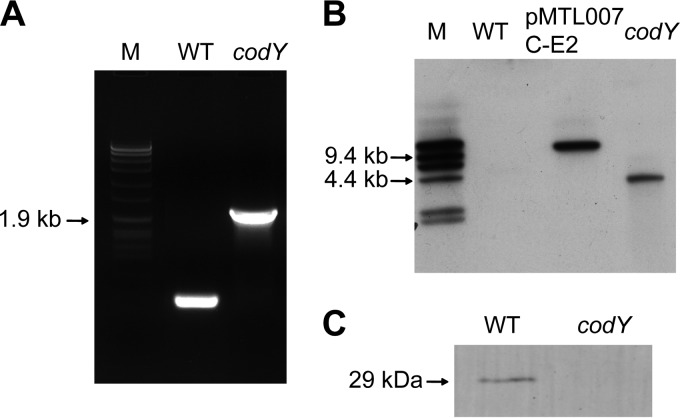
To confirm successful mutation of codY in C. botulinum group I type A1 strain ATCC 3502, the mutant DNA was screened by PCR using primers flanking the mutation target site. A 400-bp PCR product was amplified from the ATCC 3502 wild-type (WT) DNA, while the codY mutant DNA yielded a 2.2-kb amplification product demonstrating integration of a 1.8-kb fragment of the Ll.LtrB-derived intron, containing an erythromycin resistance gene (ermB), into the codY coding region (Fig. 1A). Consecutive cultures showed the codY mutant to be erythromycin resistant and stable. A single insertion of the Ll.LtrB-derived intron into the genome of the codY mutant was confirmed by Southern blotting with a probe specific for the Ll.LtrB-derived intron (Fig. 1B). Expression of CodY was analyzed using Western blotting with antibodies raised against B. subtilis CodY (32). In contrast to the WT, the mutant failed to express CodY (Fig. 1C). Finally, a slightly lower optical density at 600 nm (OD600) was observed for the codY mutant than for the WT at the transition between late exponential and early stationary growth phases (see Fig. S1 in the supplemental material). However, equal cell counts (2.3 × 10⁸/ml) measured for both cultures at 11 h suggested that inactivation of codY did not essentially affect growth.
FIG 1.
Insertional inactivation of codY in the Clostridium botulinum ATCC 3502 strain. (A) PCR analysis of codY mutation. Ll.LtrB-derived intron insertion was detected with primers flanking the insertional site yielding a 2.2-kb product in the codY mutant. M, Roche DNA molecular weight marker VII. (B) Southern blot analysis of HindIII-digested DNA from the WT, the pMTL007-C-E2 plasmid, and the codY mutant with intron-specific probe. (C) Western blot analysis of CodY expression in WT and codY mutant cultures.
CodY positively regulates botulinum neurotoxin gene expression.
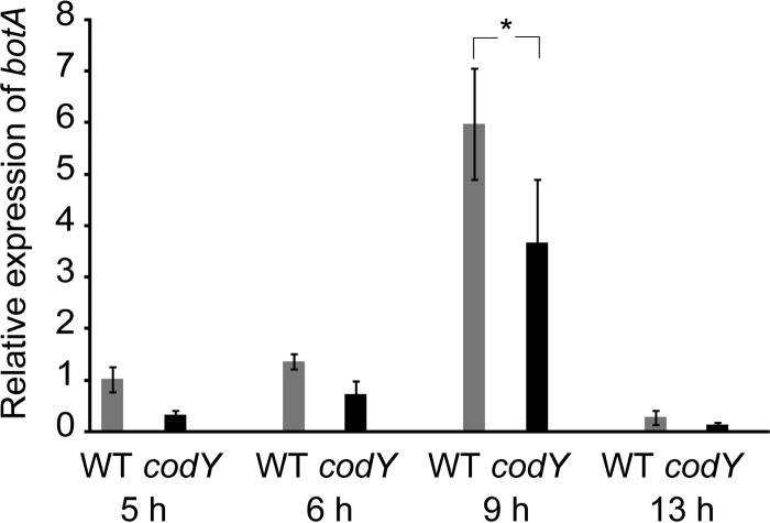
To test whether the inactivation of codY affects the transcription of the neurotoxin gene, the relative expression levels of botA was compared between the WT and the codY mutant at four time points during exponential and early stationary growth phases using RT-qPCR. Maximum botA transcript levels were observed at the transition between late exponential and early stationary growth phases (9 h) in both strains (Fig. 2), suggesting that the tightly regulated neurotoxin expression pattern was maintained despite codY mutation. The transcript levels of botA in the codY mutant were half of those observed in the WT, with most prominent differences being observed at 9 h of growth (Fig. 2).
FIG 2.
Expression of botA in the Clostridium botulinum ATCC 3502 wild-type strain and the codY mutant. RNA was isolated after 5, 6 (mid-exponential phase), 9 (transition phase), and 13 (early stationary phase) h of growth and analyzed for relative botA expression using RT-qPCR. Target gene expression was normalized to 16S rrn and calibrated to the WT at 5 h. Error bars represent standard deviations from three biological replicates. *, P < 0.05 (Student's t test).
To investigate whether the neurotoxin production is affected by the codY mutation, the amounts of neurotoxin in the culture supernatants of the WT and the codY mutant were determined in cultures grown for 5 to 96 h (24) by using ELISA. As expected, an increasing amount of neurotoxin was present in all culture supernatants collected in the exponential and late stationary growth phases (Fig. 3). In the WT culture supernatant, the neurotoxin concentrations peaked at 60 μg/ml at 48 h of growth, whereas the toxin concentrations in the codY mutant culture supernatant reached only 30 μg/ml at 48 h of growth. The half-reduced neurotoxin levels in the codY mutant compared to those in the WT were consistent with observations at the transcriptional level.
FIG 3.
Neurotoxin production by Clostridium botulinum ATCC 3502 wild-type strain and codY mutant. ELISA analysis of botulinum neurotoxin in WT and codY mutant culture supernatants after 5, 6, and 9 h (left) and 13, 24, 48, and 96 h (right) of growth. Error bars indicate standard deviations from three biological replicates. *, P < 0.05; **, P < 0.01 (Student's t test).
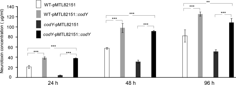
The codY mutation was complemented by introducing pMTL82151::codY, containing the codY coding sequence and its putative native promoter, into the codY mutant. The resulting codY-pMTL82151::codY strain did not show significant growth difference from the WT-pMTL82151 and codY-pMTL82151 vector control strains (see Fig. S2 in the supplemental material) but exhibited greater expression of CodY than the WT-pMTL82151strain (Fig. 4). The neurotoxin concentrations in the culture supernatant of the codY-pMTL82151::codY strain reached 3- and 2-fold-higher levels than in the codY-pMTL82151 control at 48 and 96 h of growth, respectively (Fig. 5), suggesting that complementation of the codY mutation rescued neurotoxin production. Moreover, the complementation resulted in significantly increased neurotoxin production in relation to that of the WT, probably because pMTL82151::codY replicated at a high copy number in the codY mutant and led to induced expression of the plasmid-encoded codY. To test this hypothesis, a codY overexpression strain was constructed by introducing pMTL82151::codY into the WT, generating the WT-pMTL82151::codY strain. No significant growth differences were observed between the WT-pMTL82151::codY overexpression strain, the codY-pMTL82151::codY complementation strain, and the WT-pMTL82151 and codY-pMTL82151 vector control strains (see Fig. S2 in the supplemental material). Similar to the observations in complementation, Western blotting also showed that the WT-pMTL82151::codY strain expressed CodY at a considerably higher level than the WT-pMTL82151 control (Fig. 4). Expectedly, the neurotoxin concentration in the culture supernatant of the WT-pMTL82151::codY strain was also significantly higher than that in the WT-pMTL82151 supernatant (Fig. 5).
FIG 4.
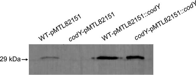
CodY expression in the complementation and overexpression strains. Western analysis of CodY expression in the codY-pMTL82151::codY complementation strain, WT-pMTL82151::codY overexpression strain, and WT-pMTL82151 and codY-pMTL82151 vector control strains.
FIG 5.
Neurotoxin production by the complementation and overexpression strains. ELISA analysis of botulinum neurotoxin in culture supernatants of the codY-pMTL82151::codY complementation strain, WT-pMTL82151::codY overexpression strain, and WT-pMTL82151 and codY-pMTL82151 vector control strains. Error bars represent standard deviations from three biological replicates. **, P < 0.01; ***, P < 0.001 (one-way ANOVA with Tukey's post hoc test).
Taken together, these data suggest that CodY plays a positive regulatory role in transcription of the botulinum neurotoxin gene and neurotoxin production.
CodY interacts with the botulinum neurotoxin gene promoter.
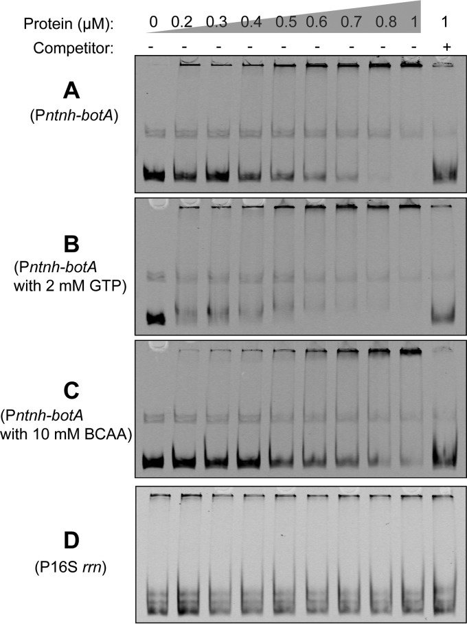
To investigate whether CodY regulates the transcription of the neurotoxin gene cluster directly, an EMSA was performed to examine the binding of recombinant CodY to a probe encompassing the upstream region of ntnh containing the promoter of the ntnh-botA operon (Pntnh-botA probe) (23). The presence of an increasing concentration of CodY caused a shift in the mobility of the Pntnh-botA probe (Fig. 6A), and the specific nature of binding was further confirmed by disappearance of both protein-DNA complexes using competition with a 200-fold excess of unlabeled probe. To test if CodY of C. botulinum responded to GTP and BCAAs, reported to enhance the binding affinity of CodY to its target gene promoters in some low-G+C Gram-positive bacteria (35, 43, 53–55), the EMSA procedure was repeated with the addition of 2 mM GTP or 10 mM BCAAs in the binding reactions. When 2 mM GTP was present, an enhanced binding affinity of CodY was observed with the Pntnh-botA probe (Fig. 6B). In contrast, the presence of 10 mM BCAAs did not enhance the binding affinity of CodY to the Pntnh-botA probe (Fig. 6C). A 16S rrn fragment (P16S rrn probe), serving as a negative control, did not show a significant shift with increased concentration of CodY (Fig. 6D). These results suggest that CodY recognizes and binds to the promoter of the ntnh-botA operon in vitro. GTP, but not BCAAs, may enhance the binding affinity of CodY to the promoter of the ntnh-botA operon, supporting the hypothesis that CodY acts in response to the intracellular GTP level in C. botulinum.
FIG 6.
Electrophoretic mobility shift assay (EMSA) for binding of CodY to the promoter of ntnh-botA operon. Pntnh-botA probe was incubated with increasing concentrations of CodY in the absence of effector (A) or with 2 mM GTP (B) or 10 mM (each) isoleucine, leucine, and valine (branched-chain amino acids [BCAAs]) (C). Specificity was confirmed using 200-fold molar excess of unlabeled competitor DNA. (D) CodY did not show significant binding to P16S rrn probe.
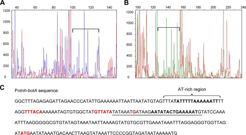
To further identify the CodY-binding site, a DNase I footprinting analysis with multiple replicates was performed using fluorescently end-labeled Pntnh-botA probe. In both strands of the Pntnh-botA sequence, a 30-bp region (bp −108 to −79 upstream of ntnh), encompassing the transcriptional start site of the ntnh-botA operon (20), was consistently found to be protected by CodY from DNase I digestion (Fig. 7A and B). Analysis of the DNA sequence of the 30-bp protection region yielded a putative CodY-binding motif, AATaTaCTGAAAAaT, with three mismatches (lowercase) to the proposed consensus CodY-binding motif, AATTTTCWGAAAATT, reported for B. subtilis (56) and Lactococcus lactis (57, 58), and with similarity to the CodY-binding sites in C. difficile (36) and in S. aureus (43). Another putative CodY-binding motif, tATTTTtAaAAAATT, similarly containing three mismatches (lowercase) to the consensus motif, was present in the probe in an AT-rich region neighboring the core promoter −35 region of the ntnh-botA operon (Fig. 7C). Immediately upstream of the 30-bp protection region, the core promoter of the ntnh-botA operon (20) and the AT-rich region showed a weak interaction with CodY with the sense strand, but no interaction with the antisense strand, of the Pntnh-botA probe (Fig. 7A and B). The results suggest that CodY interacts mainly with a 30-bp region in the promoter region of the ntnh-botA operon.
FIG 7.
DNase I footprinting assay for binding of CodY to the promoter of the ntnh-botA operon. Fragment analysis of the 5′-FAM-labeled sense strand (A) and the 5′-HEX-labeled antisense strand (B) of the Pntnh-botA probe shows DNase I digestion in the absence of (blue peaks in panel A and green peaks in panel B) or with (red peaks) 5 μM CodY. The protection region is indicated by square brackets in electropherograms, and the corresponding sequence is underlined in the Pntnh-botA sequence (C). Two putative CodY-binding motifs are indicated in bold letters. The core promoter −10 and −35 regions, transcriptional start site, and translational start site of the ntnh-botA operon are shown in red.
DISCUSSION
We suggest that CodY positively regulates botulinum neurotoxin expression in C. botulinum group I type A1 strain ATCC 3502. Positive regulation was supported by genetic and biochemical lines of evidence and adds to the slowly growing body of information on neurotoxin regulation in C. botulinum and, more generally, virulence regulation in clostridia. Direct regulation was supported by CodY interacting with a 30-bp region encompassing the transcriptional start site in the promoter region of the ntnh-botA operon (20). Analysis of the 30-bp region indicated the presence of a putative CodY-binding motif, AATaTaCTGAAAAaT, with three mismatches (lowercase) to the consensus CodY-binding motif, AATTTTCWGAAAATT (56–58). The DNA-binding domain at the C terminus of CodY is highly conserved in low-G+C Gram-positive bacteria (59, 60), supplying the CodY homologs with a general property in the recognition of target DNA. However, the degenerate sequence AATTTTCWGAAAATT is probably not a reliable guide for identifying the actual CodY-binding motif in C. botulinum. CodY has been reported to conduct its physiological function through interaction with sequences possessing up to five mismatches to the consensus motif (56, 61). The AT count of the Pntnh-botA probe is higher than 75%, which facilitates the presence of several putative CodY-binding motifs with four or five mismatches to the consensus CodY-binding motif AATTTTCWGAAAATT. However, all putative CodY-binding motifs, except the one within the 30-bp region of Pntnh-botA, showed no or little interaction with CodY in ATCC 3502. These findings suggest that CodY regulates botulinum neurotoxin gene expression mainly through interaction with the 30-bp region in the promoter of the ntnh-botA operon. The weak interaction of CodY with the putative CodY-binding motifs in the AT-rich region and other sites in the sense strand of Pntnh-botA, if at all physiologically relevant, is unlikely to represent a major mode of CodY-dependent regulation.
Immediately upstream of the proposed 30-bp CodY-binding region lies the core promoter −10 site of the ntnh-botA operon. The alternative sigma factor BotR specifically recognizes the core promoter and directs the RNA polymerase to transcribe the ntnh-botA operon (20). The close vicinity of the core promoter and the identified CodY-binding site raises the question of whether CodY interacts with BotR and/or the RNA polymerase core enzyme, thereby enhancing the transcription of the ntnh-botA operon. Another interesting question is if CodY interacts with the CBO0786 TCS response regulator, a repressor shown by us to specifically bind to the core promoter −10 region (23), thereby derepressing the transcription of the ntnh-botA operon. Interestingly, two putative CodY-binding motifs, AATTTTCAGtAgATa and AATTTTgTtAAAATa, each with three mismatches (lowercase) to the consensus CodY-binding motif, are found upstream of the translation start site of cbo0787, encoding the cognate TCS sensor kinase, implying that CodY might play a physiological function in regulating the cbo0787 expression. Further studies on the interaction of CodY with the two-component signal transduction system CBO0787/CBO0786 may offer new insights into the mechanisms controlling botulinum neurotoxin synthesis.
To facilitate the adaptation of the bacterial cell to different nutrient environments, CodY regulates multiple cellular activities by monitoring the intracellular level of GTP, BCAA, or both (32, 53, 62). The fact that GTP enhanced CodY binding to the Pntnh-botA probe suggests that CodY-mediated regulation of botulinum neurotoxin synthesis is associated with nutrition status. While availability of glucose is known to induce botulinum neurotoxin formation (27), several amino acids, such as arginine, proline, and glutamate, have been suggested to repress it (28, 29). Further characterization of the CodY regulon in C. botulinum is required to understand the relationship between cellular metabolism and botulinum neurotoxin synthesis. In many other low-G+C Gram-positive species, CodY represses the transcription of genes accounting for approximately 5% of the genome, including synthesis of several amino acids (BCAAs, histidine, and arginine) and transport of amino acids, peptides, and sugars (34). In addition to repressory effects, CodY-mediated induction has been recognized for carbon overflow metabolism. Under nutrient-rich conditions, B. subtilis uses CodY to activate the conversion of pyruvate derived from glycolysis to excreted overflow products such as acetate, lactate, and acetoin (34). In clostridia, pyruvate is metabolized by multiple anaerobic fermentation pathways and converted into a variety of fermentation end products, such as lactate, acetate, acetone, ethanol, butyrate, and butanol (49, 63, 64). Considering the temporal overlap between botulinum neurotoxin gene expression in batch cultures of C. botulinum and the pH-dependent switch from acidogenic to solventogenic metabolism described in batch cultures of Clostridium acetobutylicum (65), it is tempting to speculate that (CodY-mediated) botulinum neurotoxin production is linked to pyruvate metabolism.
The role of CodY in virulence regulation has also been documented in other clostridial pathogens. In C. perfringens type D strain CN3178, CodY was shown to positively regulate the epsilon toxin gene expression (37). Interestingly, epsilon toxin production was additionally dependent on the Agr quorum sensing system (66) also proposed to positively control neurotoxin synthesis in C. botulinum (21). As opposed to positive control of toxin production in C. perfringens and C. botulinum, CodY-mediated repression of C. difficile enterotoxin A (TcdA) and cytotoxin B (TcdB) production through transcriptional inactivation of tcdR under nutrient-rich conditions is well established (35, 36). TcdR is an alternative sigma factor that directs the transcription of tcdA and tcdB (67) and is both structurally and functionally closely related to BotR (68). Whether CodY exerts a regulatory action on botR in C. botulinum remains to be elucidated.
In the present study, we quantified neurotoxin production in C. botulinum ATCC 3502 using a commercial ELISA with type A neurotoxin as a standard. Our results suggest a 2- to 10-fold-higher neurotoxin production by C. botulinum ATCC 3502 than previously reported for C. botulinum type A strains 62A, Hall A-hyper, and NCTC 2916 with an in-house-constructed ELISA (24). Variation in neurotoxin titers between different C. botulinum strains, stocks, and culture media is recognized by laboratories working with this pathogen (24, 69) and can be poorly explained by the fragmented knowledge currently available for neurotoxin regulation. Systematic research efforts on the environmental cues and cellular mechanisms regulating neurotoxin production in different C. botulinum strains with various genetic systems encoding the neurotoxins is therefore warranted.
Supplementary Material
ACKNOWLEDGMENTS
This work was performed in the Centre of Excellence in Microbial Food Safety Research and supported by the Academy of Finland (grants 141140 and 257602), the European Community's Seventh Framework Programme (237942), and the Finnish Foundation of Veterinary Research.
We are grateful to Nigel P. Minton, University of Nottingham, for providing the ClosTron tool and pMTL82151 plasmid, Abraham L. Sonenshein, Tufts University, for providing CodY antibodies, Michel R. Popoff, Institut Pasteur, for providing purified type A botulinum neurotoxin, and Bruno Dupuy, Institut Pasteur, for providing the pBAD30 plasmid. We thank Hanna Korpunen and Esa Penttinen for technical assistance.
Footnotes
Published ahead of print 3 October 2014
Supplemental material for this article may be found at http://dx.doi.org/10.1128/AEM.02838-14.
REFERENCES
- 1.Schiavo G, Benfenati F, Poulain B, Rossetto O, Polverino de Laureto P, DasGupta BR, Montecucco C. 1992. Tetanus and botulinum-B neurotoxins block neurotransmitter release by proteolytic cleavage of synaptobrevin. Nature 359:832–835. 10.1038/359832a0. [DOI] [PubMed] [Google Scholar]
- 2.Blasi J, Chapman ER, Link E, Binz T, Yamasaki S, De Camilli P, Südhof TC, Niemann H, Jahn R. 1993. Botulinum neurotoxin A selectively cleaves the synaptic protein SNAP-25. Nature 365:160–163. 10.1038/365160a0. [DOI] [PubMed] [Google Scholar]
- 3.Johnson EA. 1999. Clostridial toxins as therapeutic agents: benefits of nature's most toxic proteins. Annu. Rev. Microbiol. 53:551–575. 10.1146/annurev.micro.53.1.551. [DOI] [PubMed] [Google Scholar]
- 4.Lim EC, Seet RC. 2010. Use of botulinum toxin in the neurology clinic. Nat. Rev. Neurol. 6:624–636. 10.1038/nrneurol.2010.149. [DOI] [PubMed] [Google Scholar]
- 5.Smith TJ, Lou J, Geren IN, Forsyth CM, Tsai R, Laporte SL, Tepp WH, Bradshaw M, Johnson EA, Smith LA, Marks JD. 2005. Sequence variation within botulinum neurotoxin serotypes impacts antibody binding and neutralization. Infect. Immun. 73:5450–5457. 10.1128/IAI.73.9.5450-5457.2005. [DOI] [PMC free article] [PubMed] [Google Scholar]
- 6.Chen Y, Korkeala H, Aarnikunnas J, Lindström M. 2007. Sequencing the botulinum neurotoxin gene and related genes in Clostridium botulinum type E strains reveals orfx3 and a novel type E neurotoxin subtype. J. Bacteriol. 189:8643–8650. 10.1128/JB.00784-07. [DOI] [PMC free article] [PubMed] [Google Scholar]
- 7.Hill KK, Smith TJ, Helma CH, Ticknor LO, Foley BT, Svensson RT, Brown JL, Johnson EA, Smith LA, Okinaka RT, Jackson PJ, Marks JD. 2007. Genetic diversity among botulinum neurotoxin-producing clostridial strains. J. Bacteriol. 189:818–832. 10.1128/JB.01180-06. [DOI] [PMC free article] [PubMed] [Google Scholar]
- 8.Dover N, Barash JR, Arnon SS. 2009. Novel Clostridium botulinum toxin gene arrangement with subtype A5 and partial subtype B3 botulinum neurotoxin genes. J. Clin. Microbiol. 47:2349–2350. 10.1128/JCM.00799-09. [DOI] [PMC free article] [PubMed] [Google Scholar]
- 9.Macdonald TE, Helma CH, Shou Y, Valdez YE, Ticknor LO, Foley BT, Davis SW, Hannett GE, Kelly-Cirino CD, Barash JR, Arnon SS, Lindström M, Korkeala H, Smith LA, Smith TJ, Hill KK. 2011. Analysis of Clostridium botulinum serotype E strains by using multilocus sequence typing, amplified fragment length polymorphism, variable-number tandem-repeat analysis, and botulinum neurotoxin gene sequencing. Appl. Environ. Microbiol. 77:8625–8634. 10.1128/AEM.05155-11. [DOI] [PMC free article] [PubMed] [Google Scholar]
- 10.Barash JR, Arnon SS. 2014. A novel strain of Clostridium botulinum that produces type B and type H botulinum toxins. J. Infect. Dis. 209:183–191. 10.1093/infdis/jit449. [DOI] [PubMed] [Google Scholar]
- 11.Johnson EA. 2014. Validity of botulinum neurotoxin serotype H. J. Infect. Dis. 210:992–993. 10.1093/infdis/jiu211. [DOI] [PubMed] [Google Scholar]
- 12.Schantz EJ, Johnson EA. 1992. Properties and use of botulinum toxin and other microbial neurotoxins in medicine. Microbiol. Rev. 56:80–99. [DOI] [PMC free article] [PubMed] [Google Scholar]
- 13.Lamanna C. 1948. Hemagglutination by botulinal toxin. Proc. Soc. Exp. Biol. Med. 69:332–336. 10.3181/00379727-69-16710. [DOI] [PubMed] [Google Scholar]
- 14.Sugii S, Sakaguchi G. 1975. Molecular construction of Clostridium botulinum type A toxins. Infect. Immun. 12:1262–1270. [DOI] [PMC free article] [PubMed] [Google Scholar]
- 15.Dineen SS, Bradshaw M, Johnson EA. 2003. Neurotoxin gene clusters in Clostridium botulinum type A strains: sequence comparison and evolutionary implications. Curr. Microbiol. 46:345–352. 10.1007/s00284-002-3851-1. [DOI] [PubMed] [Google Scholar]
- 16.Gu S, Rumpel S, Zhou J, Strotmeier J, Bigalke H, Perry K, Shoemaker CB, Rummel A, Jin R. 2012. Botulinum neurotoxin is shielded by NTNHA in an interlocked complex. Science 335:977–981. 10.1126/science.1214270. [DOI] [PMC free article] [PubMed] [Google Scholar]
- 17.Sugawara Y, Matsumura T, Takegahara Y, Jin Y, Tsukasaki Y, Takeichi M, Fujinaga Y. 2010. Botulinum hemagglutinin disrupts the intercellular epithelial barrier by directly binding E-cadherin. J. Cell Biol. 189:691–700. 10.1083/jcb.200910119. [DOI] [PMC free article] [PubMed] [Google Scholar]
- 18.Henderson I, Whelan SM, Davis TO, Minton NP. 1996. Genetic characterisation of the botulinum toxin complex of Clostridium botulinum strain NCTC 2916. FEMS Microbiol. Lett. 140:151–158. 10.1111/j.1574-6968.1996.tb08329.x. [DOI] [PubMed] [Google Scholar]
- 19.Marvaud JC, Gibert M, Inoue K, Fujinaga Y, Oguma K, Popoff MR. 1998. botR/A is a positive regulator of botulinum neurotoxin and associated non-toxin protein genes in Clostridium botulinum A. Mol. Microbiol. 29:1009–1018. 10.1046/j.1365-2958.1998.00985.x. [DOI] [PubMed] [Google Scholar]
- 20.Raffestin S, Dupuy B, Marvaud JC, Popoff MR. 2005. BotR/A and TetR are alternative RNA sigma factors controlling the expression of the neurotoxin and associated protein genes in Clostridium botulinum type A and Clostridium tetani. Mol. Microbiol. 55:235–249. 10.1111/j.1365-2958.2004.04377.x. [DOI] [PubMed] [Google Scholar]
- 21.Cooksley CM, Davis IJ, Winzer K, Chan WC, Peck MW, Minton NP. 2010. Regulation of neurotoxin production and sporulation by a putative agrBD signaling system in proteolytic Clostridium botulinum. Appl. Environ. Microbiol. 76:4448–4460. 10.1128/AEM.03038-09. [DOI] [PMC free article] [PubMed] [Google Scholar]
- 22.Connan C, Brueggemann H, Mazuet C, Raffestin S, Cayet N, Popoff MR. 2012. Two-component systems are involved in the regulation of botulinum neurotoxin synthesis in Clostridium botulinum type A strain Hall. PLoS One 7:e41848. 10.1371/journal.pone.0041848. [DOI] [PMC free article] [PubMed] [Google Scholar]
- 23.Zhang Z, Korkeala H, Dahlsten E, Sahala E, Heap JT, Minton NP, Lindström M. 2013. Two-component signal transduction system CBO0787/CBO0786 represses transcription from botulinum neurotoxin promoters in Clostridium botulinum ATCC 3502. PLoS Pathog. 9:e1003252. 10.1371/journal.ppat.1003252. [DOI] [PMC free article] [PubMed] [Google Scholar]
- 24.Bradshaw M, Dineen SS, Maks ND, Johnson EA. 2004. Regulation of neurotoxin complex expression in Clostridium botulinum strains 62A, Hall A-hyper, and NCTC 2916. Anaerobe 10:321–333. 10.1016/j.anaerobe.2004.07.001. [DOI] [PubMed] [Google Scholar]
- 25.Couesnon A, Raffestin S, Popoff MR. 2006. Expression of botulinum neurotoxins A and E, and associated non-toxin genes, during the transition phase and stability at high temperature: analysis by quantitative reverse transcription-PCR. Microbiol. 152:759–770. 10.1099/mic.0.28561-0. [DOI] [PubMed] [Google Scholar]
- 26.Chen Y, Korkeala H, Lindén J, Lindström M. 2008. Quantitative real-time reverse transcription PCR analysis reveals stable and prolonged neurotoxin complex gene activity in Clostridium botulinum type E at refrigeration temperature. Appl. Environ. Microbiol. 74:6132–6137. 10.1128/AEM.00469-08. [DOI] [PMC free article] [PubMed] [Google Scholar]
- 27.Bonventre PF, Kempe LL. 1959. Physiology of toxin production by Clostridium botulinum types A and B. II. Effect of carbohydrate source on growth, autolysis, and toxin production. Appl. Microbiol. 7:372–374. [DOI] [PMC free article] [PubMed] [Google Scholar]
- 28.Pattersoncurtis SI, Johnson EA. 1989. Regulation of neurotoxin and protease formation in Clostridium botulinum Okra B and Hall A by arginine. Appl. Environ. Microbiol. 55:1544–1548. [DOI] [PMC free article] [PubMed] [Google Scholar]
- 29.Leyer GJ, Johnson EA. 1990. Repression of toxin production by tryptophan in Clostridium botulinum type E. Arch. Microbiol. 154:443–447. 10.1007/BF00245225. [DOI] [PubMed] [Google Scholar]
- 30.Sonenshein AL. 2005. CodY, a global regulator of stationary phase and virulence in Gram-positive bacteria. Curr. Opin. Microbiol. 8:203–207. 10.1016/j.mib.2005.01.001. [DOI] [PubMed] [Google Scholar]
- 31.Stenz L, Francois P, Whiteson K, Wolz C, Linder P, Schrenzel J. 2011. The CodY pleiotropic repressor controls virulence in Gram-positive pathogens. FEMS Immunol. Med. Microbiol. 62:123–139. 10.1111/j.1574-695X.2011.00812.x. [DOI] [PubMed] [Google Scholar]
- 32.Ratnayake-Lecamwasam M, Serror P, Wong KW, Sonenshein AL. 2001. Bacillus subtilis CodY represses early-stationary-phase genes by sensing GTP levels. Genes Dev. 15:1093–1103. 10.1101/gad.874201. [DOI] [PMC free article] [PubMed] [Google Scholar]
- 33.Molle V, Nakaura Y, Shivers RP, Yamaguchi H, Losick R, Fujita Y, Sonenshein AL. 2003. Additional targets of the Bacillus subtilis global regulator CodY identified by chromatin immunoprecipitation and genome-wide transcript analysis. J. Bacteriol. 185:1911–1922. 10.1128/JB.185.6.1911-1922.2003. [DOI] [PMC free article] [PubMed] [Google Scholar]
- 34.Sonenshein AL. 2007. Control of key metabolic intersections in Bacillus subtilis. Nat. Rev. Microbiol. 5:917–927. 10.1038/nrmicro1772. [DOI] [PubMed] [Google Scholar]
- 35.Dineen SS, Villapakkam AC, Nordman JT, Sonenshein AL. 2007. Repression of Clostridium difficile toxin gene expression by CodY. Mol. Microbiol. 66:206–219. 10.1111/j.1365-2958.2007.05906.x. [DOI] [PubMed] [Google Scholar]
- 36.Dineen SS, McBride SM, Sonenshein AL. 2010. Integration of metabolism and virulence by Clostridium difficile CodY. J. Bacteriol. 192:5350–5362. 10.1128/JB.00341-10. [DOI] [PMC free article] [PubMed] [Google Scholar]
- 37.Li J, Ma M, Sarker MR, McClane BA. 2013. CodY is a global regulator of virulence-associated properties for Clostridium perfringens type D strain CN3718. mBio 4(5):e00770-13. 10.1128/mBio.00770-13. [DOI] [PMC free article] [PubMed] [Google Scholar]
- 38.Frenzel E, Doll V, Pauthner M, Lücking G, Scherer S, Ehling-Schulz M. 2012. CodY orchestrates the expression of virulence determinants in emetic Bacillus cereus by impacting key regulatory circuits. Mol. Microbiol. 85:67–88. 10.1111/j.1365-2958.2012.08090.x. [DOI] [PubMed] [Google Scholar]
- 39.van Schaik W, Château A, Dillies MA, Coppée JY, Sonenshein AL, Fouet A. 2009. The global regulator CodY regulates toxin gene expression in Bacillus anthracis and is required for full virulence. Infect. Immun. 77:4437–4445. 10.1128/IAI.00716-09. [DOI] [PMC free article] [PubMed] [Google Scholar]
- 40.Majerczyk CD, Dunman PM, Luong TT, Lee CY, Sadykov MR, Somerville GA, Bodi K, Sonenshein AL. 2010. Direct targets of CodY in Staphylococcus aureus. J. Bacteriol. 192:2861–2877. 10.1128/JB.00220-10. [DOI] [PMC free article] [PubMed] [Google Scholar]
- 41.Hendriksen WT, Bootsma HJ, Estevão S, Hoogenboezem T, de Jong A, de Groot R, Kuipers OP, Hermans PW. 2008. CodY of Streptococcus pneumoniae: link between nutritional gene regulation and colonization. J. Bacteriol. 190:590–601. 10.1128/JB.00917-07. [DOI] [PMC free article] [PubMed] [Google Scholar]
- 42.Malke H, Steiner K, McShan WM, Ferretti JJ. 2006. Linking the nutritional status of Streptococcus pyogenes to alteration of transcriptional gene expression: the action of CodY and RelA. Int. J. Med. Microbiol. 296:259–275. 10.1016/j.ijmm.2005.11.008. [DOI] [PubMed] [Google Scholar]
- 43.Lemos JA, Nascimento MM, Lin VK, Abranches J, Burne RA. 2008. Global regulation by (p)ppGpp and CodY in Streptococcus mutans. J. Bacteriol. 190:5291–5299. 10.1128/JB.00288-08. [DOI] [PMC free article] [PubMed] [Google Scholar]
- 44.Lobel L, Sigal N, Borovok I, Ruppin E, Herskovits AA. 2012. Integrative genomic analysis identifies isoleucine and CodY as regulators of Listeria monocytogenes virulence. PLoS Genet. 8:e1002887. 10.1371/journal.pgen.1002887. [DOI] [PMC free article] [PubMed] [Google Scholar]
- 45.Sebaihia M, Peck MW, Minton NP, Thomson NR, Holden MT, Mitchell WJ, Carter AT, Bentley SD, Mason DR, Crossman L, Paul CJ, Ivens A, Wells-Bennik MH, Davis IJ, Cerdeño-Tárraga AM, Churcher C, Quail MA, Chillingworth T, Feltwell T, Fraser A, Goodhead I, Hance Z, Jagels K, Larke N, Maddison M, Moule S, Mungall K, Norbertczak H, Rabbinowitsch E, Sanders M, Simmonds M, White B, Whithead S, Parkhill J. 2007. Genome sequence of a proteolytic (group I) Clostridium botulinum strain Hall A and comparative analysis of the clostridial genomes. Genome Res. 17:1082–1092. 10.1101/gr.6282807. [DOI] [PMC free article] [PubMed] [Google Scholar]
- 46.Purdy D, O'Keeffe TA, Elmore M, Herbert M, McLeod A, Bokori-Brown M, Ostrowski A, Minton NP. 2002. Conjugative transfer of clostridial shuttle vectors from Escherichia coli to Clostridium difficile through circumvention of the restriction barrier. Mol. Microbiol. 46:439–452. 10.1046/j.1365-2958.2002.03134.x. [DOI] [PubMed] [Google Scholar]
- 47.Heap JT, Kuehne SA, Ehsaan M, Cartman ST, Cooksley CM, Scott JC, Minton NP. 2010. The ClosTron: mutagenesis in Clostridium refined and streamlined. J. Microbiol. Methods 80:49–55. 10.1016/j.mimet.2009.10.018. [DOI] [PubMed] [Google Scholar]
- 48.Heap JT, Pennington OJ, Cartman ST, Minton NP. 2009. A modular system for Clostridium shuttle plasmids. J. Microbiol. Methods 78:79–85. 10.1016/j.mimet.2009.05.004. [DOI] [PubMed] [Google Scholar]
- 49.Dahlsten E, Zhang Z, Somervuo P, Minton NP, Lindström M, Korkeala H. 2014. The cold-induced two-component system CBO0366/CBO0365 regulates metabolic pathways with novel roles in group I Clostridium botulinum ATCC 3502 cold tolerance. Appl. Environ. Microbiol. 80:306–319. 10.1128/AEM.03173-13. [DOI] [PMC free article] [PubMed] [Google Scholar]
- 50.Kirk DG, Palonen E, Korkeala H, Lindström M. 2014. Evaluation of normalization reference genes for RT-qPCR analysis of spo0A and four sporulation sigma factor genes in Clostridium botulinum group I strain ATCC 3502. Anaerobe 26:14–19. 10.1016/j.anaerobe.2013.12.003. [DOI] [PubMed] [Google Scholar]
- 51.Pfaffl MW. 2001. A new mathematical model for relative quantification in real-time RT-PCR. Nucleic Acids Res. 29:e45. 10.1093/nar/29.9.e45. [DOI] [PMC free article] [PubMed] [Google Scholar]
- 52.Zianni M, Tessanne K, Merighi M, Laguna R, Tabita FR. 2006. Identification of the DNA bases of a DNase I footprint by the use of dye primer sequencing on an automated capillary DNA analysis instrument. J. Biomol. Tech. 17:103–113. [PMC free article] [PubMed] [Google Scholar]
- 53.Shivers RP, Sonenshein AL. 2004. Activation of the Bacillus subtilis global regulator CodY by direct interaction with branched-chain amino acids. Mol. Microbiol. 53:599–611. 10.1111/j.1365-2958.2004.04135.x. [DOI] [PubMed] [Google Scholar]
- 54.Handke LD, Shivers RP, Sonenshein AL. 2008. Interaction of Bacillus subtilis CodY with GTP. J. Bacteriol. 190:798–806. 10.1128/JB.01115-07. [DOI] [PMC free article] [PubMed] [Google Scholar]
- 55.Château A, van Schaik W, Joseph P, Handke LD, McBride SM, Smeets FM, Sonenshein AL, Fouet A. 2013. Identification of CodY targets in Bacillus anthracis by genome-wide in vitro binding analysis. J. Bacteriol. 195:1204–1213. 10.1128/JB.02041-12. [DOI] [PMC free article] [PubMed] [Google Scholar]
- 56.Belitsky BR, Sonenshein AL. 2008. Genetic and biochemical analysis of CodY-binding sites in Bacillus subtilis. J. Bacteriol. 190:1224–1236. 10.1128/JB.01780-07. [DOI] [PMC free article] [PubMed] [Google Scholar]
- 57.den Hengst CD, van Hijum SA, Geurts JM, Nauta A, Kok J, Kuipers OP. 2005. The Lactococcus lactis CodY regulon: identification of a conserved cis-regulatory element. J. Biol. Chem. 280:34332–34342. 10.1074/jbc.M502349200. [DOI] [PubMed] [Google Scholar]
- 58.Guédon E, Sperandio B, Pons N, Ehrlich SD, Renault P. 2005. Overall control of nitrogen metabolism in Lactococcus lactis by CodY, and possible models for CodY regulation in Firmicutes. Microbiology 151:3895–3909. 10.1099/mic.0.28186-0. [DOI] [PubMed] [Google Scholar]
- 59.Joseph P, Ratnayake-Lecamwasam M, Sonenshein AL. 2005. A region of Bacillus subtilis CodY protein required for interaction with DNA. J. Bacteriol. 187:4127–4139. 10.1128/JB.187.12.4127-4139.2005. [DOI] [PMC free article] [PubMed] [Google Scholar]
- 60.Levdikov VM, Blagova E, Joseph P, Sonenshein AL, Wilkinson AJ. 2006. The structure of CodY, a GTP- and isoleucine-responsive regulator of stationary phase and virulence in Gram-positive bacteria. J. Biol. Chem. 281:11366–11373. 10.1074/jbc.M513015200. [DOI] [PubMed] [Google Scholar]
- 61.Belitsky BR, Sonenshein AL. 2011. Contributions of multiple binding sites and effector-independent binding to CodY-mediated regulation in Bacillus subtilis. J. Bacteriol. 193:473–484. 10.1128/JB.01151-10. [DOI] [PMC free article] [PubMed] [Google Scholar]
- 62.Petranovic D, Guédon E, Sperandio B, Delorme C, Ehrlich D, Renault P. 2004. Intracellular effectors regulating the activity of the Lactococcus lactis CodY pleiotropic transcription regulator. Mol. Microbiol. 53:613–621. 10.1111/j.1365-2958.2004.04136.x. [DOI] [PubMed] [Google Scholar]
- 63.Paredes CJ, Alsaker KV, Papoutsakis ET. 2005. A comparative genomic view of clostridial sporulation and physiology. Nat. Rev. Microbiol. 3:969–978. 10.1038/nrmicro1288. [DOI] [PubMed] [Google Scholar]
- 64.Janoir C, Denève C, Bouttier S, Barbut F, Hoys S, Caleechum L, Chapetón-Montes D, Pereira FC, Henriques AO, Collignon A, Monot M, Dupuy B. 2013. Adaptive strategies and pathogenesis of Clostridium difficile from in vivo transcriptomics. Infect. Immun. 81:3757–3769. 10.1128/IAI.00515-13. [DOI] [PMC free article] [PubMed] [Google Scholar]
- 65.Jones SW, Paredes CJ, Tracy B, Cheng N, Sillers R, Senger RS, Papoutsakis ET. 2008. The transcriptional program underlying the physiology of clostridial sporulation. Genome Biol. 9:R114. 10.1186/gb-2008-9-7-r114. [DOI] [PMC free article] [PubMed] [Google Scholar]
- 66.Chen J, McClane BA. 2012. Role of the Agr-like quorum-sensing system in regulating toxin production by Clostridium perfringens type B strains CN1793 and CN1795. Infect. Immun. 80:3008–3017. 10.1128/IAI.00438-12. [DOI] [PMC free article] [PubMed] [Google Scholar]
- 67.Mani N, Dupuy B. 2001. Regulation of toxin synthesis in Clostridium difficile by an alternative RNA polymerase sigma factor. Proc. Natl. Acad. Sci. U. S. A. 98:5844–5849. 10.1073/pnas.101126598. [DOI] [PMC free article] [PubMed] [Google Scholar]
- 68.Dupuy B, Raffestin S, Matamouros S, Mani N, Popoff MR, Sonenshein AL. 2006. Regulation of toxin and bacteriocin gene expression in Clostridium by interchangeable RNA polymerase sigma factors. Mol. Microbiol. 60:1044–1057. 10.1111/j.1365-2958.2006.05159.x. [DOI] [PubMed] [Google Scholar]
- 69.Johnson EA, Bradshaw M. 2001. Clostridium botulinum and its neurotoxins: a metabolic and cellular perspective. Toxicon 39:1703–1722. 10.1016/S0041-0101(01)00157-X. [DOI] [PubMed] [Google Scholar]
Associated Data
This section collects any data citations, data availability statements, or supplementary materials included in this article.


