Abstract
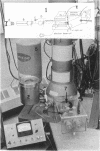
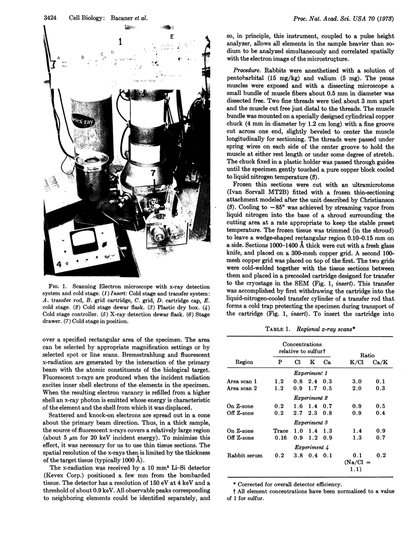
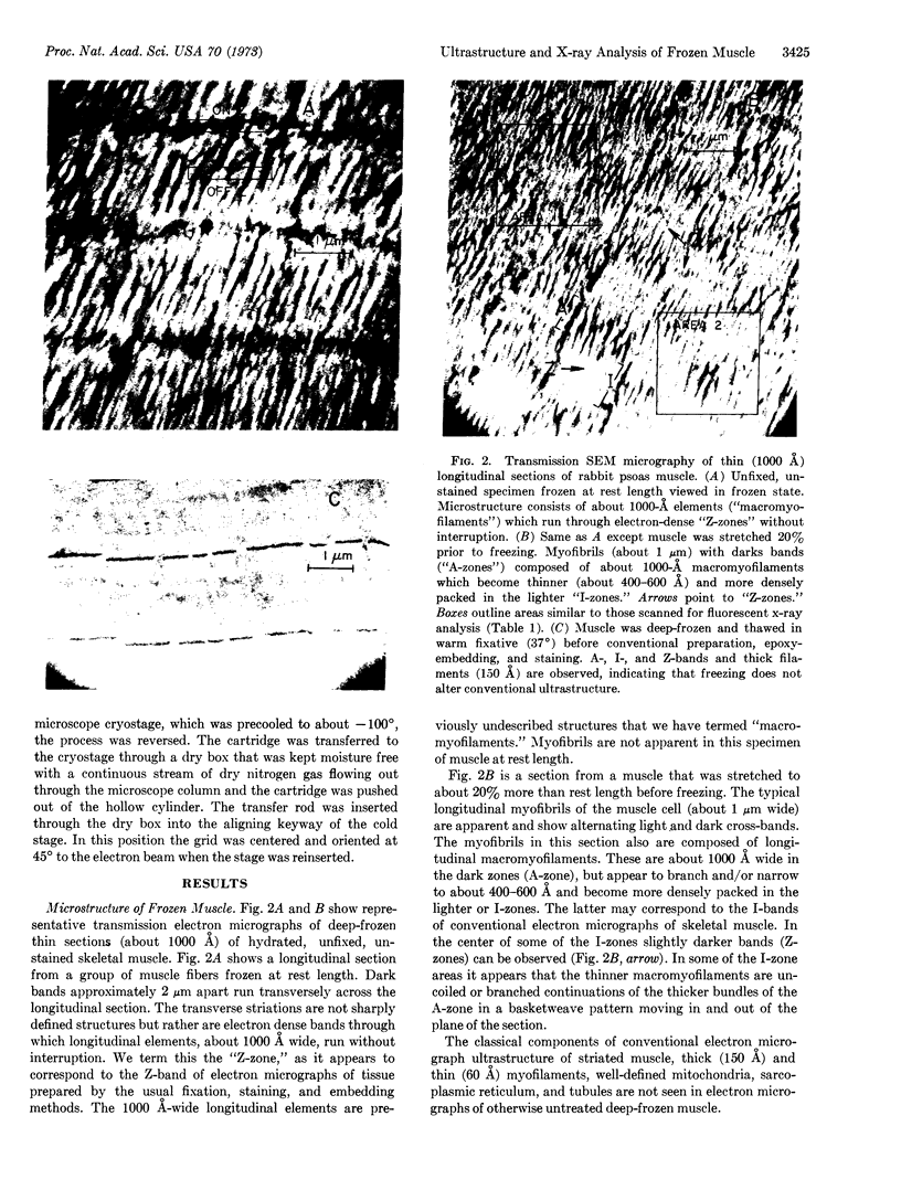
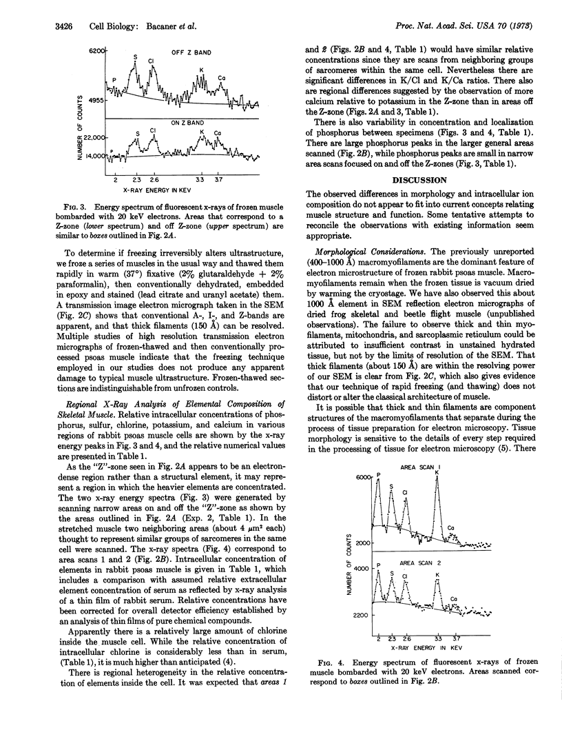
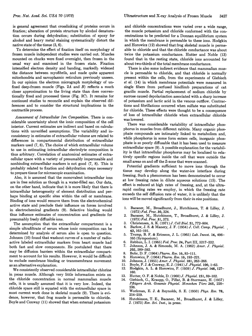
Thin sections of deep-frozen unfixed muscle were studied in a scanning electron microscope modified for transmission imaging and equipped with a “cryostage” for vacuum compatibility of hydrated tissue. With an energy-dispersive x-ray analysis system, intracellular atomic species in the scan beam path were identified by their fluorescent x-rays and spatially localized in correlation with the electron optical image of the microstructure. Marked differences are noted between the ultrastructure of deep-frozen hydrated muscle and that of fixed dehydrated muscle. In frozen muscle, myofibrils appear to be composed of previously undescribed longitudinal structures between 400-1000 Å wide (“macromyofilaments”). The usual myofilaments, mitochondria, and sarcoplasmic reticulum were not seen unless the tissue was “fixed” before examination. Fluorescent x-ray analysis of the spatial location of constituent elements clearly identified all elements heavier than Na. Intracellular Cl was relatively higher than expected.
Keywords: nondispersive x-ray analysis, cryobiology
Full text
PDF
Images in this article
Selected References
These references are in PubMed. This may not be the complete list of references from this article.
- BARLOW J. S., MANERY J. F. The changes in electrolytes, particularly chloride, which accompany growth in chick muscle. J Cell Physiol. 1954 Apr;43(2):165–191. doi: 10.1002/jcp.1030430204. [DOI] [PubMed] [Google Scholar]
- BOHR D. F. ELECTROLYTES AND SMOOTH MUSCLE CONTRACTION. Pharmacol Rev. 1964 Mar;16:85–127. [PubMed] [Google Scholar]
- Boyle P. J., Conway E. J. Potassium accumulation in muscle and associated changes. J Physiol. 1941 Aug 11;100(1):1–63. doi: 10.1113/jphysiol.1941.sp003922. [DOI] [PMC free article] [PubMed] [Google Scholar]
- Christensen A. K. Frozen thin sections of fresh tissue for electron microscopy, with a description of pancreas and liver. J Cell Biol. 1971 Dec;51(3):772–804. doi: 10.1083/jcb.51.3.772. [DOI] [PMC free article] [PubMed] [Google Scholar]
- GIEBISCH G., KRAUPP O., PILLAT B., STORMANN H. Der Ersatz von extracellulärem Natriumchlorid durch Natriumsulfat bzw. Saccharose und seine Wirkung auf die isoliert durchströmte Saugetiermuskulatur. Pflugers Arch. 1957;265(3):220–236. doi: 10.1007/BF00595649. [DOI] [PubMed] [Google Scholar]
- HODGKIN A. L., HOROWICZ P. The influence of potassium and chloride ions on the membrane potential of single muscle fibres. J Physiol. 1959 Oct;148:127–160. doi: 10.1113/jphysiol.1959.sp006278. [DOI] [PMC free article] [PubMed] [Google Scholar]
- HOROWICZ P. THE EFFECTS OF ANIONS ON EXCITABLE CELLS. Pharmacol Rev. 1964 Jun;16:193–221. [PubMed] [Google Scholar]
- HUTTER O. F., NOBLE D. The chloride conductance of frog skeletal muscle. J Physiol. 1960 Apr;151:89–102. [PMC free article] [PubMed] [Google Scholar]
- JOHNSON J. A. Kinetics of release of radioactive sodium, sulfate and sucrose from the frog sartorius muscle. Am J Physiol. 1955 May;181(2):263–268. doi: 10.1152/ajplegacy.1955.181.2.263. [DOI] [PubMed] [Google Scholar]
- JOHNSON J. A., SIMONDS M. A. Chemical and histological space determinations in rabbit heart. Am J Physiol. 1962 Mar;202:589–592. doi: 10.1152/ajplegacy.1962.202.3.589. [DOI] [PubMed] [Google Scholar]


