Abstract
Introduction: Providing desirable smile is one of the main concerns in cosmetic dentistry. Hyperpigmentation is one of the esthetic concerns especially in gummy smile patients. Lasers with different wavelength are used for oral surgery including Carbon Dioxide Laser (CO2), Neodymium-Doped Yttrium Aluminium Garnet (Nd:YAG), Erbium family and diode laser. In this case, all esthetic procedures including gingival depigmentation, caries detection and removal were done by laser technology in one session.
Case study: A 40- year-old male with a chief complaint of black gingiva in upper jaw was referred. The right side of maxillary was anesthetized and depigmentation was done by Erbium, Chromium doped Yttrium Scandium Gallium Garnet (Er-Cr: YSGG) laser. Due to scores obtained from Diagnodent which indicated caries in dentin, the cavities were prepared by Er-Cr:YSGG laser. The cavities were restored by composite resin. The patient was advised to keep oral hygiene instructions and use mouthwash.
Results: The patient reported no pain after surgery and did not use any systemic antibiotic. After 4 weeks, complete healing was observed.
Conclusion: Considering acceptable clinical outcome, Er-Cr: YSGG laser can be considered as an effective method for combination of soft and hard tissue treatment.
Keywords: gingiva, laser, YSGG Laser
Introduction
One of the factors that should be considered in cosmetic dentistry is smile window which includes the size, shape and health of teeth, gums and the curve of lips 1. Hyperpigmentation is one of the esthetic concerns especially in gummy smile patients. Several exogenous and endogenous factors are responsible for gingival discoloration2. Melanin is one of the endogenous pigments produced by melanocytes. Gingival hyperpigmentation is caused by deposition of melanin in the basal and suprabasal layers 3, 4 .
Different methods such as conventional surgery, cryosurgery, electrosurgery and chemical techniques were used. Recently, lasers with different wavelengths are used for dental surgery including Carbon Dioxide Laser (CO2), Neodymium-Doped Yttrium Aluminium Garnet (Nd:YAG), Erbium family and diode laser 5.
Erbium family lasers showed promising results for cutting soft and hard oral tissue. When Erbium laser is used for periodontal surgery, it produces minimal temperature rise in tissue and surroundings due to high absorption in water which prevents scarring and provides favorable healing. Also, by changing the parameters, hard tissue can be modified 6, 7.
On the other hand, this laser is more efficient for ablation and modification through water mediated ablation mechanism or hydrokinetic system. The laser energy is absorbed by water which causes vaporization. So, the internal pressure increases in the tissue until the explosive destruction of inorganic substances happens which produce hydrokinetic forces that can rapidly ablate the dental hard tissues 8, 9.
In this case report, all esthetic procedures including gingival depigmentation caries detection and caries removal was done by laser technology.
Case report
A 40-year-old male with a chief complaint of black gingiva in upper jaw and caries in upper right central and canine was referred to a private dental office (Figure 1).
Figure 1 .
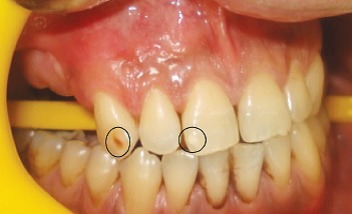
Preoperative view of hyperpigmentation and caries area in upper right central and canine
The medical history was unremarkable. He requested to finish the procedure in one session with minimal complications. In primary evaluation, the teeth were assessed visually and white spot was found on central and canine. Then, Diagnodent (Kavo, Biberach, Germany) was used to score the degree of demineralization which showed 28 and 33 for central and canine. Then, the treatment plan was designed by using Erbium, Chromium doped Yttrium Scandium Gallium Garnet (Er-Cr: YSGG) laser. First, the right side of maxillary was anesthetized and de-pigmentation was done by Er-Cr: YSGG laser (Waterlase/Biolase Co. Power: 0-6w, pulse duration: 140 μs, frequency of 20Hz, energy: 0-300 mJ), using 1.5 w power, energy of 75 mJ/pulse (35% Water, 35% air). The procedure was done in cervical-apical direction on pigmented area (Figure 2).
Figure 2 .
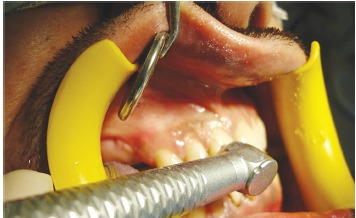
Depigmentation by Er-Cr: YSGGlaser
No packing material was applied to support wound. Due to scores obtained from Diagnodent which indicated the caries in dentin, the cavities were prepared. After that, Er-Cr: YSGG laser with power of 3W, (55% Water, 65% air), using G6 tip was used for caries removal (Figure 3). Then, tooth conditioning was done by same laser with power of 1.5 W (35% Water, 55% air) (Figure 4).
Figure 3 .
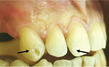
Caries removal by Er-Cr: YSGGlaser
Figure 4 .
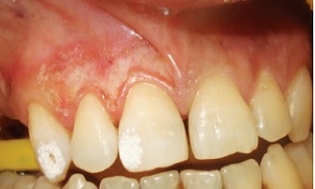
Conditioning by Er-Cr: YSGG laser
The cavity was restored by composite resin (Filtek Supreme XT, 3 M ESPE, USA) following manufacture instruction (Figure 5).
Figure 5 .
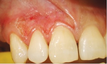
Composite restoration of teeth, gingival margin was preserved due to minimally invasive manner of laser surgery
Laser therapy was done for gingival part by diode laser 630 nm, output power of 200 mW and energy density of 2 J/cm2. All procedures were done by an expert surgeon. The patient was advised to keep oral hygiene instructions and use mouthwash. Analgesic medication was prescribed in case of pain perception.
Minimal bleeding was observed during surgery. Patient reported a mild sense of burning in first day after treatment but no pain was felt after day1.
In follow up session (Figure 6) after 4 weeks, complete healing was observed.
Figure 6 .
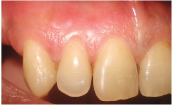
Follow up session after 4 weeks
Discussion
The demands for dental cosmetic treatments are increasing. Having attractive smile needs gingival health and good appearance. Gingival hyperpigmentation as an esthetic problem forces the patients for cosmetic treatments 10. Previous clinical studies reported different depigmentation methods. Conventional surgical methods produced bleeding during procedure that limit the surgeon’s view area. Also, the wounds after conventional methods need periodontal dressing for 7 to 10 days 11. Among different methods, laser application showed promising results. After laser surgery, postoperative pain is reduced due to protein coagulum formed on the wound surface which acts as a biological dressing. On the other hand, sealing of the ends of the sensory nerves is another factor for pain reduction after surgery 12, 13 .
Different lasers are used for gingival depigmentation including Nd:YAG, CO2, diode and Erbium family lasers. Nd:YAG and diode laser can selectively remove gingival pigmentation due to high absorption in melanin. On the other hand, CO2 laser can coagulate the site of surgery to reduce postoperative complications 12 . When diode, Nd:YAG and CO2 laser are used for depigmentation and unexpected bone exposure happened, it’s the possibility of thermal damage to underlying bone providing melting and carbonized layer on lased surface, causing delayed bone healing and bone loss. Although Erbium laser is non selective for this purpose and have low penetration depth but its application eliminate the side effects produced by other lasers such as less damage to bone 3, 14 .
Diagnodent as a caries diagnostic tool is a Diode laser with wavelength of 655nm. The mechanism of this device is through emitted fluorescence from teeth surfaces that reflects the degree of demineralization of the teeth 15 .
There are controversial results about wound healing after laser surgery. Some studies indicated that the healing happened quickly due to less scar formation compared to scalpel surgery. On the contrary, another study had stated that delay of re-epithelization in laser group resulted in late healing 13 . In this case, we used Er-Cr: YSGG laser which produced minimal thermal damage and absence of burning or charring which led to fast healing proving patient’s satisfaction.
Erbium family lasers are the best option for caries removal and cavity preparation. By application of this technology, the cavities were prepared according to minimally invasive restorative dentistry principles which resulted in preservation of tooth healthy tissues 16.
Laser irradiation on enamel surface produces irregular and microretentive patterns. Also, no smear layer production makes this surface suitable for resin composite restoration 17 .
The application of laser in this case has some advantages such as no need for injection, phosphoric acid application and antibiotic therapy after surgery. Considering acceptable clinical outcomes, Er-Cr: YSGG laser can be considered as an effective method for combination of soft and hard tissue treatment by reducing chair time and providing patient’s satisfaction in one visit.
Conclusion
The application of laser for all steps of providing desirable smile for patient was efficient and produced patient’s satisfaction which is a main goal in any treatment. Also, laser application compared to conventional methods in management of esthetic problems and designing favorable smile is more effective due to minimal bleeding and discomfort for patients even if recontouring of gingival is needed. There is need for randomized clinical trials for achieving definite evidence of efficacy of laser treatment in smile designing.
Please cite this article as follows:
Fekrazad R,Chiniforush N. One Visit Providing Desirable Smile by Laser Application. J Lasers Med Sci 2014;5(1):47-50
References
- 1.Gupta M, Pradhan S. Laser assisted smile designing. Int J Laser Dent. 2011;1(1):45–7. [Google Scholar]
- 2.Tal H, Oegiesser D, Tal M. Gingival depigmetation by Erbium:YAG laser: clinical observations and patient responses. J Periodontol. 2003;74:1660–7. doi: 10.1902/jop.2003.74.11.1660. [DOI] [PubMed] [Google Scholar]
- 3.Atsawasuwan P, Greethong K, Nimmanon V. Treatment of gingival hyperpigmentation for esthetic purposes by Nd:YAG laser: report of 4 cases. J Periodontol. 2000;71:356–60. doi: 10.1902/jop.2000.71.2.315. [DOI] [PubMed] [Google Scholar]
- 4.Rosa DS, Aranha AC, Eduardo Cde P, Aoki A. Esthetic treatment of gingival melanin hyperpigmentation with Er:YAG laser: short-term clinical observations and patient follow-up . J Periodontol. 2007;78(10):2018–25. doi: 10.1902/jop.2007.070041. [DOI] [PubMed] [Google Scholar]
- 5.Ishikawa I, Aoki A, Takasaki AA, Mizutani K, Sasaki KM, Izumi Y. Application of lasers in periodontics: True innovation or myth? Periodontology. 2009;50:90, 126. doi: 10.1111/j.1600-0757.2008.00283.x. [DOI] [PubMed] [Google Scholar]
- 6.Simsek Kaya G, Yapıcı Yavuz G, Sümbüllü MA, Dayı E. A comparison of diode laser and Er:YAG lasers in the treatment of gingival melanin pigmentation. Oral Surg Oral Med Oral Pathol Oral Radiol Endod. 2012;113(3):293–99. doi: 10.1016/j.tripleo.2011.03.005. [DOI] [PubMed] [Google Scholar]
- 7.Ishikawa I, Aoki A, Takasaki AA. Clinical application of erbium:YAG laser in periodontology. J Int Acad Periodontol. 2008;10(1):22–30. [PubMed] [Google Scholar]
- 8.Shahabi Shahabi, Chiniforush N, Bahramian H, Monzavi A, Baghalian A, Kharazifard MJ. The effect of erbium family laser on tensile bond Strengrth of composite to dentin in comparison with conventional method. Lasers Med Sci. 2013;28(1):139–42. doi: 10.1007/s10103-012-1086-3. [DOI] [PubMed] [Google Scholar]
- 9.Jafari A, Shahabi S, Chiniforush N, Shariat A. Comparison of the Shear Bond Strength of Resin Modified Glass Ionomer to Enamel in Bur-Prepared or Lased Teeth (Er: YAG) J Dent. 2013;10(2):119–23. [PMC free article] [PubMed] [Google Scholar]
- 10.Azzeh MM. Treatment of gingival hyperpigmentation by erbium-doped:yttrium, aluminum, and garnet laser for esthetic purposes. J Periodontol. 2007;78(1):177–84. doi: 10.1902/jop.2007.060167. [DOI] [PubMed] [Google Scholar]
- 11.Murthy M, Kaur J, Das R. Treatment of gingival hyperpigmentation with rotary abrasive, scalpel, and laser techniques: A case series. J Indian Soc Periodontol. 2012;16(4):614–9. doi: 10.4103/0972-124X.106933. [DOI] [PMC free article] [PubMed] [Google Scholar]
- 12.Lagdive S, Doshi Y, Marawa PP. Management of Gingival Hyperpigmentation Using Surgical Blade and Diode Laser Therapy: A Comparative Study. J Oral Laser Applications. 2009;9:41–7. [Google Scholar]
- 13.Berk G, Atici K, Berk N. Treatment of gingival pigmentation with Er-Cr: YSGGlaser. J Oral Laser Applications. 2005;5:249–53. [Google Scholar]
- 14.Wang X, Zhang C, Matsumoto K. In vivo study of the healing processes that occur in the jaws of rabbits following perforation by an Er,Cr:YSGG laser. Lasers Med Sci. 2005;20(1):21–7. doi: 10.1007/s10103-005-0329-y. [DOI] [PubMed] [Google Scholar]
- 15.Bader JD, Shugars DA. A systematic review of the performance of a laser fluorescence device for detecting caries. J Am Dent Assoc. 2004;135(10):1413–26. doi: 10.14219/jada.archive.2004.0051. [DOI] [PubMed] [Google Scholar]
- 16.Ghandehari M, Mighani G, Shahabi S, Chiniforush N, Shirmohammadi Z. Comparison of Microleakage of Glass Ionomer Restoration in Primary Teeth Prepared by Er: YAG Laser and the Conventional Method. J Dent (Tehran) 2012;9(3):215–20. [PMC free article] [PubMed] [Google Scholar]
- 17.Shahabi S, Chiniforush N, Juybanpoor N. Morphological Changes of Human Dentin after Erbium-Doped Yttrium Aluminum Garnet (Er: YAG) and Carbon Dioxide (CO2) Laser Irradiation and Acid-etch Technique: An Scanning Electron Microscopic (SEM) Evaluation . J Lasers Med Sci. 2013;4(1):48–52. [PMC free article] [PubMed] [Google Scholar]


