Abstract
In Arabidopsis suspension cells a rapid plasma membrane depolarization is triggered by abscisic acid (ABA). Activation of anion channels was shown to be a component leading to this ABA-induced plasma membrane depolarization. Using experiments employing combined voltage clamping, continuous measurement of extracellular pH, we examined whether plasma membrane H+-ATPases could also be involved in the depolarization. We found that ABA causes simultaneously cell depolarization and medium alkalinization, the second effect being abolished when ABA is added in the presence of H+ pump inhibitors. Inhibition of the proton pump by ABA is thus a second component leading to the plasma membrane depolarization. The ABA-induced depolarization is therefore the result of two different processes: activation of anion channels and inhibition of H+-ATPases. These two processes are independent because impairing one did not suppress the depolarization. Both processes are however dependent on the [Ca2+]cyt increase induced by ABA since increase in [Ca2+]cyt enhanced anion channels and impaired H+-ATPases.
Abscisic acid (ABA) induces the depolarization of the plasma membrane (Thiel et al., 1992). This depolarization has been interpreted as the consequence of the activation of anion channels in stomatal guard cells of Vicia faba (Blatt, 1990; Schroeder and Keller, 1992; Thiel et al., 1992; Ward et al., 1995), Nicotiana benthamiana (Grabov et al., 1997) and Commelina communis (Schwartz et al., 1995; Leonhardt et al., 1999). In addition, we demonstrated that the extracellular perception of ABA in Arabidopsis suspension cells was necessary for the activation of anion channels inducing the plasma membrane depolarization (Ghelis et al., 2000a), and recently we showed that this anion channel stimulation induced by extracellular ABA perception was dependent on phospholipase D activities (Hallouin et al., 2002). In guard cells that are the most studied plant cell model used for the dissection of ABA signaling pathways (Assmann, 1993; Schroeder et al., 2001), two distinct anion channels, rapid anion channels (R-type) and slow anion channels (S-type), were proposed to participate in the plasma membrane depolarization (Schroeder and Keller, 1992; Grabov et al., 1997). Both R-type and S-type channels have been suggested to contribute to an initial phase of the depolarization, while maintenance of the depolarized state of the plasma membrane was only attributed to the S-type anion channels (Schroeder and Keller, 1992). The mechanisms by which ABA activates anion channels are not entirely understood (Barbier-Brygoo et al., 1999). In V. faba guard cells, activation of anion channels by ABA can be observed without variation of the cytoplasm calcium concentration, suggesting that the ABA-induced anion efflux is calcium-independent (Schwarz and Schroeder, 1998). However, numerous data support the calcium dependence of the anion channel activation in response to ABA. Some of the anion channels involved in a long-term plasma membrane depolarization are Ca2+-sensitive and therefore are activated by an increase in cytoplasmic calcium concentration. This was shown in V. faba guard cells (Schroeder and Hagiwara, 1989; Hedrich et al., 1990; Blatt, 1999) and Arabidopsis hypocotyls (Lewis et al., 1997). However, there is no direct evidence showing that anion channels involved in ABA signaling pathways are Ca2+-dependent, although ABA is known to enhance the increase in cytoplasmic calcium by promoting calcium influxes across the plasma membrane and calcium releases from intracellular stores (Blatt, 1990; MacRobbie, 1995; Trewavas and Malho, 1998; Sanders et al., 1999). Plasma membrane depolarizations are observed in response to biotic and abiotic stimuli such as blue light, hypoosmotic stress, cold stress, Nod factors, and different elicitors like cryptogein (Spalding and Cosgrove, 1992; Lebrun-Garcia et al., 1999; Zingarelli et al., 1999; Kurkdjian et al., 2000). These depolarizations are due to modifications of plasma membrane ion channel activities. Blue light and cold stress activate anion channels following a Ca2+-dependent pathway (Cho and Spalding, 1996; Lewis et al., 1997). Nod factors induce increases in inward anion and time-dependent K+ currents (Kurkdjian et al., 2000), while cryptogein triggers chloride effluxes (Lebrun-Garcia et al., 1999). However, for most of these examples, ion channel modulation is not the single mechanism responsible for the plasma membrane depolarization. Ion channel modulation is often accompanied by the reduction of proton pump activities that also has a depolarizing effect (Serrano, 1985; Kinoshita et al., 1995; Sze et al., 1999; Roelfsema et al., 2001). The mechanisms by which these external stimuli inhibit H+-ATPases are still not completely elucidated, but several pieces of evidence suggest that an increase in cytoplasmic calcium is also involved in this process (Kinoshita et al., 1995; Lino et al., 1998; Schaller and Oecking, 1999). In Lycopersicon peruvianum cell suspensions, the oligopeptide systemin, a systemic signal of wounding, was found to block the proton pump. The systemin-induced proton-pump inhibition was abolished when calcium influx was blocked by either BAPTA, which chelates extracellular calcium or the plasma membrane calcium channel inhibitor La3+ (Schaller and Oecking, 1999). Furthermore, calcium could inhibit the H+-ATPase through a calcium-dependent phosphorylation of the proton pump. This was shown in response to blue light in V. faba guard cells and mesophyll cells (Kinoshita et al., 1995; Kinoshita and Shimazaki, 1999) as well as in reconstituted proteoliposomes integrating the plasma membrane proteins from Beta vulgaris roots (Lino et al., 1998). The reduction of proton pump activity in response to the external stimuli triggers an extracellular alkalinization. This was observed for most of the examples cited above. The consequence of such an extracellular alkalinization could be the induction of gene expression. In L. peruvianum cell suspensions, systemin induces the alkalinization of the cell culture medium, triggering the expression of several defense genes and indicating that H+-ATPases could also serve as actors of signaling pathways (Schaller and Oecking, 1999).
There is as yet no direct evidence that points to the plasma membrane H+-ATPase as a protein target of the ABA signaling cascade (Leung and Giraudat, 1998; Grill and Himmelbach, 1998; Rock, 2000). Electrogenic proton pumping activation triggered by the H+-ATPase of guard cell protoplasts of V. faba was observed with blue light stimulation (Shimazaki et al., 1986; Assmann and Shimazaki, 1999). Interestingly, this proton pumping (Assmann et al., 1985; Assmann and Shimazaki, 1999) was shown to be inhibited by 65% in the presence of ABA (Goh et al., 1996). ABA could act either by blocking one signaling event of the blue-light signaling pathway, or by inhibiting H+-ATPases. As ABA did not directly inhibit proton pumping in isolated microsomes (Blum et al., 1988; Goh et al., 1996), ABA could inhibit H+-ATPase through its own signaling pathway. Thus, H+-ATPase could participate in addition to anion channels in the plasma membrane depolarization induced in response to ABA. In this article, we examined the components necessary for ABA-induced depolarization in Arabidopsis cell suspensions where physiological cell wall functions are maintained. Our results clearly show that ABA induces plasma membrane depolarization by impairing proton pumping as well as by activating slow anion channels, and that both mechanisms are calcium dependent.
RESULTS
Anion Channel Activation Is a Component Responsible for the ABA-Induced Plasma Membrane Depolarization in Arabidopsis Suspension Cells
In order to characterize the Arabidopsis cell suspension we used for this study, we scored the membrane potentials of 132 intact cells under resting conditions. The mean membrane potential (Vm) was −42 ± 9.5 mV (n = 132). The distribution of the membrane potentials followed a Gaussian curve centered on the mean value (Fig. 1A). These cells presented inward and outward currents (Fig. 1B). The outward current is mainly due to a time-dependent outward current we previously defined as a K+-outward-rectifying current (KORC; Jeannette et al., 1999). After additions of the K+ channel blockers Cs+ and Ba2+ (10 mm each) inhibiting the K+ component, the remaining anionic current displayed slow and voltage-dependent activation and deactivation (Fig. 1C) characteristic of slow-anion channel currents (Schroeder and Keller, 1992). Upon addition of 10 mm NO3− we observed a negative shift in Erev (−20 ± 1 mV, n = 3), suggesting an anion selective channel. This current is moreover sensitive to the anion channel inhibitor anthracene-9-carboxylic acid (9-AC; ΔI = 37% ± 9.5%, n = 5; Fig. 1D). The anion currents we presented here were shown to be sensitive to ABA (Ghelis et al., 2000a). Upon ABA treatment, we observed an increase in the anion current in suspension cells (ΔI = 141% ± 13%, n = 10; Fig. 1, E and F). This increase is abolished by a subsequent addition of the anion channel inhibitor 9-AC (ΔI = 62% ± 10%, n = 5; Fig. 1H) or strongly reduced when ABA was added in the presence of 9-AC (ΔI = 16.7% ± 7%, n = 4; Fig. 1I). The consequence of the anion channel activation upon ABA treatment is the depolarization of the plasma membrane (ΔEm = 9.3% ± 3.3%, n = 6; Jeannette et al., 1999; Fig. 1, F and G). In this article, we thus focused only zon the anion currents, since we previously showed that KORC are not implicated in the plasma membrane depolarization but rather in its repolarization (Jeannette et al., 1999).
Figure 1.
Membrane potential and ionic currents in plasma membranes of Arabidopsis suspension cells under resting conditions and in response to ABA. A, Membrane potentials of 132 Arabidopsis suspension cells were scored and membrane potential frequences were plotted. The diameter of cells was between 38 and 62 μm. The theorical distribution followed a Gaussian curve centered on the mean voltage (−42 ± 9.5 mV). B, Typical whole-cell inward and outward currents recorded across the plasma membrane of intact cells polarized close to the mean membrane potential. Plasma membranes were submitted to voltage pulses from −200 to +100 mV (in 20 mV steps for 2 s), resulting currents are shown. Holding potential was −40 mV. Representative results are shown. C, Effects of K+ channel blockers on whole cell currents. Currents were activated by a depolarizing prepulse (+100 mV for 3.5 s), then, a −200 mV hyperpolarizing pulse was applied for 6.5 s before and after the addition of 10 mm CsCl and 10 mm BaCl2. Holding potential was −40 mV. The diameter of the cell was 42 μm. Representative results are shown. The blockade of the K+-outward rectifying current revealed currents displaying slow activation and deactivation typical of slow-type anion current. Note that the deactivation of KORC was completed in less than 1 s. D, Effects of 9-AC, an anion channels inhibitor, on whole cell currents. Currents were activated by a depolarizing prepulse (+100 mV for 3.5 s), then, a −200 mV hyperpolarizing pulse was applied for 9.5 s before and after addition of 200 μm 9-AC. Holding potential was −40 mV. Only the final 250 ms of the 3.5 s prepulse are shown. The diameter of the cell was 40 μm. Representative currents of 4 independent experiments are shown (ΔI = 37% ± 9.5%, n = 5). E, Typical activation of anion currents by ABA. Plasma membrane of intact cells were submitted to voltage pulses from −200 to +100 mV (in 20 mV steps for 2 s) in the absence (•) or the presence of 10 μm ABA (○). Resulting currents are shown. Holding potential was −40 mV. The diameter of the cell was 38 μm. Representative results out of 10 individual experiments are shown. F, Corresponding current-voltage relationship at 1.9 s. G, Plasma membrane depolarization monitored in response to 10 μm ABA. The resting potential was −35 mV. The diameter of the cell was 40 μm. The experiment was repeated 10 times, similar results were obtained (ΔEm = +9.2 mV ± 3.7 mV; n = 6). H, Reduction of the ABA-activated inward current by 9-AC. Cells were successively treated by 10 μm ABA and 200 μm 9-AC. Anion currents were measured at −200 mV after 1.9 s deactivation. Anion current variations are represented in percent of the control value. Data were compiled from five independent experiments for each condition. ses are indicated. I, Impairment of the ABA-induced anion current activation by 9-AC. Currents were activated by a depolarizing prepulse (+100 mV for 3.5 s), then, a −200 mV hyperpolarizing pulse was applied for 9.5 s before and after addition of 200 μm 9-AC. Holding potential was −40 mV. The diameter of the cell was 40 μm. Representative currents of eight independent experiments are shown.
As in stomata guard cells (Thiel et al., 1992), anion channel activation induced by ABA in Arabidopsis suspension cells is an event participating in the ABA-induced plasma membrane depolarization. Modulations of other ion transport systems could also lead to the plasma membrane depolarization. Inhibition of plasma membrane H+-ATPases is one of them (Serrano, 1985; Sze et al., 1999). We next examined how active plasma membrane H+-ATPases are in Arabidopsis suspension cells and if they are involved in the ABA-induced depolarization.
Arabidopsis Suspension Cells Exhibited Proton Pump Activity
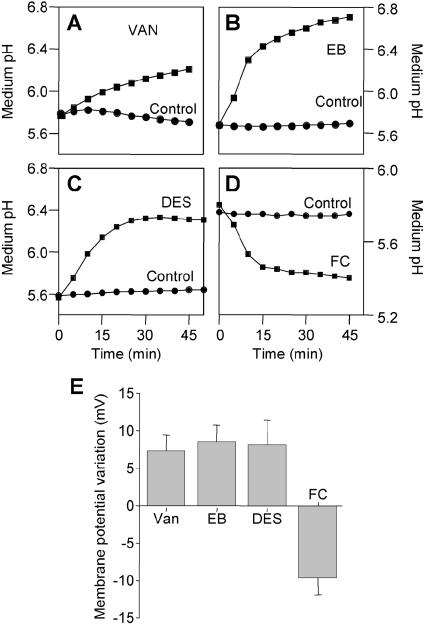
The pH of the culture medium in cultured Arabidopsis cells was monitored continuously. The pH ranged from 5.6 to 5.8. Three inhibitors of the plasma membrane H+-ATPase (vanadate [200 μm Van]; erythrosin B [25 μm EB]; and diethyl stilbestrol [100 μm DES]) rapidly induced medium alkalinization (ranged from 0.4–1 pH units in less than 30 min) as shown in Figure 2, A, B, and C, and slightly depolarized the cells (ranged from +6 mV to +12 mV) in less than 1 min (Fig. 2E). Depolarizations induced by Van (ΔEm = 7.4 ± 2.1 mV, n = 10) and EB (ΔEm = 8.6 ± 2.2 mV, n = 8) were reversible after approximately 25 min washing of the inhibitors (not shown). Fusicoccin [2 μm FC], an activator of the plant proton pump, was shown to induce both medium acidification (about 0.5 units in less than 30 min) and hyperpolarization (ΔEm = −9.7 ± 2.2 mV, n = 8) of the membrane (Fig. 2, D and E). These data revealed the electrogenic functioning of the proton pump in the plasma membrane of Arabidopsis suspension cells.
Figure 2.
pH and plasma membrane potential responses elicited by modulators of H+-ATPases in intact Arabidopsis cell suspensions. For A, B, C, and D, modulators of plasma membrane H+-ATPases were introduced into the culture medium at 0 min. The pH of the culture medium was monitored over 45 min. The initial pH ranged from 5.6 to pH 5.8. Each experiment was repeated at least three times and representative results are shown. A, B, and C show medium alkalinizations induced by three different plasma membrane H+-ATPase inhibitors: respectively 200 μm Van (after 45 min incubation ΔpH = 0.46 ± 0.03 units; n = 3), 50 μm EB (ΔpH = 0.34 ± 0.03 units; n = 3) and 100 μm DES (ΔpH = 0.26 ± 0.09 units; n = 3). D shows medium acidification induced by 2 μm fusicoccin, an activator of plasma membrane H+-ATPase (FC, ΔpH = 0.22 ± 0.12 unit; n =9). For E, maximal variations of the plasma membrane potential (mean value ± sd) elicited by plasma membrane H+-ATPases modulators were monitored after impalement of intact Arabidopsis suspension cells with microelectrodes. Variations were for 200 μm Van: ΔEm = 7.4 ± 2.1 mV (n = 10), 25 μm EB: ΔEm = 8.6 ± 2.2 mV (n = 8), 100 μm DES: ΔEm = 8.5 ± 3.3 mV (n = 6) and 10 μm FC: ΔEm = −9.7 ± 2.2 mV (n = 8). The resting membrane potential was −42 ± 9.5 mV (n = 132).
ABA Inhibits the Plasma Membrane Proton Pumping
As we have shown here, inhibition of H+-ATPase also has a depolarizing effect. Furthermore, plasma membrane H+-ATPase is proposed to be involved in the ABA transduction cascade (Irving et al., 1992; Blatt, 2000). Thus, it is tempting to hypothesize that inhibition of H+-ATPase activity could contribute to the well-known ABA-induced depolarization. To examine this hypothesis, we analyzed the effects of ABA on proton pumping. In Arabidopsis cell cultures, 10 μm ABA simultaneously induced rapid alkalinization of the medium (ΔpH = 0.06 ± 0.01, n = 6) and plasma membrane depolarization (ΔEm = 5.2 ± 1.8 mV, n = 5; Figs. 3A and 1G). 200 μm Van, shown to block the H+ extrusion (Fig. 2A), inhibited the ABA-induced alkalinization (Fig. 3B). The other H+ pump inhibitors, EB and DES, produced similar effects (not shown). ABA is thus able to inhibit the proton pump, leading to the plasma membrane depolarization.
Figure 3.
The medium alkalinization elicited by ABA coincided with the plasma membrane depolarization induced by ABA. For A and B, the pH of the culture medium was monitored over time. ABA (10 μm) was introduced into the culture medium at time 0 min. A, ABA induced a medium alkalinization by comparison to untreated cells (control). B, The medium alkalinization induced by ABA (•, ΔpH = 0.06 ± 0.01 unit; n = 6) was reduced when ABA was added in the presence of 200 μm Van (○). The initial pH ranged from 5.6 to pH 5.8. Representative results out of 9 independent experiments are shown.
Plasma Membrane Depolarization Induced by ABA Involves Both Inhibition of H+ Proton Pump and Activation of Anion Channels
In response to ABA, two different depolarizing processes are enhanced: activation of anion currents and inhibition of the proton pump. Both processes could account for the ABA-induced depolarization. In order to know if these processes are dependent or not, we analyzed the effects of H+-ATPase inhibitors on the ABA-induced anion current activation and conversely, the effects of anion channel inhibitors on the ABA-induced H+-ATPase activity.
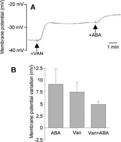
In 67% of cells pretreated for 10 min with 200 μm Van, 10 μm ABA triggered a slight depolarization of the plasma membrane ΔEm = 4.8 ± 0.7; n = 6 (Fig. 4, A and B). Similar results were obtained with 100 μm DES and 25 μm EB (not shown). To establish the cause of this depolarization, we measured the effect of ABA on anion currents in the presence of H+-ATPase inhibitors.
Figure 4.
Plasma membrane depolarization induced by ABA occurred even in the presence of H+-ATPase inhibitors. Plasma membrane potential of intact Arabidopsis suspension cells was monitored over time. Inhibitors of plasma membrane H+-ATPases were introduced into the medium before the addition of 10 μm ABA. A, 200 μm, Van was introduced 7 min before ABA. The diameter of the cell was 40 μm and the value of the resting potential was −35 mV. B, Plasma membrane potential variations elicited by 10 μm ABA, 200 μm Van, or successive additions of Van and ABA. Compilations of at least five experiments for each condition (for Van+ABA treatment: ΔEm = 4.8 ± 0.7 mV; n = 6).
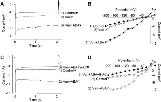
In cells pretreated with 200 μm Van, ABA treatment enhanced anion currents (Fig. 5, A and B). This ABA-induced anion current activation is similar to that which we observed in the absence of the H+-ATPase inhibitor (ΔI = 3.7 ± 1.7 nA, n = 8). The indication that this current might be due to the previously described anion currents (Ghelis et al., 2000a) was checked with 9-AC, which blocks the effect of ABA on anion channel currents (Fig. 1, H and I). A treatment with 200 μm 9-AC reduced by at least 54% the currents enhanced by ABA in the presence of Van (Fig. 5, C and D). This result showed that ABA was therefore able to trigger the activation of anion currents even if proton pumps were inhibited. That suggested that the triggering of anion current activation by ABA was independent of proton pump activity.
Figure 5.
Activation of anion currents by ABA occurred even if the H+ Pump was inhibited. A, Anion currents recorded under −200 mV pulse before (•), 5 min after the addition of 100 μm Van (○) and 4 min after a subsequent addition of 10 μm ABA (▾). Holding potential was −40 mV. The diameter of the cell was 42 μm. The mean magnitude of the anion current inhibition at −200 mV after 1.9 s of deactivation was 3.7 ± 1.7 nA (n = 8). Representative results are shown. B, Current-voltage relationships corresponding to (A). Reversal potential changes after Van addition and the subsequent addition of ABA were respectively 7 mV and 13 mV. C, Anion currents recorded under −200 mV pulses before (▿), 5 min after the conjoint addition of 100 μm Van and 10 μm ABA (Van+ABA, ○) and 3 min after a subsequent addition of 200 μm 9-AC (•). 9-AC inhibited by 54% ± 5% (n = 4) the anion currents activated by ABA. Representative results are shown. D, Current-voltage relationships corresponding to (C). Reversal potential changes after Van addition and the subsequent addition of ABA were 15 mV.
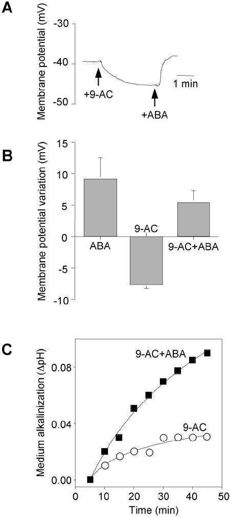
We next analyzed the effects of 9-AC on the ABA-induced inhibition of H+-ATPase. In cells pretreated with 200 μm 9-AC, 10 μm ABA was still able to provoke the depolarization of the plasma membrane ΔEm = 5.2 ± 1.8 mV; n = 5 (Fig. 6, A and B). In the presence of 200 μm 9-AC, ABA blocked the proton pump as indicated by the decreased medium alkalinization (ΔpH = 0.05 ± 0.01 unit; n = 3; Fig. 6C). This effect was similar to the one observed in the absence of the anion channel inhibitor (Fig. 3B). ABA was thus able to trigger the inhibition of proton pumps even when anion currents were inhibited. The triggering of proton pump inhibition induced by ABA was therefore independent of the anion currents.
Figure 6.
ABA induced H+-ATPase inhibition even if anion channels are blocked. A, Plasma membrane potential was monitored in response to successive additions of 200 μm 9-AC and 10 μm ABA. The resting potential was −35 mV. The diameter of the cell was 40 μm. B, Plasma membrane potential variations elicited by 10 μm ABA, 200 μm 9-AC or successive additions of 9-AC and ABA. Compilations of at least 5 experiments for each condition (for 9-AC treatment: ΔEm = −7.6 mV ±0.7 mV, n = 13; for 9-AC+ABA: ΔEm = 5.2 mV ±1.8 mV, n = 5). C, Arabidopsis cell suspensions were pretreated with 200 μm 9-AC. The pH of the culture medium was monitored over time. The medium alkalinization in the presence (<) and in the absence (○) of 10 μm ABA is shown. In the presence of 9-AC, the medium alkalinization induced by ABA was ΔpH = 0.05 ± 0.01 unit (n = 3).
The depolarization observed in response to ABA could therefore be the result of two different processes: activation of anion current and inhibition of H+-ATPase. These two processes can be independently triggered by ABA and lead to the same effect, namely the depolarization of the plasma membrane.
ABA Increases Cytoplasmic Ca2+ Concentration in Arabidopsis Suspension Cells
Inhibition of H+ proton pump and activation of anion channels in response to various stimuli were shown to be dependent on the variation of cytosolic Ca2+ (Assmann and Shimazaki, 1999; Blatt, 1999). Furthermore, ABA was shown to increase cytoplasmic calcium concentration in stomatal guard cells (MacRobbie, 1998), but the effect of ABA on cytosolic free Ca2+ levels ([Ca2+]cyt) is far less investigated for other cell types and especially for suspension cells. Before studying the implication of Ca2+ in relation with the ABA-induced plasma membrane depolarization, we investigated the action of ABA on [Ca2+]cyt in Arabidopsis suspension cells. We treated with 10 μm ABA an Arabidopsis cell suspension expressing the Ca2+-sensitive luminescent protein aequorin. Immediately after the introduction of ABA into the medium of the cells, we observed two successive transient increases in [Ca2+]cyt, 0.242 ± 0.007 μm and 0.135 ± 0.003 μm (n = 20), respectively. These are significantly larger than the ones obtained after a control treatment: 0.166 ± 0.01 μm and 0.113 ± 0.001 μm; n = 8 (Fig. 7A).
Figure 7.
ABA increased Ca2+ in cytosol of Arabidopsis cell suspension. A, Changes in [Ca2+]cyt elicited by 10 μm ABA were measured by using cell suspensions derived from Arabidopsis leaves transformed by the apoaequorin gene. At least 6 independent experiments were done and gave similar results. Means values ± se are given in the text. Representative results are shown. B, Effects of Ca2+ chelator and inhibitors of plasma membrane calcium channels on the ABA-induced changes in [Ca2+]cyt. Chelator and inhibitor concentrations were EGTA 5 mm, La3+ 100 μm, pimozide 50 μm, fluspirilene 50 μm. Each bar represents the mean [Ca2+]cyt of 5 independent experiments expressed in percentage of the control value. Standard errors are indicated.
Chelation of extracellular Ca2+ by 5 mm EGTA greatly reduced the two transient increases (Fig. 7B). This suggests that the two [Ca2+]cyt increases we observed in response to ABA mainly resulted from the mobilization of extracellular calcium. Mobilization of extracellular calcium generally occurs through calcium influx. This implied that plasma membrane calcium channels were involved. We therefore examined the effects of inhibitors of plasma membrane calcium channels on the increases in [Ca2+]cyt observed in response to ABA. We used specific inhibitors with different structures but similar effects on Ca2+ currents: La3+, which affects most of the plasma membrane Ca2+-channels, and pimozide and fluspirilene, two diphenylbutylpiperidine type Ca2+-channel inhibitors blocking L-, N-, and T-type Ca2+-channel (Pineros and Tester, 1997; Arnoult et al., 1998; Ghelis et al., 2000b). The three channel inhibitors reduced the first transient [Ca2+]cyt increase, whereas they completely blocked the second (Fig. 7B). As in guard cells, ABA increases [Ca2+]cyt in Arabidopsis suspension cells especially via the activation of plasma membrane Ca2+ channels.
ABA-Induced H+ Pump Inhibition Requires Ca2+ Influx
We therefore set out to establish whether the two processes responsible for the membrane depolarization upon ABA treatment are mediated via calcium signaling in suspension cells.
Chelation of extracellular Ca2+ by 5 mm EGTA abolished ABA-induced medium alkalinization (Fig. 8A). The ABA-induced alkalinization was decreased by about 90% after a 45 min external application of EGTA in the culture medium. Since mobilizable extracellular calcium is required for the inhibition of H+-ATPase, we therefore examined the effects of Ca2+-channel inhibitors on the ABA-induced medium alkalinization. La3+, pimozide, and fluspirilene greatly reduced the medium alkalinization induced by ABA (Fig. 8, B and C), providing clear evidence for the involvement of extracellular calcium in the ABA-induced inhibition of the H+-ATPase causing the plasma membrane depolarization.
Figure 8.
ABA-induced H+ pump inhibition requires external Ca2+ influx. Arabidopsis cell suspensions were treated with 10 μm ABA in the presence (○) or absence (•) of Ca2+ chelator or inhibitors of plasma membrane calcium channels. ABA and Ca2+ modulators were introduced into the culture medium at 0 min and the pH of the culture medium was monitored over time. The initial pH ranged from 5.6 to pH 5.8. Each experiment was repeated at least three times and representative results are shown. A, Inhibition of the ABA-induced alkalinization by 5 mm EGTA (○). B, Inhibition of the ABA-induced alkalinization by 50 μm pimozide (○). C, Inhibition of the ABA-induced alkalinization by 100 μm La3+ (○).
ABA-Induced Activation of Anion Currents Is Mediated by Calcium Signaling
We next examined whether calcium was involved in the activation of anion currents under ABA treatment.
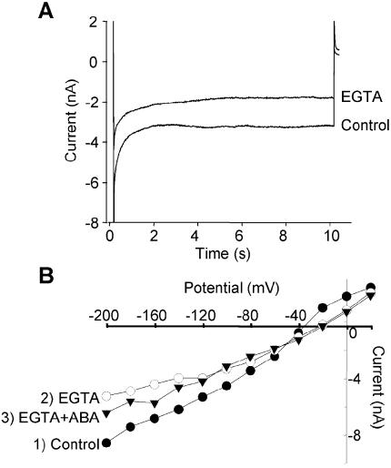
Extracellular EGTA (5 mm) reduced anionic currents by 39.2% ± 18% (n = 7) within 2 to 7 min in 7 out the 11 cells we analyzed (Fig. 9A). Figure 9B shows the reduced effect of ABA when added in the presence of EGTA. The finding that EGTA can at least partially block ABA-induced anion efflux current demonstrates that external Ca2+ is clearly involved in the anion channel modulation. We further investigated the role of calcium channels by using inhibitors shown to reduce the [Ca2+]cyt increases induced by ABA. We adopted an experimental protocol similar to the one used for EGTA. Effects of the different calcium channel inhibitors on the ABA-induced anion channel activation were summarized Figure 10.
Figure 9.
EGTA reduced ABA-induced anion current activation in intact Arabidopsis suspension cells. A, Anion currents recorded under −200 mV pulse before and 5 min after the addition of 5 mm EGTA. Holding potential was −40 mV. The diameter of the cell was 42 μm. The anion current inhibition induced by EGTA at −200 mV was 39.2% ± 18% (mean value ± sd; n =7). B, Current-voltage relationships of anion currents monitored before (•) and 15 min after (○) the addition of 5 mm EGTA and 6 min after a subsequent addition of 10 μm ABA (▾). The diameter of the cell was 35 μm. Plasma membrane currents resulted from voltage pulses from −200 to +20 mV (in 20 mV steps for 2 s) recorded at 1.9 s. Holding potential was −40 mV.
Figure 10.
Ca2+ channel blockers reduced ABA-induced anion current activation in Arabidopsis suspension cells. The effects of plasma membrane Ca2+ channel blockers on the anion currents activated by ABA were determined as described Figure 9. ABA represents the anion currents activated by 10 μm ABA. Anion currents measured in the presence of Ca2+ channel blockers were given in % with respect to the anion currents measured with ABA (100%). A total of 5 mm EGTA: ΔI = 39.2% ± 18% (mean value ± sd; n = 7); 100 μm La3+: ΔI = 77.2% ± 7.3%; n = 5; 50 μm pimozide: ΔI = 33.8% ± 5.5%; n = 5; 50 μm fluspirilene: ΔI = 40.8% ± 4.6%; n = 5.
Application of 100 μm La3+ decreased anion currents by approximately 85%. Pimozide and fluspirilene reduced anion currents by approximately 30%. Taken together, these results clearly suggest the involvement of changes in [Ca2+]cyt, triggered by Ca2+ influxes, in the ABA-induced activation of anion channels that occurred during depolarization of the plasma membrane.
DISCUSSION
The aim of this work was to identify the nature of the plasma membrane depolarization involved in the ABA signaling process in Arabidopsis suspension cells. We present experiments employing combined voltage clamping (Bouteau et al., 1999) and continuous measurements of extracellular pH during plasma membrane ABA signaling on cells where physiological cell wall functions are maintained. We extend previous data by showing that the typical early membrane depolarization triggered by ABA (Jeannette et al., 1999) might be mediated by the reduction of the H+-ATPase activity in addition to the activation of anion channels in Arabidopsis cells (Ghelis et al., 2000a). The results presented in this paper clearly show that H+-ATPase activity and slow anion channel activation have synergistic effects in promoting the early plasma membrane depolarization evoked by ABA.
Arabidopsis Suspension Cells under Resting Conditions
In suspension cells under resting conditions Em was −42 ± 9.5 mV (n = 132), and we found that the electrogenic H+ pumps contribute weakly but clearly to the transmembrane whole current. Furthermore, the medium alkalinization induced by H+-ATPase inhibitors (Fig. 2, A, B, and D) correlates reasonably well with the plasma membrane depolarization (+6 < ΔEm < +12 mV) recorded in vivo (Fig. 2E) during the pH shift in the medium (0.2 < ΔpH <0.5 units). The use of inhibitors with different structures but similar effects (Van, EB, and DES) argues for the existence of active H+-ATPase in the plasma membrane of Arabidopsis suspension cells, as pointed out by Schaller and Oecking (1999) on L. peruvianum suspension cells. In addition, good correlations exist between the rapid acidification of the medium induced by fusicoccin (FC) and the plasma membrane hyperpolarization (Fig. 2, D and E) mediated by the activation of the H+-ATPase (Blatt, 1988; Zingarelli et al., 1999).
In Arabidopsis cells, the proton pump is poorly balanced by a K+ inward current, in contrast to what is described in guard cell (Thiel et al., 1992). Our results suggest rather that most of the cells (71%) exhibited inward current mediated by an anion current (Ghelis et al., 2000a). This situation implies that the net anion efflux is a direct function of the proton pump output and contributes to the diffusion potential.
Indeed, a previous tail-current experiment on intact cells (Jeannette et al., 1999) identified that Em and Erev clearly depend on the K+ Nernst potential. However, the change in Erev was lower than the change in EK (−31 mV instead of −58 mV per decade) expected for an exclusive K+ membrane selectivity. So, the observed positive shift could be mainly due to NO3− efflux into the bathing medium used (20 mm KNO3 and 0.9 mm CaCl2) given the high permeability of plasma membrane to NO3− (PNO3−/PCl− ≫ 1; Miller and Smith, 1996; Frachisse et al., 1999). Thus, the reversal potential (≈ −27 mV) obtained in the presence of K+ inhibitors (not shown) was close to the expected value of anion equilibrium potential, i.e. ECl = −28 mV with [Cl]i = 10 mm (Grabov et al., 1997) and ENO3 = −17 mV with [NO3]i ≈ 10 mm and [NO3]o = 20 mm (Miller and Smith, 1996; Crawford and Glass, 1998). Then, the diffusion potential (Edif) for both anions was about −21 mV according to the Goldman-Hodgkin-Katz relation.
ABA Induces the Inhibition of Proton Pumps and the Activation of Anion Channels
Cell cultures of Arabidopsis respond to the addition of ABA with a rapid alkalinization of the medium (ΔpH ≈ 0.03–0.15 units, Fig. 3A) during the depolarization (Jeannette et al., 1999). This ABA effect is similar to those of the proton pump inhibitors Van, EB, and DES (Fig. 2, A–C), although the medium alkalinization promoted by the proton pump inhibitors was about 50% higher in magnitude. Since the ABA-induced alkalinization does not occur in cells pretreated with one of the proton pump inhibitors (Fig. 3B), this alkalinization is thus dependent on active proton pumps. This suggests that the target of ABA that leads to the medium alkalinization could be either the proton pump itself or a signaling pathway controlling its activity. As the ABA-induced medium alkalinization is concomitant with the ABA-induced plasma membrane depolarization, one part of the depolarization would be due to the reduction of net proton efflux via the inhibition of the plasma membrane proton pump by ABA. However, we cannot exclude that other mechanisms could also participate in the medium alkalinization induced by ABA. Activation of proton-coupled-transport system or activation of NADPH oxidase by ABA that has already been described in guard cells (Kwak et al., 2003) could account to some extent for the medium alkalinization.
In guard cells in which the membrane potential was much more negative than the K+ equilibrium potential, ABA shifted the potential to a more depolarized state (Blatt and Armstrong, 1993). It was suggested by Thiel and co-authors (1992) that the rapid depolarization recorded on Vicia stomatal guard cells was due to the activity of anion channels controlled by ABA rather than an ABA-induced fall in H+-ATPase output. However, several pieces of evidence suggest that proton pumps are involved in the stomatal activity. Stomatal opening is known to be enhanced by light (Assmann and Shimazaki, 1999), and it was reported that light-stimulated stomatal opening was inhibited by Van in a similar manner to ABA (Gepstein et al., 1982; Schwartz et al., 1991; MacRobbie, 1998). Hence, Van and ABA might have a common target. Furthermore, blue-light-induced stomatal opening is dependent on the activation of the proton pump (Assmann et al., 1985; Goh et al., 1996), and blue-light-dependent H+ pumping in V. faba was inhibited by 65% in the presence of ABA (Goh et al., 1996). ABA appears able, therefore, to modulate the H+-ATPase output in vivo.
If the proton pump is inhibited by ABA, the cytoplasmic pH should become more acidic. In response to ABA, the pH change detectable in Vicia guard cells is an alkalinization (increase about 0.2 pH units) of the cytoplasm (Blatt and Armstrong, 1993; Grabov and Blatt, 1997; MacRobbie, 1998). A possible explanation is that ABA elicits multiple actions inside and outside the cell (MacRobbie, 1995), involving also the tonoplast. The regulation of vacuolar H+-ATPase or H+-pyrophosphatase by internal ABA is suggested by several authors (Kasai et al., 1993; Barkla et al., 1999). In both cases, the stimulation of vacuolar H+-ATPase and H+-pyrophosphatase must strongly induce a cytoplasmic alkalinization in intact cells, maybe even after plasma membrane ABA-induced H+-ATPase inhibition.
Taken together, these observations suggest that the inhibition of H+-ATPase by ABA is involved in the ABA-induced depolarization in addition to the ABA-induced anion channel activation. Hence, a synergy does exist between the two mechanisms and this synergy leads to the plasma membrane depolarization in response to ABA. If these two mechanisms are independently triggered by ABA, since the inhibition of anion channels or the inhibition of the proton pump does not fully impair the ABA-induced plasma membrane depolarization, this does not exclude the possibility that the two processes could operate in concert. As shown Figure 1C, anion currents are enlarged by depolarizing conditions. Inhibition of proton pumps provoked by ABA could be one such condition. The depolarization resulting from the inhibition of proton pumps could in turn enhance anion currents leading to a rapid amplification of the signal.
Ca2+ Has a Dual Effect Leading to Plasma Membrane Depolarization: It Impairs Proton Pumping and Enhances Anion Channels
We next show that both induced depolarizing mechanisms are Ca2+-dependent. In conditions where Ca2+ influx was impaired either by using extracellular Ca2+ chelator (EGTA), or Ca2+ channel inhibitors with different structures, which have been shown to decrease stimulus-induced elevations of [Ca2+]cyt in our Arabidopsis suspension cells and other plant materials such as Commelia guard cells (Gilroy et al., 1991) and Arabidopsis seedlings (Knight et al., 1997), we observed that ABA-induced inhibition of proton pumping was abolished (Fig. 8). Hence, ABA does not inhibit proton pumping directly but through an increase in cytosolic Ca2+. This is consistent with previous observations showing that ABA had by itself no effect on proton pumping in isolated microsomes (Blum et al., 1988; Goh et al., 1996). Inhibitions of H+-ATPase by Ca2+ were previously reported for different plant materials. In L. peruvianum cell suspensions, the inhibition of Ca2+ influx by BAPTA, a calcium chelator, or La3+ impaired the systemin-induced medium alkalinization that occurs through a decrease in H+-ATPases (Schaller and Oecking, 1999). Confirmation of the sensitivity of H+-ATPases to Ca2+ was obtained in vitro. Reconstitued proteoliposomes containing plasma membrane proteins of B. vulgaris roots displayed the two H+-E1-E2-ATPase properties, namely H+-transport and ATP hydrolysis. In the presence of free Ca2+, both H+-transport and ATP hydrolysis were reduced, indicating that the two H+-ATPase properties are Ca2+-sensitive (Lino et al., 1998). In microsomal membranes of V. faba guard cells, both H+-ATPase properties were also reduced by micromolar concentrations of Ca2+, the inhibitory effect of Ca2+ being reversed by the addition of the Ca2+ chelator BAPTA (Kinoshita et al., 1995).
Allen et al. (1999) reported that anion channel activation by ABA could be mimicked by an external application of Ca2+, indicating that such an activation was Ca2+-dependent in Arabidopsis guard cells. Here, we confirmed in Arabidopsis cell suspensions this result by an opposite approach. By impairing Ca2+ influx with Ca2+ chelator or Ca2+ channel inhibitors, we reduced the anion channel activation (Fig. 10). Ca2+-dependence seems to be a common feature of Arabidopsis anion channels since the open probability of anion channels of Arabidopsis hypocotyl cells was also reported to depend on a rise in [Ca2+]cyt (Lewis et al., 1997).
The two depolarizing mechanisms induced by ABA are Ca2+-dependent and therefore dependent on an increase in [Ca2+]cyt promoted by ABA. Indeed, in V. faba guard cells, there is good evidence that ABA produces an increase in [Ca2+]cyt by promoting Ca2+ influx across the plasma membrane through the activation of nonselective and voltage-independent cation channels (Schroeder and Hagiwara, 1990; Thiel et al., 1992; White, 2000) or voltage-sensitive calcium channels (Grabov and Blatt, 1998; Hamilton et al., 2000), and also by releasing Ca2+ from intracellular stores (MacRobbie, 1997; McAinsh and Hetherington, 1998; Assmann and Shimazaki, 1999; Blatt, 2000). In Arabidopsis cells, blocking the H+-ATPase with Van or DES did not affect the ABA-induced anion channel activation (Fig. 5, A and B) and thus did not impair the ABA-induced depolarization (Fig. 4, A and B). Conversely, anion channel inhibitors did not affect the decrease in proton pump activity brought about by ABA (Fig. 6). This indicates that the depolarization initiated by the reduction of the proton pump and the activation of anion channels are not the first signals that open Ca2+-permeable channels.
ABA Enhances Two Depolarizing Processes by a Single or Multiple Signaling Pathway(s)
The finding that the two depolarizing processes are Ca2+-dependent could suggest that they may be controlled by ABA at least for the first steps by a common signaling pathway. We previously showed that induction of RAB18 gene expression by ABA was dependent on Ca2+ entry. We observed that the inductive effect of ABA on RAB18 gene expression was reduced by EGTA, pimozide, and fluspirilene (Ghelis et al., 2000b). Interestingly, the Ca2+ entry blockers that reduce the effect of ABA on anion channels and proton pumps are also efficient to reduce the effect of ABA on RAB18 gene expression, suggesting that anion channels, proton pumps, and RAB18 gene expression belong to an identical ABA-response pathway. This is consistent with our previous results showing that anion channel activation was required for RAB18 gene expression induced by ABA (Ghelis et al., 2000a). Does it also mean that proton pump inhibition would be another actor of the signaling pathway leading to the expression of RAB18 gene in response to ABA?
Role of the Plasma-Membrane Depolarization Induced by ABA
Since ABA recruits two independent depolarizing mechanisms, one can wonder whether the depolarization induced by ABA would not be, by its own, an important signal necessary for the accomplishment of the ABA responses. Such an aspect was already studied for the response to systemin, a systemic signal of wounding in tomato (Lycopersicon esculentum) plants. Systemin is known to depolarize cell plasma membranes (Moyen and Johannes, 1996) and to enhance the expression of genes encoding systemic wound-response proteins (SWRP-gene; Pearce et al., 1991). The inductive effect of systemin on SWRP-gene was mimicked by treating tomato plants with molecules able to dissipate H+ and K+ gradients, namely molecules able to depolarize plasma membranes. This could suggest a role for the plasma membrane potential in the control of gene expression (Schaller and Frasson, 2001). In previous articles (Ghelis et al., 2000a; Hallouin et al., 2002), we showed that the induction of RAB18 gene expression in response to ABA was dependent on the ABA induced activation of anion channels. Since we now know that plasma membrane depolarization induced by ABA involved both anion channel activation and proton pumping reduction, we have now to determine what signal is responsible for the induction of the RAB18 gene expression: the variation of the anion currents or the plasma membrane depolarization induced by this anion current variation.
Taken together, our results show that ABA with Ca2+ as second messenger has a dual effect; it provokes plasma-membrane depolarization both by impairing proton pumping and by stimulating anion channels. The results presented here are the first to our knowledge to identify two Ca2+-regulated components of the plasma membrane depolarization induced by ABA in cells where physiological cell wall functions were maintained. The mechanisms involved in ABA-induced plasma membrane depolarization in Arabidopsis suspension cells are similar to the ones at the origin of plasma membrane depolarization induced by ABA in guard cells. Although suspension cells do not display the ABA responses observed in guard cells, like turgor decrease for example, the signaling pathway promoted by ABA involved in both cell types similar actors: proton pumps and anion channels. This indicates that they are of general importance for early ABA signaling in the different types of plant cells (Schroeder et al., 2001).
MATERIALS AND METHODS
Arabidopsis Cell Cultures
A suspension cell culture from Arabidopsis (ecotype Columbia) was maintained as described previously (Jeannette et al., 1999). Cells were cultured at 24°C, under continuous white light (40 μE m−2 s−1) with an orbital agitation at 130 rpm, in 500-mL Erlenmeyer flasks containing 200 mL JPL culture medium (pH 5.7) (Jouanneau and Péaud-Lenoël, 1967) and transferred to fresh medium every 7 d. All the experiments were conducted on 4-d-old cells. The viability of the cells during the experimental time course was systematically checked with trypan blue tests (not shown).
Cytoplasmic Ca2+ Measurement
For determination of cytoplasmic Ca2+ variations upon ABA treatment, cell suspensions were prepared from leaves of Arabidopsis plants transformed with the apoaequorin gene (Knight et al., 1991) and maintained according to Axelos et al. (1992). For calcium measurement, aequorin was reconstituted by overnight incubation of cell suspensions in Gamborg medium (Gamborg et al., 1968) supplemented by 30 g × L−1 Suc and 2.5 μm native coelenterazine (Uptima). A total of 200 μL of cell suspension was placed in a luminometer cuvette. Treatments were performed by pipet-injection of 2 μL medium containing or not containing 10 μm ABA. Aequorin luminescence was monitored every 0.2 s with a FB12-Berthold luminometer. At the end of each experiment, the residual aequorin was discharged by addition of 10% ethanol and 1 m CaCl2 (final concentrations). The resulting luminescence was used to estimate the total amount of aequorin in each experiment. Calibration of calcium measurements was performed by using the equation: pCa = 0.332588(−logk)+5.5593, where k is a rate constant equal to luminescence counts per second divided by total remaining counts (Knight et al., 1996). To test the effects of EGTA and the different calcium channel inhibitors, cell suspensions were pretreated for 15 min before the addition of ABA.
pH Responses Measurements
Continuous measurements of extracellular pH were performed in 5 mL of cultured cells. The experiments were run simultaneously in 2 × 10-mL flasks (control and test) each containing 1gFW/5 mL of suspension medium, at 24°C, with orbital shaking at 60 rpm. For each condition, the medium pH at the beginning of the experiment was between 5.8 and 6.0. Simultaneous changes in pH were followed on both suspension medium by using two Metrohm 632 pH meters with pH sensitive combined electrodes functioning in parallel and values were monitored during 45 min. The effectors were added when a stable pH was obtained. The buffer capacity of the culture medium was 4 μeq OH−/pH unit.
Electrophysiology
The cells were equilibrated for 24 h before electrophysiological experiments in fresh culture medium (20 mM KNO3, 0.9 mm CaCl2, and 0.45 mm MgSO4, pH 6.8). Cells were immobilized by means of a microfunnel (approximately 30–80 μm o.d.) and controlled by a pneumatic micromanipulator (de Fonbrune, Ets Beaudoin, Paris). Impalements were carried out with a piezoelectric micromanipulator (PCS-5000, Burleigh, NY) on suspension cells in a continuous flow chamber (500 μL) made of perpex. The flow rate was adjusted by gravity to approximately 150 μL min−1. ABA, channels inhibitors, Van, EB, DES, and FC were diluted in the bathing medium. The bathing medium was introduced via a polyethylene catheter, and the continuous flow could be stopped at any given time. Voltage-clamp measurements of whole-cell currents from intact cells were carried out at room temperature (20–22°C) using the technique of the discontinuous single voltage-clamp microelectrode (Bouteau et al., 1999; Jeannette et al., 1999). In this technique, both current passing and voltage recording use the same microelectrode. Interactions between the two tasks are prevented by time sharing techniques. A specific software (pCLAMP 8) drives the voltage clamp amplifier (Axoclamp 2A, Axon Instruments, Foster City, CA). Microelectrodes were made from borosilicate capillary glass (Clark GC 150F, Clark Electromedical, Pangbourne Reading, UK) drawn out on a vertical puller (Narishige PEII, Japan). Their tips were 0.5 μm diameter, filled with 600 mm KCl, and had electrical resistances between 50 and 80 MΩ with the buffer solution. Voltage and current were simultaneously displayed on a dual input oscilloscope (Gould 1425, Gould Instruments, Hainault, UK), digitalized with a PC computer fitted with an acquisition board (Labmaster TL 1, Scientific Solution, Solon, OH). In whole-cell current measurements the membrane potential was held at −40 mV. Two voltage protocols were used to study the inward currents. In the first, currents were activated by a depolarizing prepulse (+100 mV for 4.5 s), then hyperpolarizing pulses ranging from −200 to +20 mV for 15 s (in 20 or 40 mV steps). In the other, currents were obtained by hyperpolarizing pulses from −200 to −20 mV (20 mV, 2 s steps). We systematically checked that cells were correctly clamped by comparing the protocol voltage values with those really imposed. Current-voltage relationships were obtained from currents, normalized at the holding potential and measured at 2000 ms for steady-state currents.
Chemicals
EB, DES, FC, 9-AC, EGTA, pimozide, fluspirilene, and verapamil were obtained from Sigma-Fluka Chemical. Stock solutions of DES, 9-AC, and fluspirilene were prepared in methanol.
Acknowledgments
We thank Drs. H. Barbier-Brygoo, J.-M. Frachisse, and C. Grigon for helpful discussions and Dr. G. Noctor for critical reading of the manuscript. We are grateful to F. Bardat, M. Convert, and R. Kiolle for cell culture and technical assistance.
Article, publication date, and citation information can be found at www.plantphysiol.org/cgi/doi/10.1104/pp.104.039255.
References
- Allen GJ, Kutchisu K, Chu SP, Murata Y, Schroeder JI (1999) Arabidopsis abi-1 and abi-2 phosphatase mutations reduce abscisic acid-induced cytoplasmic calcium rises in guard cells. Plant Cell 11: 1785–1798 [DOI] [PMC free article] [PubMed] [Google Scholar]
- Arnoult C, Villaz M, Florman HM (1998) Pharmacological properties of the T-type Ca2+ current of mouse spermatogenic cells. Mol Pharmacol 53: 1104–1111 [PubMed] [Google Scholar]
- Assmann SM (1993) Signal transduction in guard cells. Annu Rev Cell Biol 9: 345–375 [DOI] [PubMed] [Google Scholar]
- Assmann SM, Shimazaki K (1999) The multisensory guard cell. Stomatal responses to blue light and abscisic acid. Plant Physiol 119: 809–815 [DOI] [PMC free article] [PubMed] [Google Scholar]
- Assmann SM, Simoncini L, Schroeder JI (1985) Blue light activates electrogenic ion pumping in guard cell protoplasts of Vicia faba. Nature 318: 285–287 [Google Scholar]
- Axelos M, Curie C, Mazzolini L, Bardet C, Lescure B (1992) A protocol for transient gene expression in Arabidopsis thaliana protoplasts isolated from cell suspension cultures. Plant Physiol Biochem 30: 123–128 [Google Scholar]
- Barbier-Brygoo H, Frachisse J-M, Colcombet J, Thomine S (1999) Anion channels and hormone signaling in plant cells. Plant Physiol Biochem 37: 381–392 [Google Scholar]
- Barkla BJ, Vera-Estrella R, Maldonado-Gama M, Pantoja O (1999) Abscisic acid induction of vacuolar H+-ATPase activity in Mesembryanthemum crystallinum is developmentally regulated. Plant Physiol 120: 811–819 [DOI] [PMC free article] [PubMed] [Google Scholar]
- Blatt MR (1988) Mechanisms of fusicoccin action: a dominant role for secondary transport in higher-plant cell. Planta 174: 187–200 [DOI] [PubMed] [Google Scholar]
- Blatt MR (1990) Potassium channel currents in intact stomatal guard cells: rapid enhancement by abscisic acid. Planta 180: 445–455 [DOI] [PubMed] [Google Scholar]
- Blatt MR (1999) Reassessing roles for Ca2+ in guard cell signaling. J Exp Bot 50: 989–999 [Google Scholar]
- Blatt MR (2000) Cellular signaling and volume control in stomatal movements in plants. Annu Rev Cell Dev Biol 16: 221–241 [DOI] [PubMed] [Google Scholar]
- Blatt MR, Armstrong F (1993) K+ channels of stomatal guard cells: abscisic-acid-evoked control of the outward rectifier mediated by cytoplasmic pH. Planta 191: 330–341 [Google Scholar]
- Blum W, Key G, Weiler EW (1988) ATPase activity in plasmalemma-rich vesicles isolated by aqueous two-phase partitioning from Vicia faba mesophyll and epidermis: characterization and influence of abscisic acid and fusicoccin. Physiol Plant 72: 279–287 [Google Scholar]
- Bouteau F, Dellis O, Rona J-P (1999) Transient outward K+ currents through plasma membrane of laticifer from Hevea brasiliensis. FEBS Lett 458: 185–187 [DOI] [PubMed] [Google Scholar]
- Cho MH, Spalding EP (1996) An anion channel in Arabidopsis hypocotyls activated by blue light. Proc Natl Acad Sci USA 93: 8134–8138 [DOI] [PMC free article] [PubMed] [Google Scholar]
- Crawford NM, Glass ADM (1998) Molecular and physiological aspects of nitrate uptake in plants. Trends Plant Sci 3: 389–395 [Google Scholar]
- Frachisse JM, Thomine S, Colcombet J, Guern J, Barbier-Brygoo H (1999) Sulfate is both a substrate and an activator of the voltage-dependent anion channel of Arabidopsis hypocotyl cells. Plant Physiol 121: 253–261 [DOI] [PMC free article] [PubMed] [Google Scholar]
- Gamborg OL, Miller RA, Ojima K (1968) Nutrient requirement of suspensions cultures of soybean root cells. Exp Cell Res 50: 151–158 [DOI] [PubMed] [Google Scholar]
- Gepstein S, Jacobs M, Taiz L (1982) Inhibition of stomatal opening in Vicia faba epidermal tissue by vanadate and abscisic acid. Plant Sci Lett 28: 63–72 [Google Scholar]
- Ghelis T, Dellis O, Jeannette E, Bardat F, Cornel D, Miginiac E, Rona J-P, Sotta B (2000. a) Abscisic acid specific expression of Rab 18 involves activation of anion channels in Arabidopsis thaliana suspension cells. FEBS Lett 474: 43–47 [DOI] [PubMed] [Google Scholar]
- Ghelis T, Dellis O, Jeannette E, Bardat F, Miginiac E, Sotta B (2000. b) Abscisic acid plasmalemma perception triggers calcium influx essential for Rab 18 gene expression in Arabidopsis thaliana suspension cells. FEBS Lett 483: 67–70 [DOI] [PubMed] [Google Scholar]
- Gilroy S, Fricker MD, Read ND, Trewavas AJ (1991) Role of calcium in signal transduction of Commelina guard cells. Plant Cell 3: 333–344 [DOI] [PMC free article] [PubMed] [Google Scholar]
- Goh CH, Kinoshita T, Oku T, Shimazaki KI (1996) Inhibition of blue light dependent H+ pumping by abscisic acid in Vicia guard-cell protoplasts. Plant Physiol 111: 433–440 [DOI] [PMC free article] [PubMed] [Google Scholar]
- Grabov A, Blatt MR (1997) Parallel control of the inward-rectifier K+ channel by cytosolic free Ca2+ and pH in Vicia guard cells. Planta 201: 84–95 [Google Scholar]
- Grabov A, Blatt MR (1998) Membrane voltage initiate Ca2+ waves and potentiates Ca2+ increases with abscisic acid in stomatal guard cells. Proc Natl Acad Sci USA 95: 4778–4783 [DOI] [PMC free article] [PubMed] [Google Scholar]
- Grabov A, Leung J, Giraudat J, Blatt MR (1997) Alteration of anion channel kinetics in wild–type and abi1-1 transgenic Nicotiana benthamiana guard cells by abscisic acid. Plant J 12: 203–216 [DOI] [PubMed] [Google Scholar]
- Grill E, Himmelbach A (1998) ABA signal transduction. Curr Opin Plant Biol 1: 412–418 [DOI] [PubMed] [Google Scholar]
- Hallouin M, Ghelis T, Brault M, Bardat F, Cornel D, Miginiac E, Rona J-P, Sotta B, Jeannette E (2002) Plasmalemma abscisic acid perception leads to RAB 18 expression via phospholipase D activation in Arabidopsis thaliana suspension cells. Plant Physiol 130: 265–272 [DOI] [PMC free article] [PubMed] [Google Scholar]
- Hamilton DWA, Hills A, Kohler B, Blatt MR (2000) Ca2+ channels at the plasma membrane of stomatal guard cells are activated by hyperpolarization and abscisic acid. Proc Natl Acad Sci USA 97: 4967–4972 [DOI] [PMC free article] [PubMed] [Google Scholar]
- Hedrich R, Bush H, Raschke K (1990) Ca2+ and nucleotide dependent regulation of voltage dependent anion channels in the plasma membrane of guard cells. EMBO J 9: 3889–3892 [DOI] [PMC free article] [PubMed] [Google Scholar]
- Irving HR, Gehring CA, Parish RW (1992) Changes in cytosolic pH and calcium of guard cells precede stomatal movements. Proc Natl Acad Sci USA 89: 1790–1794 [DOI] [PMC free article] [PubMed] [Google Scholar]
- Jeannette E, Rona J-P, Bardat F, Cornel D, Sotta B, Miginiac E (1999) Induction of RAB18 gene expression and activation of K+ outward rectifying channels depend on an extracellular perception of ABA in Arabidopsis thaliana suspension cells. Plant J 18: 13–22 [DOI] [PubMed] [Google Scholar]
- Jouanneau J-P, Péaud-Lenoël C (1967) Growth and synthesis of proteins in cell suspensions of a kinetin dependent tobacco. Physiol Plant 20: 834–850 [Google Scholar]
- Kasai M, Tamamoto Y, Maeshima M, Matsumoto H (1993) Effects of in vivo treatment with abscisic acid and/or cytokinin on activities of vacuolar H+ pumps of tonoplast-enriched membrane vesicles prepared from barley roots. Plant Cell Physiol 34: 1107–1115 [Google Scholar]
- Kinoshita T, Shimazaki K (1999) Blue light activates the plasma membrane H+-ATPase by phosphorylation of the C-terminus in stomatal guard cells. EMBO J 18: 5548–5558 [DOI] [PMC free article] [PubMed] [Google Scholar]
- Kinoshita T, Nishimura M, Shimazaki K (1995) Cytosolic concentration of Ca2+ regulates the plasma membrane H+-ATPase in guard cells of fava bean. Plant Cell 7: 1333–1342 [DOI] [PMC free article] [PubMed] [Google Scholar]
- Knight H, Trewavas AJ, Knight MR (1996) Cold calcium signaling in Arabidopsis involves two cellular pools and a change in calcium signature after acclimatation. Plant Cell 8: 489–503 [DOI] [PMC free article] [PubMed] [Google Scholar]
- Knight H, Trewavas AJ, Knight MR (1997) Calcium signaling in Arabidopsis thaliana responding to drought and salinity. Plant J 12: 1067–1078 [DOI] [PubMed] [Google Scholar]
- Knight MR, Campbell AK, Smith SM, Trewavas AJ (1991) Transgenic plant aequorin reports the effects of touch and cold-shock and elicitors on cytoplasmic calcium. Nature 352: 524–526 [DOI] [PubMed] [Google Scholar]
- Kurkdjian A, Bouteau F, Pennarun A-M, Convert M, Cornel D, Rona J-P, Bousquet U (2000) Ion currents involved in early Nod factor response in Medicago sativa root hairs: a discontinuous single-electrode voltage-clamp study. Plant J 22: 9–17 [DOI] [PubMed] [Google Scholar]
- Kwak JM, Mori IC, Pei ZM, Leonhardt N, Torres MA, Dangl JL, Bloom RE, Bodde S, Jones JD, Schroeder JI (2003) NADPH oxidase AtrbohD and AtrbohF genes function in ROS-dependent ABA signaling in Arabidopsis. EMBO J 22: 2623–2633 [DOI] [PMC free article] [PubMed] [Google Scholar]
- Lebrun-Garcia A, Bourque S, Binet MN, Ouaked F, Wendehenne D, Chiltz A, Schaffner A, Pugin A (1999) Involvement of plasma membrane proteins in plant defense responses. Analysis of the cryptogein signal transduction in tobacco. Biochimie 81: 663–668 [DOI] [PubMed] [Google Scholar]
- Leonhardt N, Vavasseur A, Forestier C (1999) ATP binding cassette modulators control abscisic acid-regulated slow anion channels in guard cells. Plant Cell 11: 1141–1151 [DOI] [PMC free article] [PubMed] [Google Scholar]
- Leung J, Giraudat J (1998) Abscisic acid signal transduction. Annu Rev Plant Physiol Plant Mol Biol 49: 199–222 [DOI] [PubMed] [Google Scholar]
- Lewis BD, Karlin-Neumann G, Davis RW, Spalding EP (1997) Ca2+-activated anion channels and membrane depolarizations induced by blue light and cold in Arabidopsis seedlings. Plant Physiol 114: 1327–1334 [DOI] [PMC free article] [PubMed] [Google Scholar]
- Lino B, Baizabal-Aguirre VM, Gonzalez de la Vara LE (1998) The plasma membrane H+-ATPase from beet root is inhibited by a calcium-dependent phosphorylation. Planta 204: 352–359 [DOI] [PubMed] [Google Scholar]
- MacRobbie EAC (1995) ABA-induced ion efflux in stomatal guard cells: multiple actions of ABA inside and outside the cell. Plant J 7: 565–576 [Google Scholar]
- MacRobbie EAC (1997) Signaling in guard cells and regulation of ion channel activity. J Exp Bot 48: 515–528 [DOI] [PubMed] [Google Scholar]
- MacRobbie EAC (1998) Signal transduction and ion channels in guard cells. Philos Trans R Soc Lond B Biol Sci 353: 1475–1488 [DOI] [PMC free article] [PubMed] [Google Scholar]
- McAinsh MR, Hetherington AM (1998) Encoding specificity in Ca2+ signaling systems. Trends Plant Sci 3: 32–36 [Google Scholar]
- Miller AJ, Smith SJ (1996) Nitrate transport and compartmentation in cereal root cells. J Exp Bot 47: 843–854 [Google Scholar]
- Moyen C, Johannes E (1996) Systemin transiently depolarizes the tomato mesophyll cell membrane and antagonizes fusicoccin-induced extracellular acidification of mesophyll tissue. Plant Cell Environ 19: 464–470 [Google Scholar]
- Pearce G, Strydom D, Johnson S, Ryan CA (1991) A polypeptide from tomato leaves induces wound-inducible proteinase inhibitor proteins. Science 253: 895–898 [DOI] [PubMed] [Google Scholar]
- Pineros M, Tester M (1997) Calcium channels in higher plant cells: selectivity, regulation and pharmacology. J Exp Bot 48: 551–577 [DOI] [PubMed] [Google Scholar]
- Rock CD (2000) Pathways to abscisic acid-regulated gene expression. New Phytol 148: 357–396 [DOI] [PubMed] [Google Scholar]
- Roelfsema MRG, Steinmeyer R, Staal M, Hedrich R (2001) Single guard cell recordings in intact plants: light-induced hyperpolarization of the plasma membrane. Plant J 26: 1–13 [DOI] [PubMed] [Google Scholar]
- Sanders D, Brownlee C, Harper JF (1999) Communicating with calcium. Plant Cell 11: 691–706 [DOI] [PMC free article] [PubMed] [Google Scholar]
- Schaller A, Frasson D (2001) Induction of wound response gene expression in tomato leaves by ionophores. Planta 212: 431–435 [DOI] [PubMed] [Google Scholar]
- Schaller A, Oecking C (1999) Modulation of plasma membrane H+-ATPase activity differentially activates wound and pathogen defense responses in tomato plants. Plant Cell 11: 263–272 [DOI] [PMC free article] [PubMed] [Google Scholar]
- Schroeder JI, Allen GJ, Hugouvieux V, Kwak JM, Waner D (2001) Guard cell signal transduction. Annu Rev Plant Physiol Plant Mol Biol 52: 627–658 [DOI] [PubMed] [Google Scholar]
- Schroeder JI, Hagiwara S (1989) Cytosolic calcium regulates ion channels in the plasma membrane of Vicia faba guard cells. Nature 338: 427–430 [Google Scholar]
- Schroeder JI, Hagiwara S (1990) Repetitive increases in cytosolic Ca2+ of guard cells by abscisic acid activation of nonselective Ca2+ permeable channels. Proc Natl Acad Sci USA 87: 9305–9309 [DOI] [PMC free article] [PubMed] [Google Scholar]
- Schroeder JI, Keller BU (1992) Two types of anion channel currents in guard cells with distinct voltage regulation. Proc Natl Acad Sci USA 89: 5025–5029 [DOI] [PMC free article] [PubMed] [Google Scholar]
- Schwarz M, Schroeder JI (1998) Abscisic acid maintains S-type anion channel activity in ATP-depleted Vicia faba guard cells. FEBS Lett 428: 177–182 [DOI] [PubMed] [Google Scholar]
- Schwartz A, Illan N, Assmann SM (1991) Vanadate inhibition of stomatal opening in epidermal peels of Commelina communis. Cl− interferes with vanadate uptake. Planta 183: 590–596 [DOI] [PubMed] [Google Scholar]
- Schwartz A, Illan N, Schwartz M, Schaeffer J, Assmann SM, Schroeder JL (1995) Anion-channel blockers inhibit S-type anion channels and abscisic acid responses in guard cells. Plant Physiol 109: 651–658 [DOI] [PMC free article] [PubMed] [Google Scholar]
- Serrano R (1985) Plasma Membrane ATPase of Plants and Fungi. CRC Press, Boca Raton, FL
- Shimazaki K, Lino M, Zeiger E (1986) Blue light-dependent proton extrusion by guard cell protoplasts of Vicia faba. Nature 319: 324–326 [Google Scholar]
- Spalding EP, Cosgrove DJ (1992) Mechanism of blue-light-induced plasma-membrane depolarization in etiolated cucumber hypocotyls. Planta 188: 199–205 [DOI] [PubMed] [Google Scholar]
- Sze H, Li X, Palmgren MG (1999) Energization of plant cell membranes by H+ pumping ATPases: regulation and biosynthesis. Plant Cell 11: 667–689 [DOI] [PMC free article] [PubMed] [Google Scholar]
- Thiel G, MacRobbie EAC, Blatt MR (1992) Membrane transport in stomatal guard cells: the importance of voltage control. J Membr Biol 126: 1–18 [DOI] [PubMed] [Google Scholar]
- Trewavas A, Malho R (1998) Ca2+ signaling in plant cells: the big network! Curr Opin Plant Biol 1: 428–433 [DOI] [PubMed] [Google Scholar]
- Ward JM, Pei ZM, Schroeder JI (1995) Role of ion channels in initiation of signal transduction in higher plants. Plant Cell 7: 833–844 [DOI] [PMC free article] [PubMed] [Google Scholar]
- White PJ (2000) Calcium channels in higher plants. Biochim Biophys Acta 1465: 171–189 [DOI] [PubMed] [Google Scholar]
- Zingarelli L, Marre MT, Massardi F, Lado P (1999) Effects of hyper-osmotic stress on K+ fluxes, H+ extrusion, transmembrane electric potential difference and comparison with the effects of fusicoccin. Physiol Plant 106: 287–295 [Google Scholar]


