Abstract
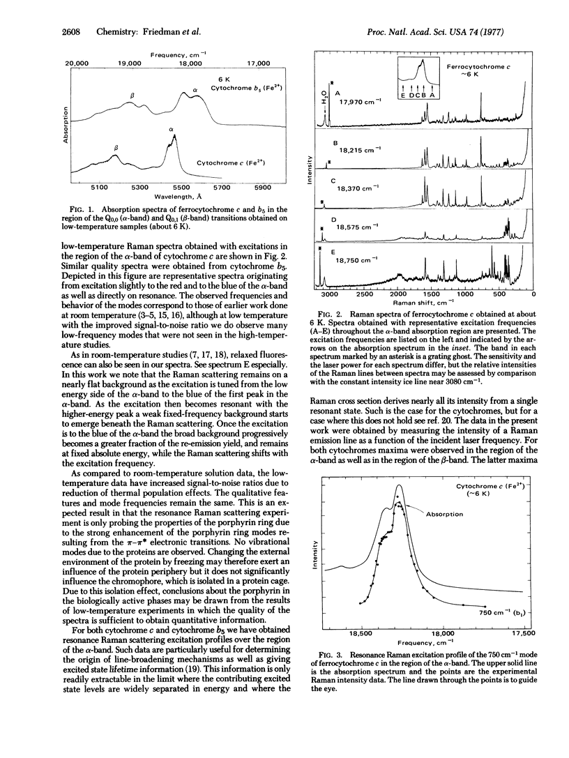
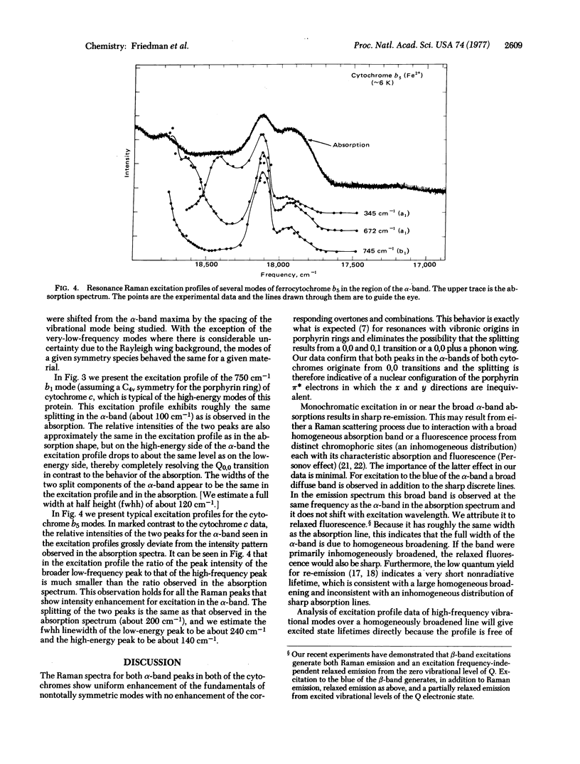
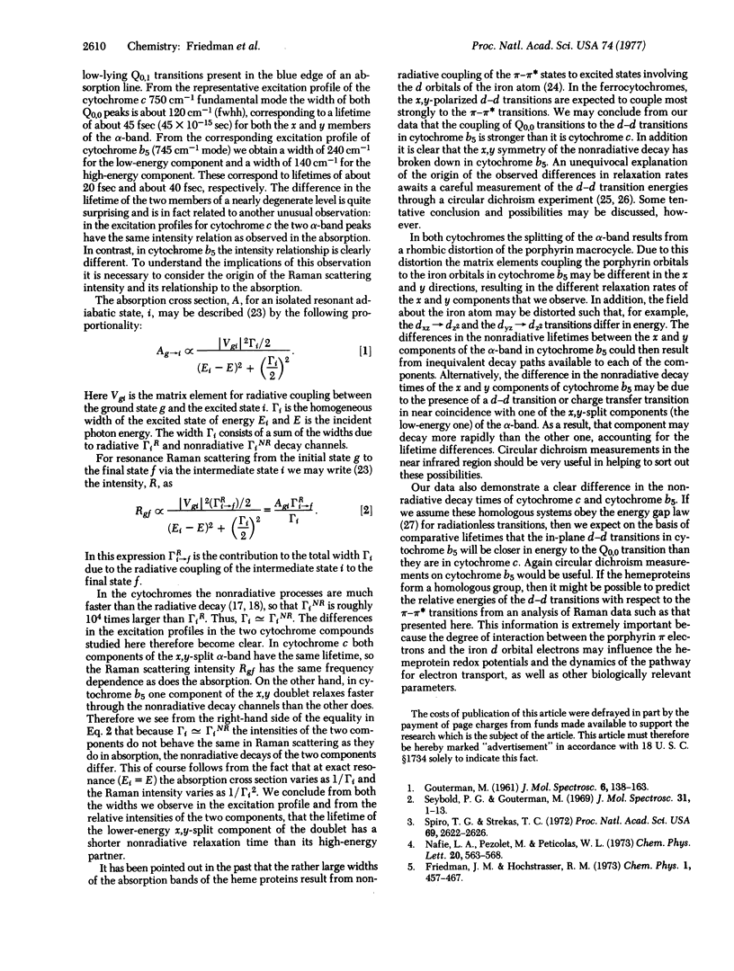
Resonance Raman scattering excitation profile data have been obtained on ferrocytochromes c and b5 in the alpha absorption band region. We observe in cytochrome c that the shape of the excitation profile agrees with the absorption band shape, while in cytochrome b5 it does not. In addition, we observe in cytochrome b5 a linewidth substantially larger than that in cytochrome c. From our data we conclude that the excited state lifetime in cytochrome c is longer than that in cytochrome b5 and that in cytochrome b5 the relaxation of the pi-pi* excited state configuration of the porphyrin ring is different in the x direction than in the y direction. Possible origins of these effects due to coupling to the d-d transitions of the iron atom are discussed.
Full text
PDF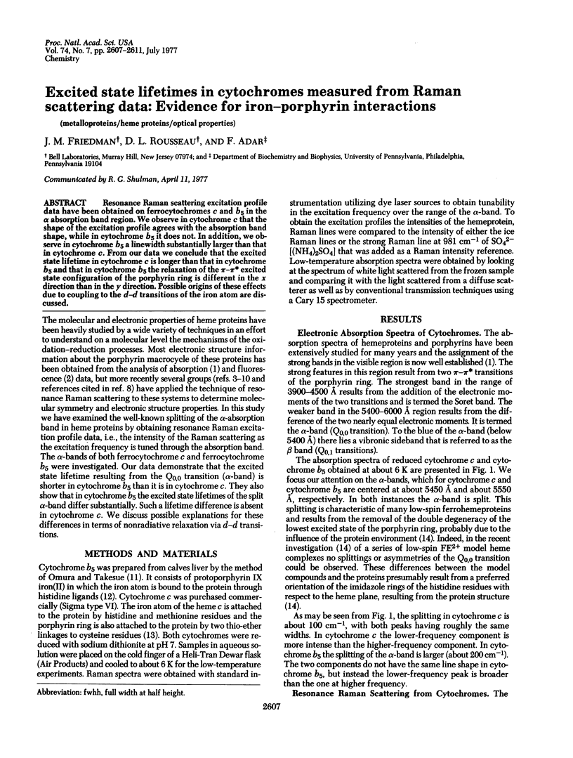
Selected References
These references are in PubMed. This may not be the complete list of references from this article.
- Adar F., Erecińska M. Resonance Raman spectra of the b- and c-type cytochromes of succinate-cytochrome c reductase. Arch Biochem Biophys. 1974 Dec;165(2):570–580. doi: 10.1016/0003-9861(74)90284-7. [DOI] [PubMed] [Google Scholar]
- Adar F. Resonance Raman spectra of cytochrome b5 and its mesoheme and deuteroheme modifications. Arch Biochem Biophys. 1975 Oct;170(2):644–650. doi: 10.1016/0003-9861(75)90160-5. [DOI] [PubMed] [Google Scholar]
- Eaton W. A., Charney E. Near-infrared absorption and circular dichroism spectra of ferrocytochrome c: d-d transitions. J Chem Phys. 1969 Nov 15;51(10):4502–4505. doi: 10.1063/1.1671818. [DOI] [PubMed] [Google Scholar]
- Kakudo M. Structure and function of bonito ferrocytochrome c at 2.3A resolution. Adv Biophys. 1975;7:163–191. [PubMed] [Google Scholar]
- Mathews F. S., Levine M., Argos P. Three-dimensional Fourier synthesis of calf liver cytochrome b 5 at 2-8 A resolution. J Mol Biol. 1972 Mar 14;64(2):449–464. doi: 10.1016/0022-2836(72)90510-4. [DOI] [PubMed] [Google Scholar]
- Omura T., Takesue S. A new method for simultaneous purification of cytochrome b5 and NADPH-cytochrome c reductase from rat liver microsomes. J Biochem. 1970 Feb;67(2):249–257. doi: 10.1093/oxfordjournals.jbchem.a129248. [DOI] [PubMed] [Google Scholar]
- Spiro T. G., Strekas T. C. Resonance Raman spectra of hemoglobin and cytochrome c: inverse polarization and vibronic scattering. Proc Natl Acad Sci U S A. 1972 Sep;69(9):2622–2626. doi: 10.1073/pnas.69.9.2622. [DOI] [PMC free article] [PubMed] [Google Scholar]
- Wagner G. C., Kassner J. Spectroscopic properties of low-spin ferrous heme complexes and hemeproteins at 77degrees K. Biochem Biophys Res Commun. 1975 Mar 17;63(2):385–391. doi: 10.1016/0006-291x(75)90700-7. [DOI] [PubMed] [Google Scholar]


