Abstract
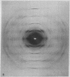
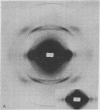
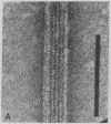
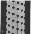
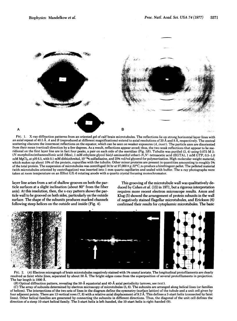
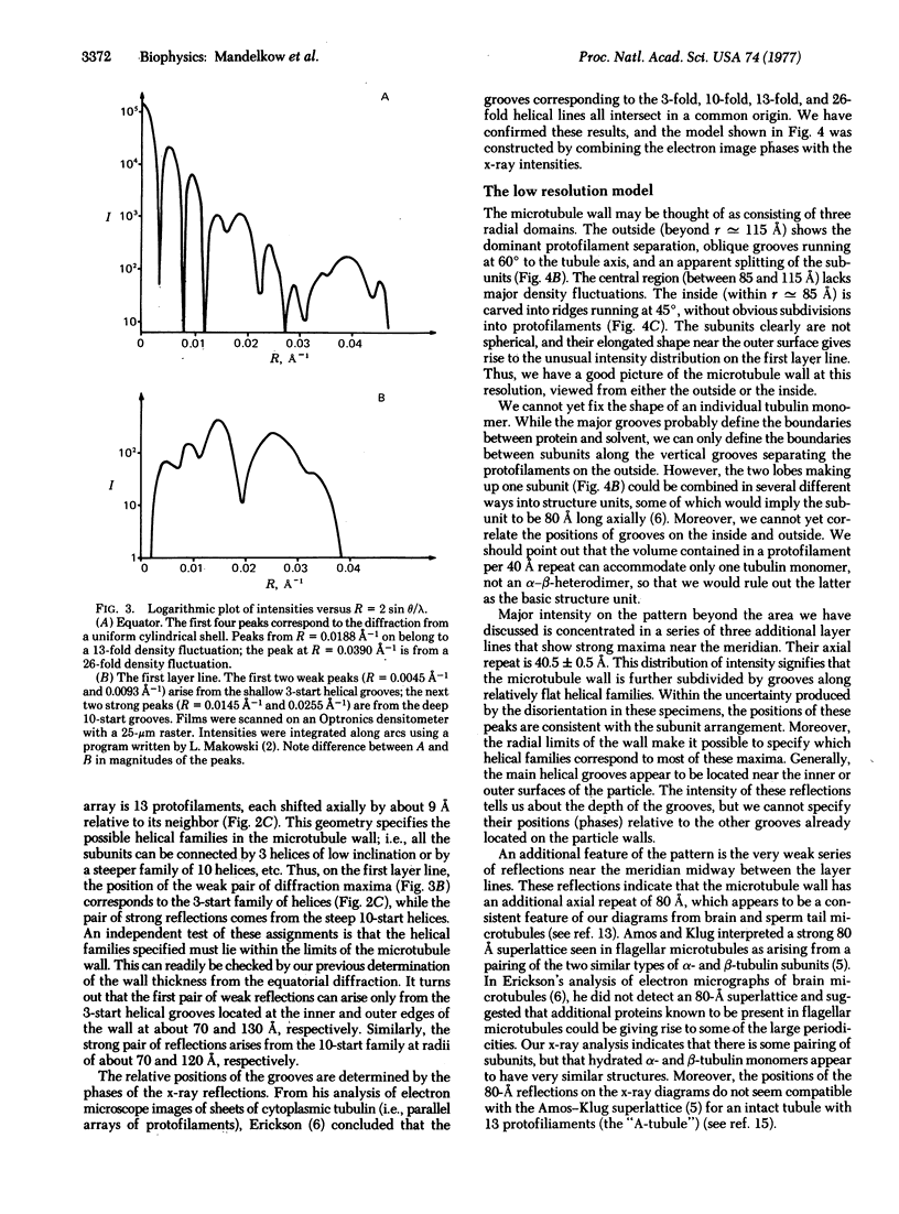
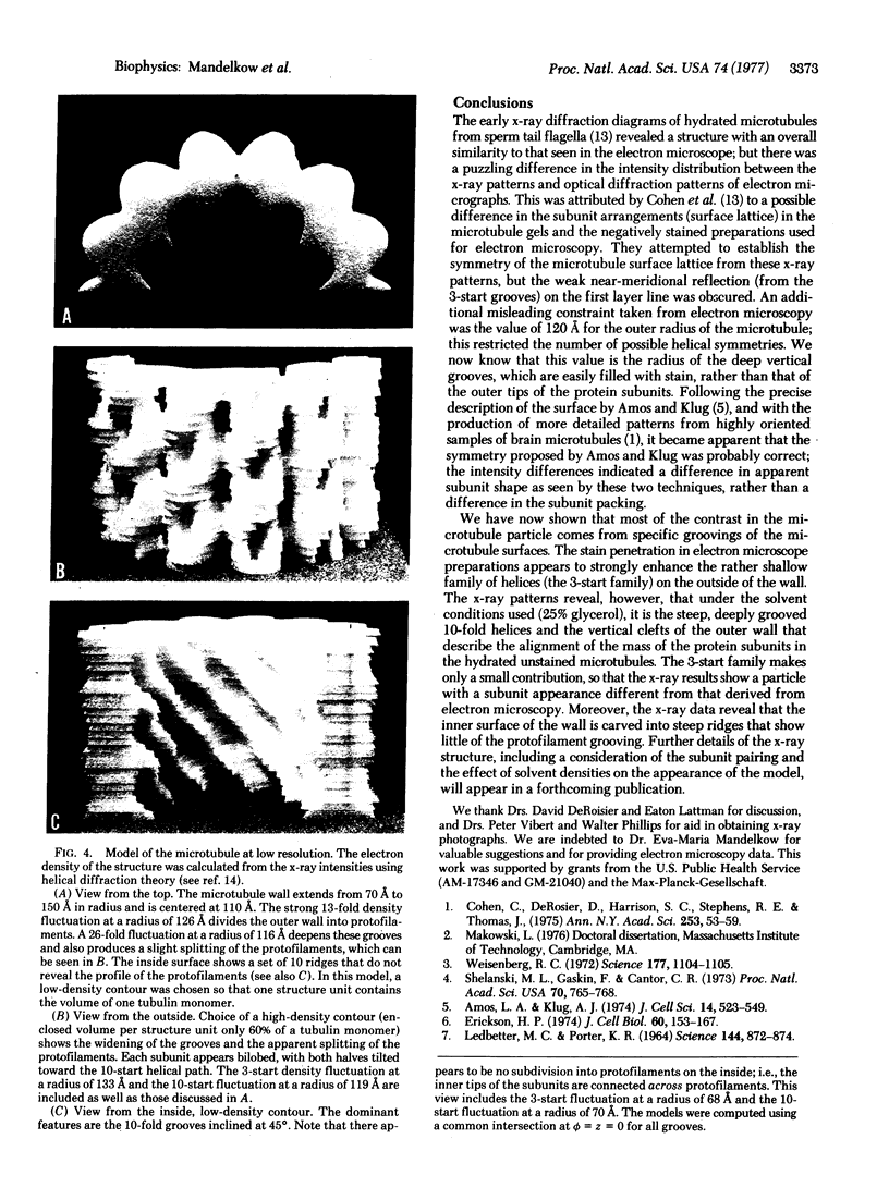
Analysis of x-ray diagrams of oriented hydrated cytoplasmic microtubules shows that the tubule wall extends from about 70 to 150 A radially. The central region of the wall appears homogeneous, but the outside surface is subdivided by vertical grooves separating the 13 protofilaments and by a steep 10-fold family of grooves. The inside surface is dominated by the 10-start grooves with no clear subdivision between the protofilaments.
Full text
PDF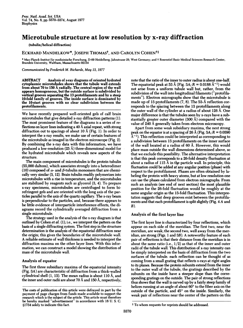
Images in this article
Selected References
These references are in PubMed. This may not be the complete list of references from this article.
- Amos L., Klug A. Arrangement of subunits in flagellar microtubules. J Cell Sci. 1974 May;14(3):523–549. doi: 10.1242/jcs.14.3.523. [DOI] [PubMed] [Google Scholar]
- Bryan J., Wilson L. Are cytoplasmic microtubules heteropolymers? Proc Natl Acad Sci U S A. 1971 Aug;68(8):1762–1766. doi: 10.1073/pnas.68.8.1762. [DOI] [PMC free article] [PubMed] [Google Scholar]
- Cohen C., DeRosier D., Harrison S. C., Stephens R. E., Thomas J. X-ray patterns from microtubules. Ann N Y Acad Sci. 1975 Jun 30;253:53–59. doi: 10.1111/j.1749-6632.1975.tb19192.x. [DOI] [PubMed] [Google Scholar]
- Cohen C., Harrison S. C., Stephens R. E. X-ray diffraction from microtubules. J Mol Biol. 1971 Jul 28;59(2):375–380. doi: 10.1016/0022-2836(71)90057-x. [DOI] [PubMed] [Google Scholar]
- Erickson H. P. Microtubule surface lattice and subunit structure and observations on reassembly. J Cell Biol. 1974 Jan;60(1):153–167. doi: 10.1083/jcb.60.1.153. [DOI] [PMC free article] [PubMed] [Google Scholar]
- Ledbetter M. C., Porter K. R. Morphology of Microtubules of Plant Cell. Science. 1964 May 15;144(3620):872–874. doi: 10.1126/science.144.3620.872. [DOI] [PubMed] [Google Scholar]
- Linck R. W., Amos L. A. The hands of helical lattices in flagellar doublet microtubules. J Cell Sci. 1974 May;14(3):551–559. doi: 10.1242/jcs.14.3.551. [DOI] [PubMed] [Google Scholar]
- Mandelkow E. M., Mandelkow E., Unwin N., Cohen C. Tubulin hoops. Nature. 1977 Feb 17;265(5595):655–657. doi: 10.1038/265655a0. [DOI] [PubMed] [Google Scholar]
- Olmsted J. B., Witman G. B., Carlson K., Rosenbaum J. L. Comparison of the microtubule proteins of neuroblastoma cells, brain, and Chlamydomonas flagella. Proc Natl Acad Sci U S A. 1971 Sep;68(9):2273–2277. doi: 10.1073/pnas.68.9.2273. [DOI] [PMC free article] [PubMed] [Google Scholar]
- Shelanski M. L., Gaskin F., Cantor C. R. Microtubule assembly in the absence of added nucleotides. Proc Natl Acad Sci U S A. 1973 Mar;70(3):765–768. doi: 10.1073/pnas.70.3.765. [DOI] [PMC free article] [PubMed] [Google Scholar]
- Shelanski M. L., Taylor E. W. Properties of the protein subunit of central-pair and outer-doublet microtubules of sea urchin flagella. J Cell Biol. 1968 Aug;38(2):304–315. doi: 10.1083/jcb.38.2.304. [DOI] [PMC free article] [PubMed] [Google Scholar]
- Tilney L. G., Bryan J., Bush D. J., Fujiwara K., Mooseker M. S., Murphy D. B., Snyder D. H. Microtubules: evidence for 13 protofilaments. J Cell Biol. 1973 Nov;59(2 Pt 1):267–275. doi: 10.1083/jcb.59.2.267. [DOI] [PMC free article] [PubMed] [Google Scholar]
- Weisenberg R. C. Microtubule formation in vitro in solutions containing low calcium concentrations. Science. 1972 Sep 22;177(4054):1104–1105. doi: 10.1126/science.177.4054.1104. [DOI] [PubMed] [Google Scholar]


