

Abstract
Exosomes are 30–120 nm endocytic membrane-derived vesicles that participate in cell-to-cell communication and protein and RNA delivery. Exosomes harbor a variety of proteins, nucleic acids, and lipids and are present in many and perhaps all bodily fluids. A significant body of literature has demonstrated that molecular constituents of exosomes, especially exosomal proteins and microRNAs (miRNAs), hold great promise as novel biomarkers for clinical diagnosis. In this minireview, we summarize recent advances in the research of exosomal biomarkers and their potential application in clinical diagnostics. We also provide a brief overview of the formation, function, and isolation of exosomes.
1. Introduction
Exosomes are small cell-derived vesicles of 30–120 nm that are present in many and perhaps all biological fluids. Exosomes were first discovered in the mid-1980s by the Johnstone group, who found that, in maturing mammalian reticulocytes, the transferrin receptor and some other membrane-associated elements are selectively released in multivesicular body- (MVB-) derived circulating vesicles, which they named exosomes [1–3]. Since Valadi et al. first reported in 2007 that exosomes also contain RNAs [4], the composition and function of exosomes have been intensely investigated. Now we know that exosomes carry various molecular constituents of their cell of origin, including proteins, lipids, mRNAs, and microRNAs (miRNAs). They are released from many cell types, such as dendritic cells (DCs), lymphocytes, platelets, mast cells, epithelial cells, endothelial cells, and neurons, and can be found in most bodily fluids including blood, urine, saliva, amniotic fluid, breast milk, hydrothoracic fluid, and ascitic fluid, as well as in culture medium of most cell types [5]. Exosomes were initially thought to serve simply as “garbage bags” for cells to get rid of unwanted constituents. However, an increasing body of evidence has demonstrated that exosomes play an important role in cell-to-cell communication and influence both physiological and pathological processes. Additionally, molecular constituents in exosomes have been found to be associated with certain diseases and treatment responses, indicating that they may also serve as a diagnostic tool. A PubMed search generated a list of over 1,000 exosome-related articles published in the past 5 years, showing the increasing level of interest in the biomedical research community. In this review, we summarize recent progress in the study of exosomes as novel biomarkers for clinical diagnosis. We also provide a brief overview of the formation, function, and isolation of exosomes.
2. Formation and Composition of Exosomes
MVBs are late endosomes that carry many “intraluminal endosomal vesicles.” Some MVBs are destined for degradation in lysosomes, while other MVBs merge with the cell membrane and release the internal vesicles into the extracellular space. Of the released vehicles, the smaller ones with a diameter of 30–120 nm are called exosomes; and the larger vehicles with a diameter of 120–1000 nm are called microvesicles [6–8]. Exosome formation involves the endosomal sorting complex required for transport (ESCRT), which recognizes ubiquitylated proteins. In addition to ESCRT, other ESCRT-independent mechanisms operate to generate exosomes of certain biochemical compositions [9]. Exosomes isolated by differential ultracentrifugation have a cup-shaped morphology as revealed by electron microscopy imaging [10].
Exosomes have a unique and complex composition. According to Exocarta (Version 4; http://www.exocarta.org), the latest exosome content database, 4,563 proteins, 194 lipids, 1,639 mRNAs, and 764 miRNAs have been identified in exosomes from multiple organisms [11]. The proteins most frequently identified in exosomes are membrane transporters and fusion proteins (e.g., GTPases, annexins, and flotillin), heat shock proteins (e.g., HSC70), tetraspanins (e.g., CD9, CD63, and CD81), MVB biogenesis proteins (e.g., alix and TSG101), and lipid-related proteins and phospholipases [10, 12]. Several proteins are recognized as specific exosomal markers, among which the tetraspanins, CD63 and CD81, are the most commonly used. Exosomes are also rich in lipids, which are predominantly cholesterol, sphingolipids, phospholipids, and bisphosphates. The exosomal lipid composition has been thoroughly analyzed in exosomes secreted from several cell types including DCs and mast cells [13], reticulocytes [14], and B-lymphocytes [15]. Several reports have suggested that certain lipid components of exosomes, such as phosphatidylserine [16] and prostaglandins [17], may play an important role in exosomal functions. The discovery that exosomes also contain mRNAs and miRNAs indicates that exosomes could be a carrier of genetic information. Although the majority of RNAs found in exosomes are degraded RNA fragments with a length of less than 200 nucleotides, some full length RNAs might be present and may be shuttled to a recipient cell via endocytosis and potentially affect protein production in the recipient cell. Meanwhile, exosomal miRNAs have been found to be associated with certain diseases. For instance, several studies have noted that miRNA contents of circulating exosomes are similar to those of their originating cancer cells, suggesting that exosomal miRNAs have potential for cancer diagnostics [18–20]. Also, an increasing number of studies have reported that miRNAs can be detected in exosomes isolated from noninvasively obtained bodily fluids such as saliva [21], showing potential advantages of exosomal miRNAs as novel biomarkers.
3. Function of Exosomes
Exosomes can merge with and release their contents into recipient cells (Figure 1). By transferring cellular constituents of proteins, RNAs, and lipids from one cell to another, exosomes play an important role in cell-to-cell communication [22–24]. A substantial body of literature has demonstrated that exosomes exhibit a broad range of functions, depending on their cell or tissue of origin. Particularly, exosomes from certain types of immune cells, such as DCs and B cells, may mediate adaptive immune responses to pathogens and tumors [25]. Tumor cell-derived exosomes play an active role in tumorigenesis, metastasis, and response to therapy through the transfer of oncogenes and onco-miRNAs between cancer cells and between cancer cells and the tumor stroma [26]. Exosomes shed from stimulated blood cells and the vascular endothelium is involved in neurological disorders such as multiple sclerosis, transient ischemic attacks, and antiphospholipid syndrome [27]. Interestingly, a few recent studies have shown that exosomes are also exploited by pathogens such as prions or viruses to transfer molecules of pathogenic origin between host cells and are thereby implicated in viral spread and immune evasion [28, 29]. Furthermore, since the molecular composition of exosomes is reflective of physiological or pathophysiological changes in their cell or tissue of origin, exosomes have significant potential as biomarkers for disease diagnosis.
Figure 1.
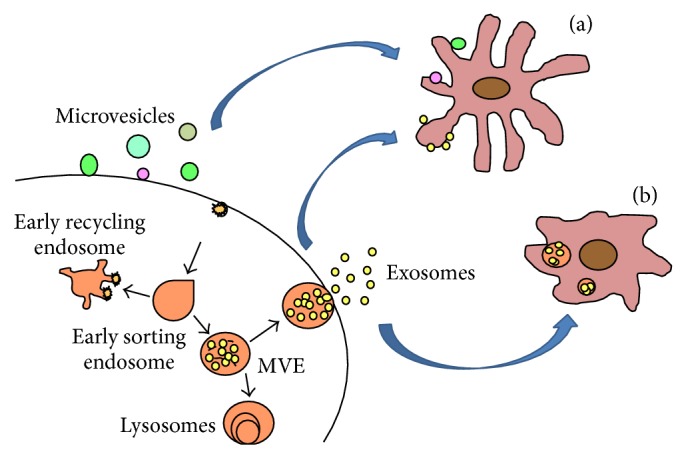
Exosomes are released from host cell and uptook by recipient cells. Exosomes are generated in host cell by merging MVBs with the cell membrane and releasing into the extracellular space. These exosomes can be fused with the plasma membrane (a) or be internalized (b) by recipient cells.
4. Isolation of Exosomes
Owing to their small size and low density, exosomes are usually isolated from bodily fluids and cell culture media by differential ultracentrifugation [44, 45]. Briefly, the collected biofluid is centrifuged at 300 ×g followed by a second centrifugation at 10,000 ×g to remove dead cells and cell debris. The supernatant is collected and subjected to ultracentrifugation at 100,000 ×g for 1 h or more. The pellet containing crude exosomes is subsequently washed with phosphate buffer solution to remove remaining proteins and other contaminants. The sample is subjected to a second ultracentrifugation at 100,000 ×g to yield purified exosomes. Several studies have also shown that exosomes may be isolated with higher purity using ultracentrifugation in a continuous density gradient of sucrose [46, 47].
Ultracentrifugation not only is labor-intensive and time-consuming but also requires expensive laboratory equipment, making it unsuitable for clinical applications. However, several recent technical advantages have made exosome isolation easier and faster and thus more cost-efficient. Cheruvanky et al. [48] and Merchant et al. [49] have successfully used ultrafiltration and microfiltration, respectively, for rapid isolation of urinary exosomes. Exosomes have also been isolated by immunoaffinity capture methods using lectins or antibodies against exosomal markers such as CD63, CD81, EpCAM, or Rab5 [45, 50, 51]. Precipitation followed by centrifugation is another method that has been explored for rapid exosome isolation. Nowadays, exosomes can be isolated in a one-step precipitation procedure using commercial reagents such as ExoQuick (System Biosciences, Mountain View, CA, USA). The Rekker group has demonstrated that ExoQuick is as efficient as ultracentrifugation in isolating serum exosomes for exosomal miRNA profiling and may be more efficient than ultracentrifugation in the context of exosomal RNA analysis [52].
5. Exosomes in Diagnostics
Exosomes are shed by cells under both normal and pathological conditions. They carry nucleic acids and proteins from their host cells that are indicative of pathophysiological conditions, and they are widely considered to be crucial for biomarker discovery for clinical diagnostics. For instance, tumor cells release exosomes containing tumor-specific RNAs that can be potentially used for cancer diagnosis. Over the past few years, numerous studies have demonstrated that exosomes contain nucleic acids and proteins implicated in cancer as well as neurodegenerative, metabolic, infectious, and other diseases. Furthermore, exosomes can be isolated from easily attainable biofluids such as blood and urine, making them very attractive targets for diagnostic application. In this review, we briefly summarize the main research advances reported to date in the context of diagnostic applications of exosomes.
5.1. Exosomal Proteins as Diagnostic Biomarkers
Exosomes contain diverse types of proteins including common membrane and cytosolic proteins as well as origin-specific subsets of proteins reflective of cell functions and conditions. Recently, an increasing number of exosomal proteins have been found to be potential biomarkers for a variety of diseases including cancer as well as liver and kidney diseases.
Tetraspanins, a family of scaffolding membrane proteins, are highly enriched in exosomes. The exosomal marker CD63 is a member of the tetraspanin family. Logozzi and coworkers reported in 2009 that plasma CD63+ exosomes are significantly increased in melanoma patients compared with healthy controls [31]. Most recently in 2013, Yoshioka and coworkers performed a comparative analysis of exosomal protein markers in different human cancer types and found that CD63 is present at higher levels in exosomes derived from malignant cancer cells than those derived from noncancer cells, providing further evidence that exosomal CD63 could be a protein marker for cancer [65]. CD81, another exosomal marker from the tetraspanin family, plays a critical role in hepatitis C attachment and/or cell entry. In addition, Welker and coworkers reported in 2012 that the level of serum exosomal CD81 is elevated in patients with chronic hepatitis C and seems to be associated with inflammation and severity of fibrosis, suggesting that exosomal CD81 may be a potential marker for hepatitis C diagnosis and treatment response [30].
A number of exosomal protein biomarkers have been found to be potentially useful in the diagnosis of central nervous system diseases. In 2008, Skog and coworkers detected glioblastoma-specific epidermal growth factor receptor vIII (EGFRvIII) in serum exosomes isolated from 7 out of 25 glioblastoma patients, indicating that exosomal EGFRvIII may provide diagnostic information for glioblastoma [33]. A year later, in line with Skog's findings, Graner et al. reported that serum exosomes from patients with brain tumors possess EGFR, EGFRvIII, and TGF-beta [66]. It has also been reported that exosomal amyloid peptides accumulate in the brain plaques of Alzheimer's disease (AD) patients [67]; and tau phosphorylated at Thr-181, an established biomarker for AD, is present at elevated levels in exosomes isolated from cerebrospinal fluid specimens of AD patients with mild symptoms [68]. These findings highlight the potential value of exosomes in the early diagnosis of AD, which is very important in sabotaging disease progression but currently difficult to achieve. Studies have also shown that α-synuclein, whose aggregation plays a central role in Parkinson's disease pathology, is released in exosomes in an in vitro model system of Parkinson's disease [69]; and prion proteins, biomarkers for transmissible spongiform encephalopathies, are packaged into exosomes released from prion-infected neuronal cells [70]. These exosomal proteins may have great potential in clinical diagnostics and should be further explored.
Proteins in urinary exosomes, which are easily attainable by noninvasive means, have also been exploited for potential utility in diagnostics, especially for urinary tract diseases. In 2006, Zhou et al. found that urinary exosomal fetuin-A is increased in intensive care unit patients with acute kidney injury (AKI) compared with patients without AKI [36]. Two years later, the same group reported that activating transcription factor 3 was found in exosomes isolated from patients with AKI but not from patients with chronic kidney disease or controls [37]. The authors thus concluded that measurement of these urinary exosomal proteins might offer diagnostic information for AKI. Urinary exosomal proteins have also been investigated as potential biomarkers for bladder cancer and prostate cancer. In 2008, Smalley et al. compared the protein profile of urinary exosomes between patients with bladder cancer and healthy controls and identified eight urinary exosomal proteins as potential biomarkers for bladder cancer, including five proteins associated with the EGFR pathway, the alpha subunit of Gs protein, resistin, and retinoic acid-induced protein 3 [40]. In 2009, Nilsson and coworkers demonstrated the presence of two known prostate cancer biomarkers, PCA-3 and TMPRSS2:ERG, in exosomes isolated from the urine of prostate cancer patients [41]. In 2012, Chen et al. identified that 24 urinary exosomal proteins were presented at significantly different levels between bladder cancer and hernia (control) patients (P < 0.05) [71]. These urinary exosomal proteins hold great promise as new diagnostic tools and wait to be further explored. Table 1 summarizes candidate exosomal protein biomarkers reported to date for diagnostic applications.
Table 1.
Summary of exosomal proteins for clinical diagnostic applications.
| Biofluid | Disease | Associated proteins | Reference |
|---|---|---|---|
| Plasma | Chronic hepatitis C | CD81 | [30] |
| Melanoma | CD63, caveolin-1, TYRP2, VLA-4, HSP70, HSP90 | [31, 32] | |
| Glioblastoma | Epidermal growth factor receptor VIII | [33] | |
| Prostate cancer | Survivin | [34] | |
| Plasma cell dyscrasias | c-src | [35] | |
|
| |||
| Urine | Acute kidney injury | Fetuin-A, ATF 3 | [36, 37] |
| Liver injury | CD26, CD81, S1c3A1, CD10 | [38] | |
| Bartter syndrome type 1 | NKCC2 | [39] | |
| Bladder cancer | EGF, α subunit of Gs, resisitin, retinoic acid-induced protein 3, and so forth. | [40] | |
| Prostate cancer | PSA, PCA3 | [41] | |
|
| |||
| Plasma, cell culture medium, and ascites | Human ovarian cancer | L1CAM, CD24, ADAM10, EMMPRIN, claudin | [42, 43] |
5.2. Exosomal Nucleic Acids as Diagnostic Biomarkers
Valadi's discovery in 2007 that exosomes contain RNAs [4] has intrigued great interest in the research of exosomal RNAs, especially miRNAs as diagnostic biomarkers. Recent studies have shown that exosomal miRNAs are protected from RNase-dependent degradation and thus can be stably detected in circulating plasma and serum [20, 54, 72], making them “ideal” biomarkers for clinical diagnostic applications.
Exosomal miRNAs have been most frequently exploited as biomarkers for cancer diagnosis. In 2008, a year after Valadi's discovery on exosomal miRNA, Taylor and Gercel-Taylor reported that eight miRNAs previously demonstrated as diagnostic markers for ovarian cancer are found at similar levels in biopsy specimens of ovarian cancer and serum exosomes isolated from the same ovarian cancer patients; however, these exosomal miRNAs could not be detected in normal controls, suggesting that exosomal miRNAs, which are easily attainable, could potentially be used as surrogate diagnostic markers for biopsy profiling [20]. In 2009, Rabinowits and coworkers performed a similar study in lung adenocarcinoma, in which they compared circulating levels of tumor-derived exosomes, exosomal small RNA, and specific exosomal miRNAs between lung adenocarcinoma patients and control subjects. They found similar miRNA patterns in circulating exosomes and tumor biopsies from lung adenocarcinoma patients, both significantly different from those detected in control subjects, suggesting that circulating exosomal miRNA might be useful as a screening test of lung adenocarcinoma [19].
Early detection and diagnosis of prostate cancer may be achieved using the prostate-specific antigen (PSA) test; however, the PSA test suffers from low specificity and a high false-positive rate, which may lead to overtreatment of indolent prostate cancers. Therefore, new markers with a higher diagnostic accuracy are much needed for prostate cancer. In 2008, Mitchell et al. reported that the level of circulating miR-141 is a robust diagnostic marker for prostate cancer [54]. Furthermore, later work by Brase and coworkers showed that serum levels of miR-141 and miR-375 are correlated with tumor progression in prostate cancer [55]. Given that exosomal miRNAs are a major component of RNase-resistant miRNAs in serum or plasma specimens [54, 72], it is plausible to speculate that circulating exosomal miR-141 and miR-375 may be valuable markers for prostate cancer diagnosis.
Exosomal miRNAs also show potential as biomarkers for the diagnosis of esophageal squamous cell cancer (ESCC). In 2013, Tanaka et al. reported that the exosomal miR-21 level is elevated in serum from patients with ESCC versus serum from patients who have benign tumors without systemic inflammation. In addition, exosomal miR-21 is positively correlated with tumor progression and aggressiveness. Importantly, Tanaka et al. claimed that miRNA-21 was not detected in serum that remained after exosome extraction, suggesting that exosomal miRNA-21 is the exclusive source of circulating miRNA-21 [56]. Moreover, Takeshita and coworkers reported in 2013 that serum miRNA-1246 shows a sensitivity of 71.3% and a specificity of 73.9% for ESCC diagnosis. Serum miRNA-1246 is also significantly correlated with the tumor, lymph node, and metastasis stage and has been found to be a strong independent risk factor for poor survival [57]. Interestingly, Takeshita et al. found that although miRNA-1246 is elevated in serum exosomes from ECSS patients, it is not upregulated in ESCC tissue samples, suggesting that serum exosomal miRNA-1246, but not biopsy derived-miRNA-1246, has strong potential as a novel diagnostic and prognostic biomarker in ESCC [57].
Exosomal miRNAs also demonstrate potential as diagnostic biomarkers for cardiovascular diseases and renal fibrosis [59, 60, 63, 64]. Besides, a few studies have shown that, in addition to miRNAs, exosomal mRNAs may be potentially used as biomarkers in clinical diagnostics [62, 64]. Table 2 summarizes candidate exosomal RNA biomarkers reported to date for diagnostic applications.
Table 2.
Summary of exosomal RNAs for clinical diagnostic applications.
| Biofluid | Disease | Associated RNAs | Reference |
|---|---|---|---|
| Plasma | Ovarian cancer | miR-21, -141, -200a, -200b, -200c, -203, -205, -214 | [20] |
| Lung cancer | miR-17, -3p, -21, -20b, -223, -301, let-7f | [19, 53] | |
| Prostate cancer | miR-141, miR-375 | [54, 55] | |
| Esophageal squamous cell cancer (ESCC) | miR-21, miR-1246 | [56, 57] | |
| Breast cancer | miR-21 | [58] | |
| Cardiovascular disease | miR-1, miR-133a | [59, 60] | |
|
| |||
| Cell culture medium | Gastric cancer | Let-7 family miRNAs | [61] |
| Colorectal cancer | mRNAs | [62] | |
|
| |||
| Urine | Renal fibrosis | miR-29c, CD2APmRNA | [63, 64] |
5.3. Exosomes from Other Biofluids as Diagnostic Biomarkers
There is emerging evidence that bodily fluids other than serum and urine may serve as alternate sources for diagnostic exosomes. For instance, Palanisamy et al. reported in 2010 that human saliva contains hundreds of stable mRNA core transcripts, which may be exploited as a possible resource for disease diagnostics [73]. In 2013, Lau et al. showed that saliva exosomes may provide discriminatory biomarkers for pancreatic cancer [74]. Amniotic fluid is another bodily fluid that has been investigated as a potential source of diagnostic exosomal markers. In 2007, Keller et al. isolated exosomes from amniotic fluid and demonstrated for the first time that fetal exosomes are present in amniotic fluid [75], suggesting that exosomes from amniotic fluid may potentially be used in early prenatal diagnostics. Gilad et al. reported in 2008 that miRNAs associated with the human placenta (miR-526a, -527, -515-5p, and -R521) are detectable in both the serum and amniotic fluid of pregnant women and are correlated with pregnancy stage [76]. Further research towards these directions should broaden the application range of exosomal biomarkers.
6. Conclusions
Research on the biology, function, and potential application of exosomes has increased exponentially over the past decade. A significant body of literature has demonstrated that one or perhaps the most important biomedical utility of exosomes is their potential application as biomarkers in clinical diagnostics. Compared with biomarkers detected in conventional specimens such as serum or urine, exosomal biomarkers provide comparable or higher specificity and sensitivity attributed to their excellent stability. Particularly, exosomal biomarkers from easily obtainable biofluids such as saliva would be very suitable for clinical applications. Recent technical advances in exosome isolation not only facilitated exosome research but also made exosomal diagnostics more cost-efficient. In addition to exosomal proteins and RNAs, exosomal lipids have also been shown to have diagnostic potential [77]. The utility of exosomes may be further expanded, since they are found not only in mammalian cells but also in diverse pathological microorganisms such as gram-negative bacteria, eukaryotic parasites of the kinetoplast lineage, and opportunistic fungal pathogens [78]. In general, exosomal biomarkers are still in the early discovery/development stage and their potential value in clinical diagnostics waits to be fully explored.
Acknowledgment
This study was supported by Grants from the National Science Foundation of China (no. 81360083).
Abbreviations
- AD:
Alzheimer's disease
- AKI:
Acute kidney injury
- DCs:
Dendritic cells
- EGFRvIII:
Epidermal growth factor receptor vIII
- ESCC:
Esophageal squamous cell cancer
- miRNAs:
MicroRNAs
- MVB:
Multivesicular body
- PSA:
Prostate-specific antigen.
Conflict of Interests
The authors declare no conflict of interests.
References
- 1.Johnstone R. M. The Jeanne Manery-Fisher Memorial Lecture 1991. Maturation of reticulocytes: formation of exosomes as a mechanism for shedding membrane proteins. Biochemistry and Cell Biology. 1992;70(3-4):179–190. doi: 10.1139/o92-028. [DOI] [PubMed] [Google Scholar]
- 2.Pan B. T., Johnstone R. M. Fate of the transferrin receptor during maturation of sheep reticulocytes in vitro: selective externalization of the receptor. Cell. 1983;33(3):967–977. doi: 10.1016/0092-8674(83)90040-5. [DOI] [PubMed] [Google Scholar]
- 3.Pan B.-T., Teng K., Wu C., Adam M., Johnstone R. M. Electron microscopic evidence for externalization of the transferrin receptor in vesicular form in sheep reticulocytes. Journal of Cell Biology. 1985;101(3):942–948. doi: 10.1083/jcb.101.3.942. [DOI] [PMC free article] [PubMed] [Google Scholar]
- 4.Valadi H., Ekström K., Bossios A., Sjöstrand M., Lee J. J., Lötvall J. O. Exosome-mediated transfer of mRNAs and microRNAs is a novel mechanism of genetic exchange between cells. Nature Cell Biology. 2007;9(6):654–659. doi: 10.1038/ncb1596. [DOI] [PubMed] [Google Scholar]
- 5.Théry C., Zitvogel L., Amigorena S. Exosomes: composition, biogenesis and function. Nature Reviews Immunology. 2002;2(8):569–579. doi: 10.1038/nri855. [DOI] [PubMed] [Google Scholar]
- 6.Bang C., Thum T. Exosomes: new players in cell-cell communication. The International Journal of Biochemistry & Cell Biology. 2012;44(11):2060–2064. doi: 10.1016/j.biocel.2012.08.007. [DOI] [PubMed] [Google Scholar]
- 7.Sun D., Zhuang X., Zhang S., et al. Exosomes are endogenous nanoparticles that can deliver biological information between cells. Advanced Drug Delivery Reviews. 2013;65(3):342–347. doi: 10.1016/j.addr.2012.07.002. [DOI] [PubMed] [Google Scholar]
- 8.Vlassov A. V., Magdaleno S., Setterquist R., Conrad R. Exosomes: current knowledge of their composition, biological functions, and diagnostic and therapeutic potentials. Biochimica et Biophysica Acta: General Subjects. 2012;1820(7):940–948. doi: 10.1016/j.bbagen.2012.03.017. [DOI] [PubMed] [Google Scholar]
- 9.El Andaloussi S., Mäger I., Breakefield X. O., Wood M. J. A. Extracellular vesicles: biology and emerging therapeutic opportunities. Nature Reviews Drug Discovery. 2013;12(5):347–357. doi: 10.1038/nrd3978. [DOI] [PubMed] [Google Scholar]
- 10.Conde-Vancells J., Rodriguez-Suarez E., Embade N., et al. Characterization and comprehensive proteome profiling of exosomes secreted by hepatocytes. Journal of Proteome Research. 2008;7(12):5157–5166. doi: 10.1021/pr8004887. [DOI] [PMC free article] [PubMed] [Google Scholar]
- 11.Mathivanan S., Fahner C. J., Reid G. E., Simpson R. J. Exocarta 2012: database of exosomal proteins, RNA and lipids. Nucleic Acids Research. 2012;40(1):D1241–D1244. doi: 10.1093/nar/gkr828. [DOI] [PMC free article] [PubMed] [Google Scholar]
- 12.Subra C., Grand D., Laulagnier K., et al. Exosomes account for vesicle-mediated transcellular transport of activatable phospholipases and prostaglandins. Journal of Lipid Research. 2010;51(8):2105–2120. doi: 10.1194/jlr.m003657. [DOI] [PMC free article] [PubMed] [Google Scholar]
- 13.Laulagnier K., Motta C., Hamdi S., et al. Mast cell- and dendritic cell-derived display a specific lipid composition and an unusual membrane organization. Biochemical Journal. 2004;380(1):161–171. doi: 10.1042/BJ20031594. [DOI] [PMC free article] [PubMed] [Google Scholar]
- 14.Olver C., Vidal M. Proteomic analysis of secreted exosomes. Subcellular Biochemistry. 2007;43:99–131. doi: 10.1007/978-1-4020-5943-8_7. [DOI] [PubMed] [Google Scholar]
- 15.Wubbolts R., Leckie R. S., Veenhuizen P. T. M., et al. Proteomic and biochemical analyses of human B cell-derived exosomes: potential implications for their function and multivesicular body formation. Journal of Biological Chemistry. 2003;278(13):10963–10972. doi: 10.1074/jbc.m207550200. [DOI] [PubMed] [Google Scholar]
- 16.Zakharova L., Svetlova M., Fomina A. F. T cell exosomes induce cholesterol accumulation in human monocytes via phosphatidylserine receptor. Journal of Cellular Physiology. 2007;212(1):174–181. doi: 10.1002/jcp.21013. [DOI] [PubMed] [Google Scholar]
- 17.Xiang X., Poliakov A., Liu C., et al. Induction of myeloid-derived suppressor cells by tumor exosomes. International Journal of Cancer. 2009;124(11):2621–2633. doi: 10.1002/ijc.24249. [DOI] [PMC free article] [PubMed] [Google Scholar]
- 18.Feng D.-Q., Huang B., Li J., et al. Selective miRNA expression profile in chronic myeloid leukemia K562 cell-derived exosomes. Asian Pacific Journal of Cancer Prevention. 2013;14(12):7501–7508. doi: 10.7314/apjcp.2013.14.12.7501. [DOI] [PubMed] [Google Scholar]
- 19.Rabinowits G., Gerçel-Taylor C., Day J. M., Taylor D. D., Kloecker G. H. Exosomal microRNA: a diagnostic marker for lung cancer. Clinical Lung Cancer. 2009;10(1):42–46. doi: 10.3816/clc.2009.n.006. [DOI] [PubMed] [Google Scholar]
- 20.Taylor D. D., Gercel-Taylor C. MicroRNA signatures of tumor-derived exosomes as diagnostic biomarkers of ovarian cancer. Gynecologic Oncology. 2008;110(1):13–21. doi: 10.1016/j.ygyno.2008.04.033. [DOI] [PubMed] [Google Scholar]
- 21.Michael A., Bajracharya S. D., Yuen P. S. T., et al. Exosomes from human saliva as a source of microRNA biomarkers. Oral Diseases. 2010;16(1):34–38. doi: 10.1111/j.1601-0825.2009.01604.x. [DOI] [PMC free article] [PubMed] [Google Scholar]
- 22.Lakkaraju A., Rodriguez-Boulan E. Itinerant exosomes: emerging roles in cell and tissue polarity. Trends in Cell Biology. 2008;18(5):199–209. doi: 10.1016/j.tcb.2008.03.002. [DOI] [PMC free article] [PubMed] [Google Scholar]
- 23.Schorey J. S., Bhatnagar S. Exosome function: from tumor immunology to pathogen biology. Traffic. 2008;9(6):871–881. doi: 10.1111/j.1600-0854.2008.00734.x. [DOI] [PMC free article] [PubMed] [Google Scholar]
- 24.van Niel G., Porto-Carreiro I., Simoes S., Raposo G. Exosomes: a common pathway for a specialized function. Journal of Biochemistry. 2006;140(1):13–21. doi: 10.1093/jb/mvj128. [DOI] [PubMed] [Google Scholar]
- 25.Li X. B., Zhang Z. R., Schluesener H. J., Xu S. Q. Role of exosomes in immune regulation. Journal of Cellular and Molecular Medicine. 2006;10(2):364–375. doi: 10.1111/j.1582-4934.2006.tb00405.x. [DOI] [PMC free article] [PubMed] [Google Scholar]
- 26.Kharaziha P., Ceder S., Li Q., Panaretakis T. Tumor cell-derived exosomes: a message in a bottle. Biochimica et Biophysica Acta: Reviews on Cancer. 2012;1826(1):103–111. doi: 10.1016/j.bbcan.2012.03.006. [DOI] [PubMed] [Google Scholar]
- 27.Horstman L. L., Jy W., Minagar A., et al. Cell-derived microparticles and exosomes in neuroinflammatory disorders. International Review of Neurobiology. 2007;79:227–268. doi: 10.1016/s0074-7742(07)79010-4. [DOI] [PubMed] [Google Scholar]
- 28.Belting M., Wittrup A. Nanotubes, exosomes, and nucleic acid-binding peptides provide novel mechanisms of intercellular communication in eukaryotic cells: implications in health and disease. Journal of Cell Biology. 2008;183(7):1187–1191. doi: 10.1083/jcb.200810038. [DOI] [PMC free article] [PubMed] [Google Scholar]
- 29.Pegtel D. M., van de Garde M. D. B., Middeldorp J. M. Viral miRNAs exploiting the endosomal-exosomal pathway for intercellular cross-talk and immune evasion. Biochimica et Biophysica Acta. 2011;1809(11-12):715–721. doi: 10.1016/j.bbagrm.2011.08.002. [DOI] [PubMed] [Google Scholar]
- 30.Welker M. W., Reichert D., Susser S., et al. Soluble serum CD81 is elevated in patients with chronic hepatitis c and correlates with alanine aminotransferase serum activity. PLoS ONE. 2012;7(2) doi: 10.1371/journal.pone.0030796.e30796 [DOI] [PMC free article] [PubMed] [Google Scholar]
- 31.Logozzi M., de Milito A., Lugini L., et al. High levels of exosomes expressing CD63 and caveolin-1 in plasma of melanoma patients. PLoS ONE. 2009;4(4) doi: 10.1371/journal.pone.0005219.e5219 [DOI] [PMC free article] [PubMed] [Google Scholar]
- 32.Peinado H., Alečković M., Lavotshkin S., et al. Melanoma exosomes educate bone marrow progenitor cells toward a pro-metastatic phenotype through MET. Nature Medicine. 2012;18(6):883–891. doi: 10.1038/nm.2753. [DOI] [PMC free article] [PubMed] [Google Scholar]
- 33.Skog J., Würdinger T., van Rijn S., et al. Glioblastoma microvesicles transport RNA and proteins that promote tumour growth and provide diagnostic biomarkers. Nature Cell Biology. 2008;10(12):1470–1476. doi: 10.1038/ncb1800. [DOI] [PMC free article] [PubMed] [Google Scholar]
- 34.Khan S., Jutzy J. M. S., Valenzuela M. M. A., et al. Plasma-derived exosomal survivin, a plausible biomarker for early detection of prostate cancer. PLoS ONE. 2012;7(10) doi: 10.1371/journal.pone.0046737.e46737 [DOI] [PMC free article] [PubMed] [Google Scholar]
- 35.di Noto G., Paolini L., Zendrini A., Radeghieri A., Caimi L., Ricotta D. C-src enriched serum microvesicles are generated in malignant plasma cell dyscrasia. PLoS ONE. 2013;8(8) doi: 10.1371/journal.pone.0070811.e70811 [DOI] [PMC free article] [PubMed] [Google Scholar]
- 36.Zhou H., Pisitkun T., Aponte A., et al. Exosomal Fetuin-A identified by proteomics: a novel urinary biomarker for detecting acute kidney injury. Kidney International. 2006;70(10):1847–1857. doi: 10.1038/sj.ki.5001874. [DOI] [PMC free article] [PubMed] [Google Scholar]
- 37.Zhou H., Cheruvanky A., Hu X., et al. Urinary exosomal transcription factors, a new class of biomarkers for renal disease. Kidney International. 2008;74(5):613–621. doi: 10.1038/ki.2008.206. [DOI] [PMC free article] [PubMed] [Google Scholar]
- 38.Conde-Vancells J., Rodriguez-Suarez E., Gonzalez E., et al. Candidate biomarkers in exosome-like vesicles purified from rat and mouse urine samples. PROTEOMICS—Clinical Applications. 2010;4(4):416–425. doi: 10.1002/prca.200900103. [DOI] [PMC free article] [PubMed] [Google Scholar]
- 39.Gonzales P. A., Pisitkun T., Hoffert J. D., et al. Large-scale proteomics and phosphoproteomics of urinary exosomes. Journal of the American Society of Nephrology. 2009;20(2):363–379. doi: 10.1681/ASN.2008040406. [DOI] [PMC free article] [PubMed] [Google Scholar]
- 40.Smalley D. M., Sheman N. E., Nelson K., Theodorescu D. Isolation and identification of potential urinary microparticle biomarkers of bladder cancer. Journal of Proteome Research. 2008;7(5):2088–2096. doi: 10.1021/pr700775x. [DOI] [PubMed] [Google Scholar]
- 41.Nilsson J., Skog J., Nordstrand A., et al. Prostate cancer-derived urine exosomes: a novel approach to biomarkers for prostate cancer. British Journal of Cancer. 2009;100(10):1603–1607. doi: 10.1038/sj.bjc.6605058. [DOI] [PMC free article] [PubMed] [Google Scholar]
- 42.Keller S., König A.-K., Marmé F., et al. Systemic presence and tumor-growth promoting effect of ovarian carcinoma released exosomes. Cancer Letters. 2009;278(1):73–81. doi: 10.1016/j.canlet.2008.12.028. [DOI] [PubMed] [Google Scholar]
- 43.Li J., Sherman-Baust C. A., Tsai-Turton M., Bristow R. E., Roden R. B., Morin P. J. Claudin-containing exosomes in the peripheral circulation of women with ovarian cancer. BMC Cancer. 2009;9, article 244 doi: 10.1186/1471-2407-9-244. [DOI] [PMC free article] [PubMed] [Google Scholar]
- 44.Raposo G., Nijman H. W., Stoorvogel W., et al. B lymphocytes secrete antigen-presenting vesicles. Journal of Experimental Medicine. 1996;183(3):1161–1172. doi: 10.1084/jem.183.3.1161. [DOI] [PMC free article] [PubMed] [Google Scholar]
- 45.Théry C., Amigorena S., Raposo G., Clayton A. UNIT 3.22 isolation and characterization of exosomes from cell culture supernatants and biological fluids. Current Protocols in Cell Biology. 2006 doi: 10.1002/0471143030.cb0322s30. [DOI] [PubMed] [Google Scholar]
- 46.Escola J.-M., Kleijmeer M. J., Stoorvogel W., Griffith J. M., Yoshie O., Geuze H. J. Selective enrichment of tetraspan proteins on the internal vesicles of multivesicular endosomes and on exosomes secreted by human B-lymphocytes. The Journal of Biological Chemistry. 1998;273(32):20121–20127. doi: 10.1074/jbc.273.32.20121. [DOI] [PubMed] [Google Scholar]
- 47.van Niel G., Mallegol J., Bevilacqua C., et al. Intestinal epithelial exosomes carry MHC class II/peptides able to inform the immune system in mice. Gut. 2003;52(12):1690–1697. doi: 10.1136/gut.52.12.1690. [DOI] [PMC free article] [PubMed] [Google Scholar]
- 48.Cheruvanky A., Zhou H., Pisitkun T., et al. Rapid isolation of urinary exosomal biomarkers using a nanomembrane ultrafiltration concentrator. American Journal of Physiology: Renal Physiology. 2007;292(5):F1657–F1661. doi: 10.1152/ajprenal.00434.2006. [DOI] [PMC free article] [PubMed] [Google Scholar]
- 49.Merchant M. L., Powell D. W., Wilkey D. W., et al. Microfiltration isolation of human urinary exosomes for characterization by MS. PROTEOMICS—Clinical Applications. 2010;4(1):84–96. doi: 10.1002/prca.200800093. [DOI] [PubMed] [Google Scholar]
- 50.Barrès C., Blanc L., Bette-Bobillo P., et al. Galectin-5 is bound onto the surface of rat reticulocyte exosomes and modulates vesicle uptake by macrophages. Blood. 2010;115(3):696–705. doi: 10.1182/blood-2009-07-231449. [DOI] [PubMed] [Google Scholar]
- 51.Chen C., Skog J., Hsu C.-H., et al. Microfluidic isolation and transcriptome analysis of serum microvesicles. Lab on a Chip. 2010;10(4):505–511. doi: 10.1039/b916199f. [DOI] [PMC free article] [PubMed] [Google Scholar]
- 52.Rekker K., Saare M., Roost A. M., et al. Comparison of serum exosome isolation methods for microRNA profiling. Clinical Biochemistry. 2014;47(1-2):135–138. doi: 10.1016/j.clinbiochem.2013.10.020. [DOI] [PubMed] [Google Scholar]
- 53.Silva J., García V., Zaballos Á., et al. Vesicle-related microRNAs in plasma of nonsmall cell lung cancer patients and correlation with survival. European Respiratory Journal. 2011;37(3):617–623. doi: 10.1183/09031936.00029610. [DOI] [PubMed] [Google Scholar]
- 54.Mitchell P. S., Parkin R. K., Kroh E. M., et al. Circulating microRNAs as stable blood-based markers for cancer detection. Proceedings of the National Academy of Sciences of the United States of America. 2008;105(30):10513–10518. doi: 10.1073/pnas.0804549105. [DOI] [PMC free article] [PubMed] [Google Scholar]
- 55.Brase J. C., Johannes M., Schlomm T., et al. Circulating miRNAs are correlated with tumor progression in prostate cancer. International Journal of Cancer. 2011;128(3):608–616. doi: 10.1002/ijc.25376. [DOI] [PubMed] [Google Scholar]
- 56.Tanaka Y., Kamohara H., Kinoshita K., et al. Clinical impact of serum exosomal microRNA-21 as a clinical biomarker in human esophageal squamous cell carcinoma. Cancer. 2013;119(6):1159–1167. doi: 10.1002/cncr.27895. [DOI] [PubMed] [Google Scholar]
- 57.Takeshita N., Hoshino I., Mori M., et al. Serum microRNA expression profile: miR-1246 as a novel diagnostic and prognostic biomarker for oesophageal squamous cell carcinoma. British Journal of Cancer. 2013;108(3):644–652. doi: 10.1038/bjc.2013.8. [DOI] [PMC free article] [PubMed] [Google Scholar]
- 58.Corcoran C., Friel A. M., Duffy M. J., Crown J., O'Driscoll L. Intracellular and extracellular microRNAs in breast cancer. Clinical Chemistry. 2011;57(1):18–32. doi: 10.1373/clinchem.2010.150730. [DOI] [PubMed] [Google Scholar]
- 59.Chen J. F., Mandel E. M., Thomson J. M., et al. The role of microRNA-1 and microRNA-133 in skeletal muscle proliferation and differentiation. Nature Genetics. 2006;38(2):228–233. doi: 10.1038/ng1725. [DOI] [PMC free article] [PubMed] [Google Scholar]
- 60.Kuwabara Y., Ono K., Horie T., et al. Increased microRNA-1 and microRNA-133a levels in serum of patients with cardiovascular disease indicate myocardial damage. Circulation: Cardiovascular Genetics. 2011;4(4):446–454. doi: 10.1161/circgenetics.110.958975. [DOI] [PubMed] [Google Scholar]
- 61.Ohshima K., Inoue K., Fujiwara A., et al. Let-7 microRNA family is selectively secreted into the extracellular environment via exosomes in a metastatic gastric cancer cell line. PLoS ONE. 2010;5(10) doi: 10.1371/journal.pone.0013247.e13247 [DOI] [PMC free article] [PubMed] [Google Scholar]
- 62.Hong B. S., Cho J.-H., Kim H., et al. Colorectal cancer cell-derived microvesicles are enriched in cell cycle-related mRNAs that promote proliferation of endothelial cells. BMC Genomics. 2009;10, article 556 doi: 10.1186/1471-2164-10-556. [DOI] [PMC free article] [PubMed] [Google Scholar]
- 63.Lv L.-L., Cao Y.-H., Ni H.-F., et al. MicroRNA-29c in urinary exosome/microvesicle as a biomarker of renal fibrosis. The American Journal of Physiology—Renal Physiology. 2013;305(8):F1220–F1227. doi: 10.1152/ajprenal.00148.2013. [DOI] [PubMed] [Google Scholar]
- 64.Lv L. L., Cao Y. H., Pan M. M., et al. CD2AP mRNA in urinary exosome as biomarker of kidney disease. Clinica Chimica Acta. 2014;428:26–31. doi: 10.1016/j.cca.2013.10.003. [DOI] [PubMed] [Google Scholar]
- 65.Yoshioka Y., Konishi Y., Kosaka N., Katsuda T., Kato T., Ochiya T. Comparative marker analysis of extracellular vesicles in different human cancer types. Journal of Extracellular Vesicles. 2013;2 doi: 10.3402/jev.v2i0.20424. [DOI] [PMC free article] [PubMed] [Google Scholar]
- 66.Graner M. W., Alzate O., Dechkovskaia A. M., et al. Proteomic and immunologic analyses of brain tumor exosomes. FASEB Journal. 2009;23(5):1541–1557. doi: 10.1096/fj.08-122184. [DOI] [PMC free article] [PubMed] [Google Scholar]
- 67.Rajendran L., Honsho M., Zahn T. R., et al. Alzheimer's disease β-amyloid peptides are released in association with exosomes. Proceedings of the National Academy of Sciences of the United States of America. 2006;103(30):11172–11177. doi: 10.1073/pnas.0603838103. [DOI] [PMC free article] [PubMed] [Google Scholar]
- 68.Saman S., Kim W., Raya M., et al. Exosome-associated tau is secreted in tauopathy models and is selectively phosphorylated in cerebrospinal fluid in early Alzheimer disease. Journal of Biological Chemistry. 2012;287(6):3842–3849. doi: 10.1074/jbc.M111.277061. [DOI] [PMC free article] [PubMed] [Google Scholar]
- 69.Alvarez-Erviti L., Seow Y., Schapira A. H., et al. Lysosomal dysfunction increases exosome-mediated alpha-synuclein release and transmission. Neurobiology of Disease. 2011;42(3):360–367. doi: 10.1016/j.nbd.2011.01.029. [DOI] [PMC free article] [PubMed] [Google Scholar]
- 70.Vella L. J., Sharples R. A., Lawson V. A., Masters C. L., Cappai R., Hill A. F. Packaging of prions into exosomes is associated with a novel pathway of PrP processing. The Journal of Pathology. 2007;211(5):582–590. doi: 10.1002/path.2145. [DOI] [PubMed] [Google Scholar]
- 71.Chen C.-L., Lai Y.-F., Tang P., et al. Comparative and targeted proteomic analyses of urinary microparticles from bladder cancer and hernia patients. Journal of Proteome Research. 2012;11(12):5611–5629. doi: 10.1021/pr3008732. [DOI] [PubMed] [Google Scholar]
- 72.Hunter M. P., Ismail N., Zhang X., et al. Detection of microRNA expression in human peripheral blood microvesicles. PLoS ONE. 2008;3(11) doi: 10.1371/journal.pone.0003694.e3694 [DOI] [PMC free article] [PubMed] [Google Scholar]
- 73.Palanisamy V., Sharma S., Deshpande A., Zhou H., Gimzewski J., Wong D. T. Nanostructural and transcriptomic analyses of human saliva derived exosomes. PLoS ONE. 2010;5(1) doi: 10.1371/journal.pone.0008577.e8577 [DOI] [PMC free article] [PubMed] [Google Scholar]
- 74.Lau C., Kim Y., Chia D., et al. Role of pancreatic cancer-derived exosomes in salivary biomarker development. The Journal of Biological Chemistry. 2013;288(37):26888–26897. doi: 10.1074/jbc.m113.452458. [DOI] [PMC free article] [PubMed] [Google Scholar]
- 75.Keller S., Rupp C., Stoeck A., et al. CD24 is a marker of exosomes secreted into urine and amniotic fluid. Kidney International. 2007;72(9):1095–1102. doi: 10.1038/sj.ki.5002486. [DOI] [PubMed] [Google Scholar]
- 76.Gilad S., Meiri E., Yogev Y., et al. Serum microRNAs are promising novel biomarkers. PLoS ONE. 2008;3(9) doi: 10.1371/journal.pone.0003148.e3148 [DOI] [PMC free article] [PubMed] [Google Scholar]
- 77.del Boccio P., Raimondo F., Pieragostino D., et al. A hyphenated microLC-Q-TOF-MS platform for exosomal lipidomics investigations: application to RCC urinary exosomes. Electrophoresis. 2012;33(4):689–696. doi: 10.1002/elps.201100375. [DOI] [PubMed] [Google Scholar]
- 78.Silverman J. M., Reiner N. E. Exosomes and other microvesicles in infection biology: organelles with unanticipated phenotypes. Cellular Microbiology. 2011;13(1):1–9. doi: 10.1111/j.1462-5822.2010.01537.x. [DOI] [PubMed] [Google Scholar]