Abstract
Studies have identified chemicals within the stony coral genus Montipora that have significant biological activities. For example, Montiporic acids A and B and other compounds have been isolated from the adult tissue and eggs of Montipora spp. and have displayed antimicrobial activity and cytotoxicity in cultured cells. The ecological role of these toxic compounds is currently unclear. This study examines the role these toxins play in reproduction. Toxins were found in the eggs and larvae of the coral Montipora capitata. Releasing these toxins by crushing both the eggs and larvae resulted in irreversible inhibition of photosynthesis in endogenous and exogenous zooxanthellae within minutes. Moreover, these toxins were stable, as frozen storage of eggs and larvae did not affect toxicity. Photosynthetic competency of Porites compressa zooxanthellae treated with either frozen or fresh, crushed eggs was inhibited similarly (P > 0.05, ANCOVA). Addition of toxic eggs plugs to live P. compressa fragments caused complete tissue necrosis under the exposed area on the fragments within 1 week. Small volumes of M. capitata crushed eggs added to sperm suspensions reduced in vitro fertilization success by killing the sperm. After 30 min, untreated sperm maintained 90 ± 1.9% SEM motility while those treated with crushed eggs were rendered immotile, 4 ± 1.4% SEM. Flow cytometry indicated membrane disruption of the immotile sperm. Fertilization success using untreated sperm was 79 ± 4% SEM, whereas the success rate dropped significantly after exposure to the crushed eggs, 1.3 ± 0% SEM. Unlike the eggs and the larvae, M. capitata sperm did not reduce the photosynthetic competency of P. compressa zooxanthellae, suggesting the sperm was nontoxic. The identity of the toxins, cellular mechanism of action, advantage of the toxins for M. capitata and their role on the reef are still unknown.
Introduction
Adaptation is a key component to species survival. The field of marine chemical ecology continues to unravel the complex ecological functions of marine natural products. These compounds feature heavily in predator–prey interactions, trophic cascades, competition, prey capture, reproduction, and larval recruitment, and have added to ecological theory [1]. Competition for space is an important ecological process on coral reefs, and Pawlik et al. [2] found that sponge compounds may stress corals by adversely influencing their symbiotic zooxanthellae.
Allelopathy is one such adaptation where natural chemical defenses, such as toxins, inhibit the growth of another cell. Allelopathic compounds may have complex chemical structures and significant biological activities. They have been isolated from almost all phyla of marine organisms [1,3]. The most commonly known producers of toxins in the marine environment are marine algae, soft corals, sponges and gorgonians [1,2,4]. Little is known about allelopathy in stony corals, but some studies have identified a variety of chemicals within the genus Montipora that have significant biological activities. For example, Montiporic acids A and B and other compounds have been isolated from the eggs of Montipora spp. and have displayed antimicrobial activity and cytotoxicity in cultured cells [5–9]. In addition, Gochfeld and Aeby [10] tested Montipora capitata extracts for their antimicrobial activity and found that extracts of this coral inhibited up to 54% of bacterial strains, including known coral pathogens found in the surrounding seawater. Montipora [11] is the second largest genus in the family Acroporidae and has a cosmopolitan distribution [12], so if these adaptations are shared by many of the species within the genus, it could have broad implications for survival and adaptation on reefs around the world.
The broad antimicrobial capability within Montipora tissue was known, however, in preliminary studies on the physiology of zooxanthellae from M. capitata [13], we observed something far more deadly. Specifically, the zooxanthellae from the tissue of three Hawaiian coral species were successfully harvested from host tissues and remained viable for days, whereas when M. capitata zooxanthellae were extracted, photosynthesis ceased within minutes. Moreover, when either fresh, boiled (100°C for 15 min) or frozen adult M. capitata tissue was added to samples of the viable exogenous zooxanthellae from other species, photosynthesis ceased in minutes. This suggested the presence of toxic compounds within M. capitata tissue.
M. capitata is found in the shallow areas of Kaneohe Bay, Hawaii, and assumes a plate-like form (Fig. 1A). Adult M. capitata transfer zooxanthellae horizontally into their eggs prior to spawning (Fig. 1B and C). During the massive spawns, M. capitata egg/sperm bundles form large slicks, which can be easily damaged by wind and waves potentially releasing a toxic cocktail. To examine this further, we investigated the role toxins might play in M. capitata reproduction over the 3-month spawning period. In this paper, we examined: (1) the toxic affects within the eggs, sperm and larvae on endogenous and exogenous zooxanthellae; (2) the stability of the toxins to freezing; (3) whether the toxins can adversely affect live zooxanthellae in intact Porites compressa fragments; (4) the impact the toxic compounds in the eggs might have on sperm motility, viability and in vitro fertilization; and, (5) the toxicity of various coral-derived organic chemical fractions against zooxanthellae.
Fig 1. Morphology of M. capitata.
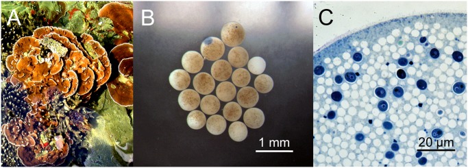
(A) Plate-like adult M. capitata found in Hawaii. (B) Light micrograph of M. capitata eggs demonstrating size and appearance of vertically transmitted intracellular zooxanthellae. (C) Transmission electron micrograph of a single M. capitata egg demonstrating the size and appearance of white yolk droplets and deep blue membrane-bound endogenous zooxanthellae.
Materials and Methods
Coral Collection and Husbandry
M. capitata, P. compressa and Fungia scutaria colonies were collected from various shallow reef flats around Coconut Island in Kaneohe Bay, Hawaii throughout the summer, and then maintained in shallow running seawater tables at the Hawaii Institute of Marine Biology, University of Hawaii. Care was taken to choose colonies from different locations in the bay, so as to try to ensure as much genetic diversity as possible. Colonies were kept in flowing seawater tables, supplied by water directly drawn from Kaneohe Bay. M. capitata spawns on the new moon throughout the summer in Hawaii [14–16], and the captive M. capitata fragments were used mainly for collecting gametes. Collection was performed with the appropriate permits from the state of Hawaii’s Department of Land and Natural Resources (Special Activity Permit # SAP 2011–1). No ethical approval was required for any of the experimental research described herein.
Egg/sperm bundles were collected in June, July and August of 2011 and June and July of 2012 and either used fresh or flash frozen in liquid nitrogen, then stored frozen at-80°C. To rear larvae, two egg/sperm bundles from different individuals were placed into 10 ml of filtered seawater (FSW) in 20 ml glass vials and allowed to develop for 3 days. Each vial produced about 100 larvae, and many vials were combined to produce enough tissue for experiments.
Microscopy
Samples for transmission electron microscopy were processed according to Padilla-Gamiño et al. [17]. In brief, specimens were fixed with 4% glutaraldehyde in 0.1 M sodium cacodylate buffer for 48 h, washed in 0.1 M sodium cacodylate, followed by post-fixation with 1% OsO4 in 0.1 M sodium cacodylate buffer for 1h. Tissue was dehydrated in a graded ethanol series and embedded in LX112 epoxy resin. Ultrathin (60–80 nm) sections were cut with a Reichert Ultracut E ultramicrotome, double stained with uranyl acetate and lead citrate, viewed on a LEO 912 EFTEM at 100 kV, and photographed with a Proscan frame-transfer CCD.
Monitoring Zooxanthellae
The presence of intracellular zooxanthellae is critical to the health of the coral polyp. Therefore, zooxanthellae number and robustness of photosynthetic performance were monitored over time using a Pulse Amplitude Modulated fluorometer (PAM, Walz, Germany) to determine relative levels of photosynthesis in photosystems I and II between the treatments and controls. Briefly stated, the PAM measured the initial fluorescence (recorded as “F”), then administered a saturating pulse of light, recording the maximal fluorescence (recorded as “M”). A ratio of these, F/M, yielded the effective quantum yield, recorded as the “Y-value”. We reported the effective quantum yield or Y-value throughout. The samples were not dark acclimated, but recorded under fluorescent lighting in the laboratory at mean light level of 4.08 ± 0.29 µmols/m2/s SEM (n = 5). Zooxanthellae were harvested from their host as described in Hagedorn et al. [13]. Unless stated otherwise, all solutions were made with 0.2 µm- FSW for these studies. A reduction in the effective quantum yield (Y) correlated with dying zooxanthellae [13]. These experiments mixed dead and live zooxanthellae to produce a known live/dead yield curve [13].
Exogenous zooxanthellae extracted from two different coral species (F. scutaria and P. compressa) were used for these experiments. M. capitata zooxanthellae could not be extracted successfully, so the only physiological examination of M. capitata zooxanthellae was of the endogenous zooxanthellae vertically transmitted into the eggs and larvae.
Experiments
Experiment 1: Assessing the effect of damaged M. capitata eggs and larvae on endogenous zooxanthellae. Two equal sub-samples (0.5 ml) of egg-sperm bundles were taken from each M. capitata colony (n = 7) diluted with FSW to a total volume of 1.0 ml per sample. For each pair, one sub-sample was designated as the control (no treatment) and the other as the experimental (crushed).
The viability of both samples was evaluated with the PAM fluorometer for effective quantum yield at t = 0. Then, the experimental sample was homogenized with ten strokes in a glass homogenizer. The time count was started at the moment that the homogenization of the sample began. Fluorescence was measured in both the control and the experimental samples at t = 1, 2, 3, 4, 5, and 10 min. PAM data were acquired with the fiber optic tip held in the center of the 1 ml sample in a 1.5 ml microcentrifuge tube at 26.5°C.
To determine whether the toxicity remains during development, parallel crushing experiments were done with 4 day-old M. capitata larvae. Thousands of larvae were reared in 20 ml glass scintillation vials for 4 days from 4 specific crosses. The larvae from identical crosses were consolidated then divided into two sub-samples, diluted in FSW yielding a final volume of 500 µl.
Experiment 2: Assessing stability of the toxins after frozen storage. Small fragments (10 x 3 cm; n = 10 individuals) of P. compressa were collected from around the Hawaii Institute of Marine Biology, placed in a flowing seawater table for a few days, and then their zooxanthellae were extracted following previous methods [13]. The extracted zooxanthellae sample from each colony was split into two sub-samples for parallel exposure to fresh or frozen/thawed egg tissue. One ml samples of M. capitata eggs with as little seawater as possible were frozen in liquid nitrogen, then thawed in a 30°C water bath; this process was repeated three times. Both the fresh and thawed eggs were crushed in a glass homogenizer, using 10 to 15 strokes. Each P. compressa subsample consisted of 500 µl of P. compressa zooxanthellae at ~106 cells/ml to which 75 µl of either fresh or frozen crushed M. capitata eggs were added. The effect on the quantum yield was monitored over time (t = 5, 10, 15 and 30 min) by PAM.
To establish the toxicity threshold, a dose response curve was determined using frozen/thawed eggs from mixed M. capitata colonies (n = 10 individuals). Eggs from M. capitata were crushed (as described above) and 25, 50, 100 or 250 µl of tissue added to 500 µl of P. compressa zooxanthellae at ~106 cells/ml (n = 1 individual). PAM measurements were taken over 60 min.
To confirm the optimal dose determined above, paired, matched samples (as described above) were collected for 8 different P. compressa colonies, and 100 µl of crushed eggs was added to one sub-sample while 100 µl of FSW was added to the second sub-sample and their quantum yield was monitored over 30 min with a PAM.
Experiment 3: Assessing the effect of frozen, crushed M. capitata eggs, sperm and larvae on exogenous zooxanthellae. Zooxanthellae were extracted and cleaned from eight ~5 cm individual fragments of P. compressa (as described above). Zooxanthellae samples from each fragment were diluted to 2 x 10⁶ and divided into five 1 ml sub-samples.
Prior to treatment, all P. compressa zooxanthellae samples were examined with a Junior-PAM (Walz) to obtain baseline effective quantum yield values (time 0). Samples from each individual were then treated with the following matrices: 1) 100 μl of FSW; 2) 100 μl of crushed F. scutaria sperm at 3x10⁸; 3) 100 μl of crushed M. capitata sperm at 3x10⁸; 4) 100 μl of M. capitata eggs packed as tightly as possible that were mixed 1:1 with filtered seawater and crushed; and, 5) 100 μl of M. capitata larvae that were also packed as tightly as possible and mixed 1:1 with filtered seawater and crushed. After solutions were added, each sub-sample was monitored using PAM with quantum yield values recorded at t = 1, 2, 5, 10, 20, 30 and 40 min.
Experiment 4: Assessing the effect of frozen M. capitata eggs on the viability of live zooxanthellae and tissue in P. compressa fragments. To investigate whether the toxins in M. capitata eggs and larvae might assist in larval settlement by clearing space for growth on the reef, P. compressa fragments were exposed to small volumes of frozen, crushed M. compressa eggs, frozen crushed F. scutaria larvae (negative control) and FSW (negative control). Small P. compressa fragments (10 cm x 3 cm; n = 15 individuals) were collected around the Hawaii Institute of Marine Biology and held in flowing seawater tables. Specifically, 400 µl of crushed eggs, larvae or FSW were added to the caps of 1.5 ml plastic Eppendorf tubes, flash frozen in the caps, stored at-80°C for 1 to 3 weeks and then thawed at 30°C prior to use. Prior to use, each cap was pierced on two opposite sides with a dremel tool, and a plastic ziptie tightly inserted preventing any solution leakage. One of each type of cap was strapped onto each P. compressa fragment (n = 15 individuals). Each fragment with its 3 tethered caps was placed back into a flowing seawater table. After 24 h, the caps were removed and viability of the P. compressa tissue and zooxanthellae under each cap was assessed. Digital images were taken of each of the 15 fragments 24 h after treatment with a Wild M3 dissecting microscope at 10x. The change in the coral tissue was assessed using computer image analysis with NIH Image J software examining the relative color change under the treatment area or cap, as compared to a nearby-untreated area (control). An average intensity of red, green and blue colors was taken for a specific sized area (measured in pixels) under the treatment caps and an equal sized area of untreated adjacent tissue. The difference between the color intensity of treated tissue and untreated tissue was calculated. Additionally the change in the viability of the P. compressa zooxanthellae was assessed with a PAM. The mean control viability of the zooxanthellae of each fragment was assessed in 5 untreated areas by measuring the mean quantum yield. This control value was compared with those measured in the treated areas under each cap.
Experiment 5: Assessing the effect of damaged M. capitata eggs on in vitro fertilization. Two equal samples of egg-sperm bundles were taken from each M. capitata colony (n = 18) and placed into 1 ml micro-centrifuge tubes. Each sample comprised 10 egg-sperm bundles, plus FSW to a total volume of 500 µl per sample. The egg-sperm bundles were allowed to fall apart without agitation, and the sperm was gently removed from the bottom of the tube and placed into a clean 1 ml micro-centrifuge tube, and divided in two. One sub-sample received no treatment. The other sub-sample was treated with 5 µl crushed eggs (M. capitata eggs were crushed with as little seawater as possible) at t = 0, then the motility of both samples monitored at t = 15 and 30 min, following the methods of Hagedorn et al. [18].
After 30 min, the sperm viability was assessed with a flow cytometer, viability being defined as those cells with intact cell membranes, following the methods of Hagedorn et al. [18]. To determine whether the sperm was membrane intact, sperm from 4 different colonies were treated in three different ways, either with 5 µl FSW (positive control), killed by 3 cycles of freeze/thawing (negative control), or treated with 5 µl of crushed eggs for 15 or 30 min (experimental treatment). We used a standard propidium iodide (Invitrogen) assay that exposed cells to a fluorescent dye that intercalates with DNA nucleotides and is usually excluded by intact cells (Garner et al., 1994). Stained cells were analyzed by flow cytometry (Accuri C6, Accuri Cytometers, Inc. Ann Arbor, MI USA), and 10,000 events were analyzed per sample. Additionally, cell numbers were counted to ensure that no drop in numbers occurred during the treatments.
To determine the impact of the toxins on fertilization, we tested untreated sperm and egg-extract exposed sperm in fertilization assays. M. capitata does not self-fertilize [15], therefore after motility assessments, untreated or egg-treated sperm at 5 x 10⁶ cells/ml was added to a single egg/sperm bundle from a different colony (n = 18 individuals) in 5 ml of FSW in a 20 ml glass scintillation vial. Fertilization was assessed as the number of developing larvae successfully achieving the prawn-chip stage 12 h later. The mean number of eggs in a M. capitata bundle (15 ± 5) was used to evaluate the percent fertilization success [17]. As a positive control for in vitro fertilization, two bundles, each from a different colony, were placed into 5 ml of seawater in a 20 ml glass scintillation vial and allowed to gently fall apart without agitation (n = 39). As a negative control, 1 egg-sperm bundle from a single colony was placed into 5 ml of seawater in a 20 ml glass scintillation vial and allowed to gently fall apart without agitation (n = 15).
Experiment 6: Chemical analysis of M. capitata eggs. Over 80 ml of concentrated M. capitata eggs were collected during four mass-spawning events in 2011. Most of the water was removed from the eggs, which were then frozen in liquid nitrogen, and stored at-80°C, then combined and freeze-dried. The freeze dried material (13.4 g) was extracted with ethyl acetate (EtOAc)-methanol (MeOH) (1:1) followed by MeOH- water (H2O) (4:1). The combined organic and aqueous extract (6.950 g) was partitioned between EtOAc and H2O. The aqueous portion was subsequently partitioned between n-butanol (n-BuOH) and H2O. Concentration of these extracts furnished 6.09 g of EtOAc-soluble fraction, 0.069 g of n-BuOH-soluble fraction and 0.994 g of H2O-soluble fraction. These fractions were fully lyophilized until a yellowish resin or oil remained. All fractions were analyzed by proton nuclear magnetic resonance spectroscopy to determine the presence of secondary metabolites and to assess the structures of known compounds. Sufficient dimethyl sulfoxide was added to bring all the fractions to ~ 3% solutions (wt/vol) in FSW. F. scutaria zooxanthellae (n = 2) were removed from adult tissue, cleaned and resuspended in 1 ml sub-samples in FSW at 107 cells/ml. Then 3, 30 or 300 µl of zooxanthellae solution was removed and replaced with either dimethyl sulfoxide (control for carrier), one of the three fractions, frozen, crushed M. capitata eggs (negative positive control for activity) or FSW (positive negative control for activity). The quantum yield (Y) was monitored over 40 min, and the data were normalized for comparison.
Statistics
All analyses in this study were performed using Graphpad Prism 5.0 (San Diego, CA) and Microsoft Excel (version 2007). All correlation analyses, ANOVAs were done with a Tukey’s Multiple Comparison Test or Dunnett’s post-test on various data sets. ANCOVA and linear regression was used to determine the difference between the slopes and Y-intercepts of experimental lines. These tests were identified specifically in the results when reporting the P-value, and those values ≤ 0.05 were considered significant.
Results
Experiment 1: Crushing M. capitata eggs or larvae damaged endogenous zooxanthellae
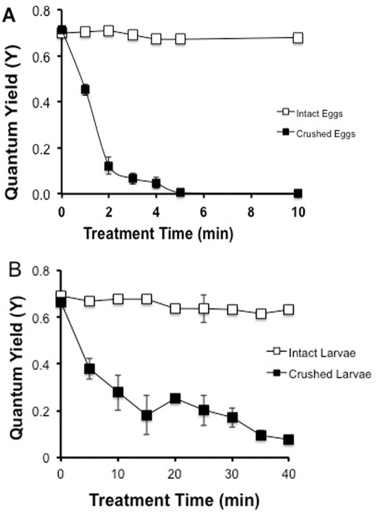
The zooxanthellae of the M. capitata egg/sperm bundles produced a high quantum yield (Y-value); inferring that they were fully functional. Once the eggs were crushed and their internal zooxanthellae released, the eggs’ own zooxanthellae displayed a Y-value close to zero within 5 min (Fig. 2A). A Y-value close to zero indicated that most of the zooxanthellae in the population were severely inhibited or dead [13]. A similar pattern was observed for M. capitata larvae, whereby the M. capitata zooxanthellae displayed a Y-value close to zero within 15 to 30 min (Fig. 2B). Samples (50 to 500 larvae) from 4 different crosses were split into two equal sub-samples and were either entirely crushed or left intact as an untreated control. The untreated group showed no change in its quantum yield over time (P> 0.05, Linear Regression, ANCOVA), whereas the crushed group showed a 90% loss in their quantum yield within 40 min; the two groups have different slopes (P <0.05, ANCOVA, F = 37.7). The time to full inhibition of M. capitata’s endogenous zooxanthellae was slightly longer for the larvae than the crushed eggs, perhaps due to the difference in the concentration of the eggs versus the larvae in the samples. Even though the times to inhibition were slightly different, it was concluded that damaged eggs and larvae were both capable of inhibiting or damaging endogenous zooxanthellae within minutes, and that the toxins persisted in the larvae prior to settlement.
Fig 2. Toxicity of intact and crushed M. capitata (A) eggs and (B) larvae against endogenous zooxanthellae determined by quantum yield.
Bars indicate SEM.
Experiment 2: Frozen storage does not affect stability of toxins
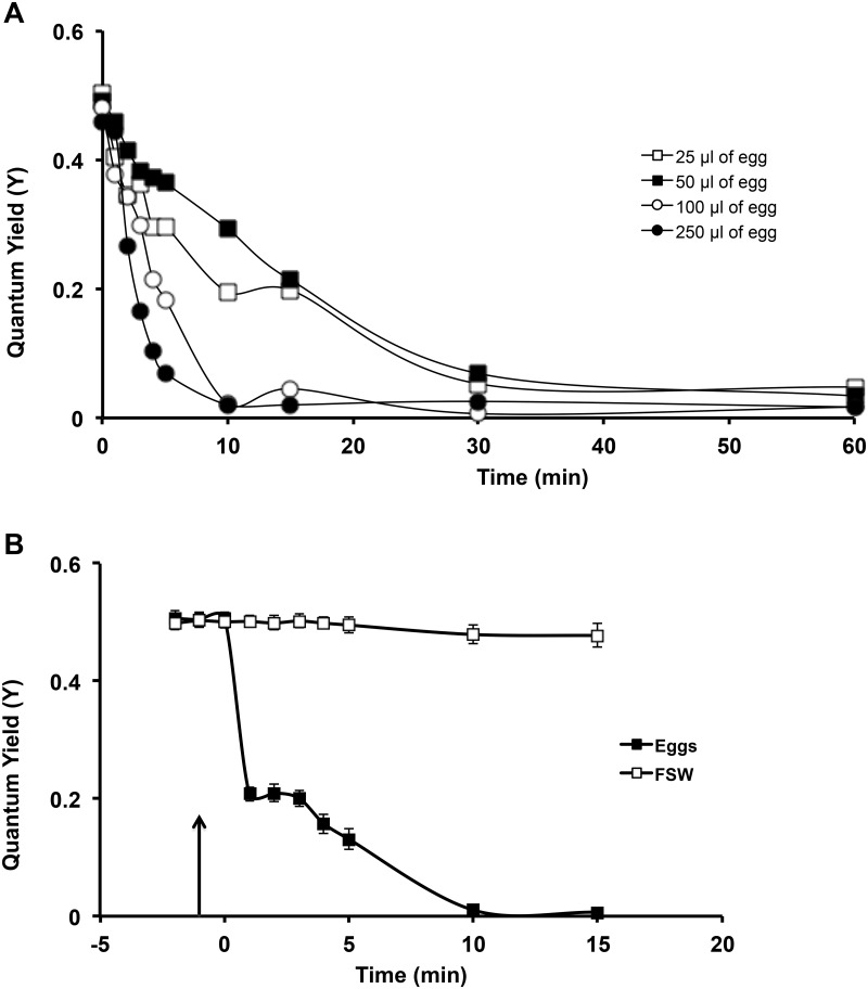
A dose response curve of the toxins found in M. capitata eggs were conducted against P. compressa zooxanthellae from a single colony. Four previously frozen egg volumes (25, 50, 100 and 250 µl) were added to each volume of fresh P. compressa zooxanthellae, and the effect on the quantum yield was monitored over 60 min. Both 100 and 250 µl of egg volume reduced the P. compressa zooxanthellae Y-values within 10 min, whereas the smaller volumes took 30 min to do the same (Fig. 3A). These experiments were repeated with more P. compressa zooxanthellae samples from10 individuals that were split into two sub-samples and their quantum yield monitored for 2 min, then 100 µl of M. capitata eggs were crushed and added to one of the subsamples with the other subsample treated with 100 µl of FSW added. After 10 min the zooxanthellae subsamples treated with M. capitata eggs had a 100% reduction in their Y-values, whereas the FSW-treated were unchanged (Fig 3B).
Fig 3. Dose response curve of M. capitata eggs on exogenous zooxanthellae.
(A) Preliminary experiments on the dose response curve of the toxins found in M. capitata eggs were conducted against P. compressa zooxanthellae. Both 100 and 250 µl of egg volume reduced the P. compressa zooxanthellae y-values within 10 min, whereas the smaller volumes took 30 min to do the same. (B) These experiments were repeated with a larger number of P. compressa zooxanthellae samples (n = 10 individuals) split into two sub-samples treated with either 100 µl of crushed M. capitata (at the 0 min mark indicated by the arrow) or 100 µl of FSW. Only the M. capitata eggs reduced the Y-values. Bars indicate SEM.
When parallel sub-samples from the same individuals (n = 10) were tested, both the frozen and the fresh crushed M. capitata eggs fully inhibited the quantum yield of P. compressa zooxanthellae within 30 min (data not shown). There was no difference between the slopes (ANCOVA; P > 0.05), demonstrating that the activity of the toxins affected the exogenous zooxanthellae similarly, whether in the fresh or frozen/thawed state.
Experiment 3: M. capitata sperm was not toxic to exogenous zooxanthellae
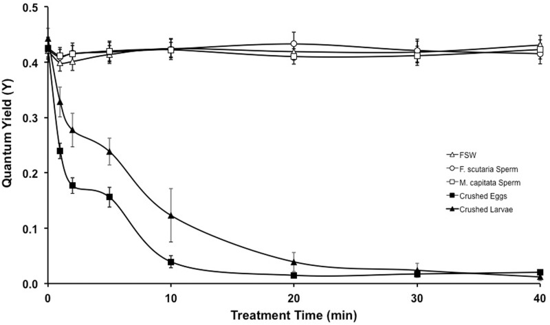
P. compressa zooxanthellae were treated with frozen crushed sperm, eggs, larvae or FSW (Fig. 4). None of these treatments, except the crushed M. capitata eggs and larvae, affected the quantum yield within the 40 min time period. Specifically, M. capitata sperm-treated zooxanthellae (n = 8 individuals, P>0.05; Linear Regression, F = 0.004) and the F. scutaria sperm-treated zooxanthellae (n = 8 individuals, P>0.05; Linear Regression, F = 0.037) were not different than the FSW-treated zooxanthellae (n = 8 individuals, P>0.05; Linear Regression, F = 1.319). In addition, the quantum yield reduced below 0.1 within 20 min for the egg-treated zooxanthellae (n = 8 individuals, P<0.05; Linear Regression, F = 67.93) and the larvae-treated zooxanthellae (n = 4 individuals, P<0.05; Linear Regression, F = 73.01). Although the sperm cells are smaller than the egg cells and not as easily crushed, the repeated freeze/thaw process in combination with the crushing and high cell number should have released some toxins, if present within the cells. These results indicate that the toxins are not located in M. capitata’s sperm or the concentration is so low that it was not observed in this assay. The reason the F. scutaria sperm was used in these experiments was to act as a control for cell degradation components from the M. capitata sperm that might have produced negative effects. Neither sperm addition produced these effects.
Fig 4. M. capitata sperm does not contain the same toxic properties as its eggs.
P. compressa zooxanthellae from 8 individuals were split into 5 sub-samples and exposed to five different treatments: 1) frozen/thawed F. scutaria sperm, 2) frozen/thawed M. capitata sperm, 3) toxic eggs: frozen/thawed crushed M. capitata eggs, 4) frozen/thawed crushed M. capitata larvae, and 5) FSW. After 20 min, the crushed M. capitata eggs and larvae reduced quantum yield greater than 90% (P < 0.05), while the other treatments remained unchanged at 0.4 (P > 0.05).
Experiment 4: Crushed M. capitata eggs adversely affected the tissue and zooxanthellae of live P. compressa
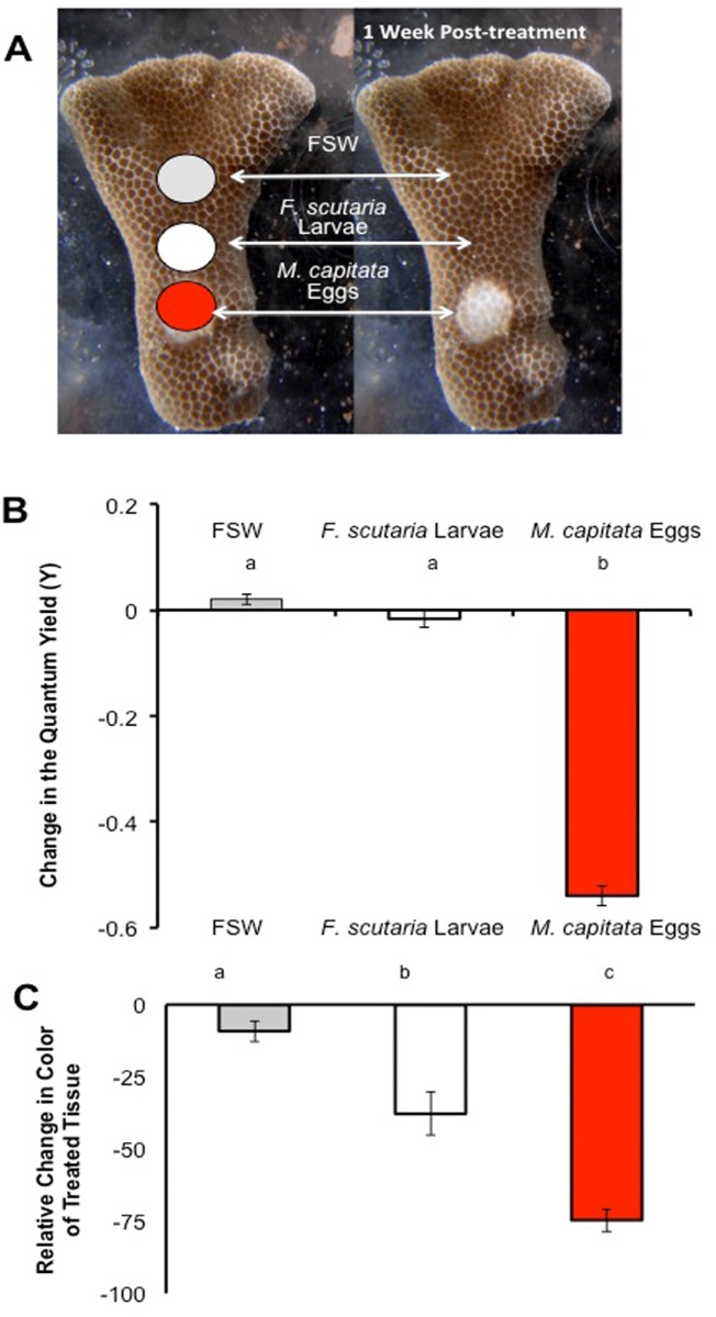
A possible ecological role of the toxins might be to reduce predation; however, this coral is not avoided by coralivores [19]. An alternative hypothesis is that it provides a competitive edge. To test this hypothesis, ‘mock competition’ trials were done by filling Eppendorf caps with either frozen, crushed F. scutaria larvae, M. capitata eggs or frozen FSW (Fig. 5A), and attaching one of each of these caps to live P. compressa fragments (n = 15). Within 24 h the caps with the crushed M. capitata eggs appeared to damage both the tissue and the zooxanthellae, as measured by a reduction in quantum yield (P< 0.05, F = 386, ANOVA, Tukey’s Multiple Comparison Test, Fig. 5B) and by a color loss under the caps (P< 0.05, F = 44.5, ANOVA, Tukey’s Multiple Comparison Test). This led to tissue necrosis within a week, indicating that the toxins could cause damage to competitors. In contrast, the other treatments caused no long-term damage to the fragments (P< 0.05, F = 45, ANOVA, Tukey’s Multiple Comparison Test, Fig. 5C). The addition of the F. scutaria larvae did cause a transient color change, but the P. compressa tissue appeared completely normal 6 days later.
Fig 5. Toxins with the frozen, crushed M. capitata eggs destroyed the tissue and the zooxanthellae of living P. compressa fragments.
(A) Only the addition of the caps with the M. capitata eggs caused tissue necrosis. (B, C) Twenty-four h later, only the caps with the M. capitata eggs reduced the quantum yield, indicating that the zooxanthellae in those areas were impaired (P< 0.05). Both the F. scutaria larvae and the M. capitata eggs caused a changed color on the fragment (P< 0.05), but only addition of the M. capitata eggs led to complete tissue necrosis under the cap 6 days later. Bars with the same letter indicate (P > 0.05), but bars with different letters indicate (P < 0.05). Bars indicate SEM.
Experiment 5: M. capitata eggs adversely affected fertilization success
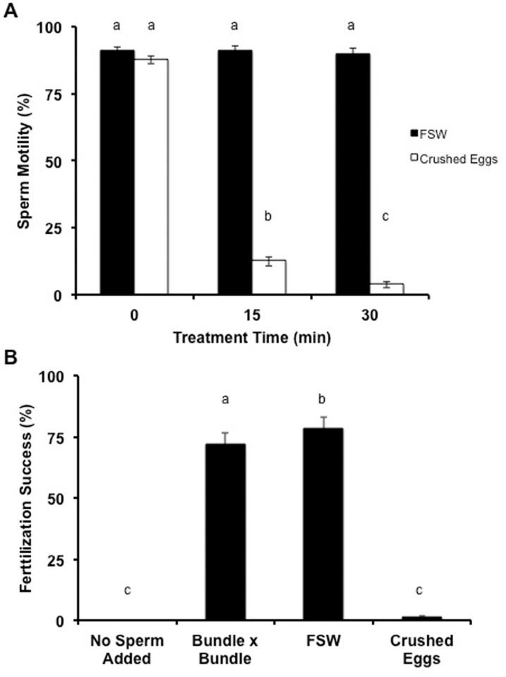
Fig. 6A demonstrated that when 5 µl of FSW was added to pair-matched samples of M. capitata sperm, sperm motility remained constant over time at 90% (n = 12 individuals, P > 0.05, ANOVA, F = 0.2, Tukey’s Multiple Comparison Test); however, when 5 µl of crushed M. capitata eggs were added to M. capitata sperm, the motility decreased by more than 90% after 15 and 30 min (n = 12 individuals, P < 0.05, ANOVA, F = 620.9, Tukey’s Multiple Comparison Test). Addition of crushed M. capitata eggs negatively impacted sperm motility.
Fig 6. The addition of crushed M. capitata eggs negatively impacted sperm motility and in vitro fertilization success.
(A) When 5 µl of FSW was added to M. capitata sperm, its motility remained constant over time, however when 5 µl of crushed M. capitata eggs were added to M. capitata sperm, the motility decreased greater than 90% after 15 and 30 min. (B) After 30 min, the uncrushed- and crushed egg-treated sperm were tested in in vitro fertilization assays. The sperm exposed to the crushed eggs showed little fertilization success (1.3%), whereas the freshly collected bundles from two individuals (positive control) and the uncrushed egg-treated sperm had excellent fertilization success at 72 and 79%. Bars indicate SEM.
To determine whether the sperm was just immotile or whether their membranes were disrupted, crushed egg-treated and FSW-treated sperm were exposed to propidium iodide and examined with a flow cytometer. At 30 min, only 1.2% of the crushed egg-treated sperm cells were membrane intact, whereas 85% of the FSW-treated sperm cells were membrane intact with a mean mortality of only 8.3%. Cell numbers were calculated in each sample. From these data, it was concluded that reduction in viability was not due to an overall loss of cell numbers, but a loss of membrane integrity.
When egg-treated sperm (described above) were used in in vitro fertilization assays (Fig. 6B), the sperm exposed to the crushed eggs showed little fertilization success (1.3%), whereas the freshly collected bundles from two individuals (positive control) and the uncrushed egg-treated sperm had excellent fertilization success at 72 and 79% at 30 min, respectively (n = 12 individuals, P < 0.05, ANOVA, F = 4.54, Tukey’s Multiple Comparison Test). Vials with freshly collected bundles from one individual had no fertilization success (negative control).
Experiment 6: Toxic components are extractable
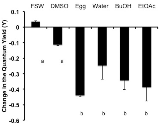
Many known compounds previously reported from Montipora spp. could be tentatively identified in the extracts based on 1H-NMR. The ethyl acetate extract contained large amounts of the diacetylenes (S1 Fig.), many of which have been previously reported [6,7], and the butanol fraction appeared to contain Montiporic acids A and B, based on the polarity and characteristic signals in the 1H-NMR (S2 Fig.) [5]. These compounds have all been reported to have cytotoxic activity. The aqueous extract did not contain these known compounds (S3 Fig.). Methyl montiporate A (S4 Fig.) and montiporyne G (S5 Fig.) were isolated from the complex mixture of diacetylenes in the ethyl acetate fraction. All fractions from the M. capitata eggs were toxic (Fig. 7), suggesting multiple toxic compounds of different polarities. These showed similar reductions in photosynthetic yields of exogenous zooxanthellae as did the frozen M. capitata eggs and larvae. Dimethyl sulfoxide was used as a solvent for all the fractions, however at the highest volume tested (300 µl), the solvent alone produced adverse effects on the Y-values of the zooxanthellae (data not shown). While in the 3-µl test volume, neither the solvent nor the coral extracts produced an adverse impact on the zooxanthellae (data not shown). Only the 30-µl test volume produced a reduction in the quantum yield related to the coral extracts (Fig. 7).
Fig 7. The reduction in the quantum yield of F. scutaria zooxanthellae due to M. capitata egg chemical fractions.
The zooxanthellae from two individuals were divided up into 5 sub-samples and exposed to 30 µl of FSW, dimethyl sulfoxide (DMSO), frozen crushed M. capitata eggs, water fraction, butanol fraction (BuOH) or ethyl acetate fraction (EtOAc). The quantum yield of FSW and DMSO, were not different (P>0.05, ANOVA, Dunnett’s Multiple Comparison Test). But adding 30 µl of each of the chemical fractions or frozen eggs caused a reduction of 25 to 44% in the quantum yield over 40 min over the FSW values (P> 0.05, ANOVA, Dunnett’s Multiple Comparison Test). Bars with different letters were (P < 0.05). Bars indicate SEM.
Discussion
Extracts from a number of scleractinian species adversely affect potential pathogens, competitors, and conspecifics, and interestingly, some of these compounds may be seasonally produced [20–22]. For example, extracts of 46 species of adult stony coral from the families of Mussidae, Merulinidae, Siderastreidae, Oculinidae and Dendrophylliidae from the Great Barrier Reef demonstrated bioactivity such as toxicity to mice and fish and antimicrobial activity; however, the antimicrobial activity was only observed prior to reproduction [20]. In contrast, when the adult tissue of six species of soft and stony adult coral were tested in the Red Sea, only soft corals exhibited antimicrobial activity [23]. Additionally, studies have identified antimicrobial activity in the eggs and adult tissue of at least two montiporid species, M. capitata and M. digitata [5–10,24]. Both Montiporid species have horizontally transferred zooxanthellae, and one theory is that these toxins may be of plant origin.
Our studies revealed that the toxic components are located in M. capitata eggs, larvae and adult tissue (data on adults not shown) of M. capitata. Once the eggs or larvae were crushed, the released toxins were capable of compromising both the zooxanthellae of M. capitata and those of other species within minutes. But it is not known whether this toxin production is broadly shared throughout the Montiporid complex. The toxins were resistant to freezing and boiling and found in every chemical fraction tested. Coral fragments with zooxanthellae treated with these toxins, produced tissue necrosis within a week’s time. Clearly, the toxins could lead to cell death. Strict parallel experimental exposures on the P. compressa fragments were not done because we used F. scutaria larvae and M. capitata eggs. This was due to timing of the reproduction of the species and availability of material during the fragment experiments. Sperm treated with crushed eggs displayed reduced motility over time and finally membrane disruption at 30 min. This characterization suggests a stable cocktail of chemicals present with M. capitata adults and offspring capable of producing adverse effects on plant and animal cells.
While the ecological role of the M. capitata toxins was unclear, the literature points to interspecific competition [19,25]. Toxins may provide a defense against aggressive predation by fish and parasitism by other organisms, but we hypothesize that if the toxins were released, then interspecific competition, and not predator deterrence, may be its major function. Studies on Hawaiian reefs lend support to this hypothesis. They have revealed that coralivorous reef fishes feed freely on Montipora capitata with no adverse response [19,25], but when predators were excluded from reef cages, M. capitata overgrew and outcompeted P. compressa [19,25]—the dominant species on the reefs in Kaneohe Bay, Hawaii. This edge in competition could be faster growth, or faster growth coupled with chemicals that may inhibit growth in the competing species. Supporting this idea, Gunthorpe and Cameron [21] and Fearon and Cameron [22] found that an aqueous extract from Goniopora tenuidens killed the swimming larvae of four different scleractinian corals. Additionally, octocoral allelopathy reduced recruitment and settlement of other corals nearby [26].
Our studies determined that the toxins affected the viability of the sperm and reproductive success of the coral during in vitro fertilization, which is commonly done in the investigation of coral reproduction. However, the effects of these toxins may be inferred from previous in vitro reproductive studies in M. capitata. Typically, during in vitro fertilization, egg/sperm bundles are collected, and gametes are separated, cleaned, counted and fertilized with typical values of 60 to 80% fertilization success [14]. However, Babcock and Heyward’s method has not worked well for M. capitata [27]. Instead, only intact bundles from separate individuals placed in small volumes of seawater and allowed to gently fall apart without agitation yielded the typical high fertilization success of 60 to 80% [27]. Even though these toxins inhibited the coral, it is not likely that they might actually impact natural reproduction on the reef. Wind-generated waves can break up the eggs, thus releasing the toxins onto the reef killing sperm and eggs in small areas, but the overall impact to reproductive success over the years would be minimal. Finally, chemical extracts from M. digitata eggs produced species-specific chemotaxis to sperm. So, the eggs may produce a wide variety of chemicals that have numerous roles in reproduction [28] and defense, potentially leading to greater survival.
An intriguing question is, how does the coral keep these potent toxins from damaging its own tissues and zooxanthellae? We hypothesize that the toxins are stored either in vesicles or diffusely in the cytoplasm, where it may become toxic only after its release. Padilla-Gamiño et al. [17] described a number of types of vesicles within the hydrated eggs of M. capitata that could potentially sequester these toxins, but more studies would be needed to determine whether, in fact, these toxins are stored there. Other examples of these types of “activated defenses” can be found in the chemical ecology literature for green macroalgae [29,30] and sponges [31].
Reproduction and adaptation are key contributors to species survival. Throughout the world, coral reefs are being degraded at unprecedented rates. Locally, reefs are damaged by pollution, nutrients and sedimentation from outdated land-use, fishing and mining practices [32]. Globally, increased greenhouse gases are warming and acidifying oceans, making corals more susceptible to stress, bleaching and newly emerging diseases [33–35]. The coupling of climate change and anthropogenic stressors has caused a widespread and well-recognized reef crisis [33–38]. However, the types of adaptations that animal populations develop are key to surviving such stressors and depend upon both growth rates and reproduction. The toxic components need to be isolated, identified and then tested to determine which ones elicit which response; only then will their roles become clearer.
Supporting Information
1H NMR (600 MHz, CDCl3) spectrum of ethyl acetate soluble less polar fraction from Montipora capitata, indicating the presence of less polar diacetylenes.
(PDF)
1H NMR (600 MHz, CD3OD) spectrum of n-Butanol-soluble polar fraction from Montipora capitata, indicating the presence of polar diacetylenic montiporic acids.
(PDF)
1H NMR (600 MHz, CD3OD) spectrum of water-soluble polar fraction from Montipora capitata.
(PDF)
1H NMR (600 MHz, CDCl3) spectrum of montiporyne G isolated from the ethyl acetate soluble fraction from Montipora capitata.
(PDF)
1H NMR (600 MHz, CDCl3) spectrum of methyl montiporate A isolated from the ethyl acetate soluble fraction from Montipora capitata.
(PDF)
Acknowledgments
We would like to thank the Gates and Smith laboratories at the University of Hawaii for the use of the Diving PAM and the PAM Jr, respectively, to assess the viability of the zooxanthellae samples. In particular, we like to thank Ned Busch, Andre Burnier and John Burnier for their assistance in collection and John Burnier and Billie Jean Marks for their stimulating discussion. Finally, the comments from the anonymous reviewers of this manuscript were extremely helpful in improving the manuscript. It has been approved for publication by the Hawaii Institute of Marine Biology as contribution # 1523 and as Smithsonian Marine Station at Fort Pierce contribution # 970.
Data Availability
All relevant data are within the paper.
Funding Statement
These experiments were funded through grants from the Roddenberry Foundation (http://roddenberryfoundation.org/), the Anela Kolohe Foundation, Hawaii Community Foundation (www.hawaiicommunityfoundation.org) and the Taronga Conservation Society Australia (http://taronga.org.au/conservation). No individuals were employed or contracted by the funders (other than the named authors), played any role in: study design, data collection and analysis, decision to publish, or preparation of the manuscript. The funders had no role in study design, data collection and analysis, decision to publish, or preparation of the manuscript.
References
- 1. Paul VJ, Ritson-Williams R, Sharp K (2011a) Marine chemical ecology in benthic environments. Nat Prod Rep 28: 345–387. 10.1039/c0np00040j [DOI] [PubMed] [Google Scholar]
- 2. Pawlik JR, Steindler L, Henkel TP, Beer S, Ilan M (2007) Chemical warfare on coral reefs: sponge metabolites differentially affect coral symbiosis in situ. Limnol Oceanogr 52: 907–911. [Google Scholar]
- 3. Paul VJ, Ritson-Williams R (2008) Marine chemical ecology. Nat Prod Rep 25: 662–695. 10.1039/b702742g [DOI] [PubMed] [Google Scholar]
- 4. Rasher DB, Hay ME (2014) Competition induces allelopathy but suppresses growth and anti-herbivore defence in a chemically rich seaweed. Proc R Soc Lond B Biol Sci 281: 20132615. [DOI] [PMC free article] [PubMed] [Google Scholar]
- 5. Fusetani N, Toyoda T, Asai N, Matsunaga S, Maruyama T (1996) Montiporic acids A and B, cytotoxic and antimicrobial polyacetylene carboxylic acids from eggs of the scleractinian coral Montipora digitata . J Nat Prod 59: 796–797. [DOI] [PubMed] [Google Scholar]
- 6. Bae BH, Im KS, Choi WC, Hong J, Lee CO, et al. (2000) New acetylenic compounds from the stony coral Montipora sp . J Nat Prod 63: 1511–1514. [DOI] [PubMed] [Google Scholar]
- 7. Alam N, Bae BH, Hong J, Lee CO, Im KS, et al. (2001a) Cytotoxic diacetylenes from the stony coral Montipora species. J Nat Prod 64: 1059–1063. [DOI] [PubMed] [Google Scholar]
- 8. Alam N, Hong J, Lee CO, Choi JS, Im KS, et al. (2002) Additional cytotoxic diacetylenes from the stony coral Montipora sp . Chem Pharm Bull (Tokyo) 50: 661–662. [DOI] [PubMed] [Google Scholar]
- 9. Marquis CP, Baird AH, de Nys R, Holmstrom C, Koziumi N (2005) An evaluation of the antimicrobial properties of the eggs of 11 species of scleractinian corals. Coral Reefs 24: 248–253. [Google Scholar]
- 10. Gochfeld DJ, Aeby GS (2008) Antibacterial chemical defenses in Hawaiian corals provide possible protection from disease. Mar Ecol Prog Ser 362: 119–128. [Google Scholar]
- 11.Hoeksema B (2014) Montipora Blainville, 1830. World Register of Marine Species.
- 12. Blainville HMd, editor (1830) Zoophytes. Paris: Le Normat; 548 p. [Google Scholar]
- 13. Hagedorn M, Carter VL, Leong JC, Kleinhans FW (2010) Physiology and cryosensitivity of coral endosymbiotic algae (Symbiodinium). Cryobiology 60: 147–158. 10.1016/j.cryobiol.2009.10.005 [DOI] [PubMed] [Google Scholar]
- 14. Babcock RC, Heyward AJ (1986) Larval development of certain gamete-spawning scleractinian corals. Coral Reefs 5: 111–116. [Google Scholar]
- 15. Kolinski SP, Cox EF (2003) An update on modes and timing of gamete and planula release in Hawaiian scleractinian corals with implications for conservation and management. Pac Sci 57: 17–27. [Google Scholar]
- 16. Padilla-Gamiño JL, Gates RD (2012) Spawning dynamics in the Hawaiian reef-building coral Montipora capitata . Mar Ecol Prog Ser 449: 145–160. [Google Scholar]
- 17. Padilla-Gamiño JL, Weatherby TM, Waller RG, Gates RD (2011) Formation and structural organization of the egg-sperm bundle of the scleractinian coral Montipora capitata . Coral Reefs 30: 371–380. [Google Scholar]
- 18. Hagedorn M, Carter V, Martorana K, Paresa MK, Acker J, et al. (2012) Preserving and using germplasm and dissociated embryonic cells for conserving Caribbean and Pacific coral. PLoS One 7: e33354 10.1371/journal.pone.0033354 [DOI] [PMC free article] [PubMed] [Google Scholar]
- 19. Cox EF (1994) Resource use by corallivorous butterflyfishes (Family Chaetodontidae) in Hawaii. Bull Mar Sci 54: 535–545. [Google Scholar]
- 20. Gunthorpe L, Cameron AM (1990) Intracolonial variation in toxicity in scleractinian corals. Toxicon 28: 1347–1350. [DOI] [PubMed] [Google Scholar]
- 21. Gunthorpe L, Cameron AM (1990) Toxic exudate from the hard coral Goniopora tenuidens . Toxicon 28: 1347–1350. [DOI] [PubMed] [Google Scholar]
- 22. Fearon RJ, Cameron AM (1996) Larvotoxic extracts of the hard coral Goniopora tenuidens: allelochemicals that limit settlement of potential competitors? Toxicon 34: 361–367. [DOI] [PubMed] [Google Scholar]
- 23. Kelman D, Kashman Y, Rosenberg E, Kushmaro A, Loya Y (2006) Antimicrobial activity of Red Sea corals. Mar Biol 149: 357–363. [Google Scholar]
- 24. Alam N, Hong J, Lee CO, Im KS, Son BW, et al. (2001. b) Montipyridine, a new pyridinium alkaloid from the stony coral Montipora species. J Nat Prod 64: 956–957. [DOI] [PubMed] [Google Scholar]
- 25. Cox EF (1986) The effects of a selective corallivore on growth rates and competition for space between two species of Hawaiian corals. J Exp Mar Biol Ecol 101: 161–174. [Google Scholar]
- 26. Maida M, Sammarco PW, Coll JC (1995) Effects of soft corals on scleractinian coral recruitment. i: directional allelopathy and inhibition of settlement. Mar Ecol Prog Ser 121: 191–202. [Google Scholar]
- 27. Kolinski SP (2004) Sexual reproduction and the early life history of Montipora capitata in Kaneohe Bay, Oahu Hawaii: University of Hawaii. [Google Scholar]
- 28. Coll JC, Bowden BF, Meehan GV, Konig GM, Carroll AR, et al. (1994) Chemical aspects of mass spawning in corals. I. Sperm-attractant molecules in the eggs of the scleractinian coral Montipora digitata . Mar Biol 118: 177–182. [Google Scholar]
- 29. Paul VJ, Van Alstyne KL (1992) Activation of chemical defenses in the tropical green algae Halimeda spp.. J Exp Mar Bio Ecol 160: 191–203. [Google Scholar]
- 30. Jung VJ, Pohnert G (2001) Rapid wound-activated transformation of the green algal defensive metabolite caulerpenyne. Tetrahedron 57: 7169–7172. [Google Scholar]
- 31. Thoms C, Schupp PJ (2008) Activated chemical defense in marine sponges—a case study on Aplysinella rhax . J Chem Ecol 34: 1242–1252. 10.1007/s10886-008-9518-z [DOI] [PubMed] [Google Scholar]
- 32. Bellwood DR, Hughes TP, Folke C, Nystrom M (2004) Confronting the coral reef crisis. Nature 429: 827–833. [DOI] [PubMed] [Google Scholar]
- 33. Hoegh-Guldberg O (1999) Climate change, coral bleaching and the future of the world’s coral reefs. Mar Freshwater Res 50: 839–866. [Google Scholar]
- 34. Goreau TJ, Hayes RL, McClanahan T (2000) Conservation of coral reefs after the 1998 global bleaching event. Conserv Biol 14: 5–15. [Google Scholar]
- 35. Hughes TP, Baird AH, Bellwood DR, Card M, Connolly SR, et al. (2003) Climate change, human impacts, and the resilience of coral reefs. Science 301: 929–933. [DOI] [PubMed] [Google Scholar]
- 36. Glynn PW, D’Crox L (1990) Experimental evidence for high temperature stress as the cause of El Nino-coincident coral mortality. Coral Reefs 8: 181–191. [Google Scholar]
- 37. Glynn PW (1996) Coral reef bleaching: facts, hypotheses and implications. Glob Chang Biol 2: 495–509. [Google Scholar]
- 38. Veron JE, Hoegh-Guldberg O, Lenton TM, Lough JM, Obura DO, et al. (2009) The coral reef crisis: the critical importance of <350 ppm CO2. Mar Pollut Bull 58: 1428–1436. 10.1016/j.marpolbul.2009.09.009 [DOI] [PubMed] [Google Scholar]
Associated Data
This section collects any data citations, data availability statements, or supplementary materials included in this article.
Supplementary Materials
1H NMR (600 MHz, CDCl3) spectrum of ethyl acetate soluble less polar fraction from Montipora capitata, indicating the presence of less polar diacetylenes.
(PDF)
1H NMR (600 MHz, CD3OD) spectrum of n-Butanol-soluble polar fraction from Montipora capitata, indicating the presence of polar diacetylenic montiporic acids.
(PDF)
1H NMR (600 MHz, CD3OD) spectrum of water-soluble polar fraction from Montipora capitata.
(PDF)
1H NMR (600 MHz, CDCl3) spectrum of montiporyne G isolated from the ethyl acetate soluble fraction from Montipora capitata.
(PDF)
1H NMR (600 MHz, CDCl3) spectrum of methyl montiporate A isolated from the ethyl acetate soluble fraction from Montipora capitata.
(PDF)
Data Availability Statement
All relevant data are within the paper.


