β-Lactamases represent the major cause of bacterial resistance against β-lactam antibiotics, and they have been divided into four classes (A to D) on the basis of their amino acid sequences (21). The class B enzymes have no sequence or structural similarity to the active-site serine enzymes of classes A, C, and D (6); require a bivalent metal ion (Zn2+) for activity; and constitute group 3 in the Bush-Jacoby-Medeiros functional classification (2). The identification of Zn-β-lactamase-producing pathogenic strains of Aeromonas, Bacteroides, Flavobacterium, Legionella, Serratia, and Stenotrophomonas has greatly increased interest in this class of enzymes (2). The fact that they hydrolyze almost all β-lactam antibiotics, including carbapenems, underlines their clinical relevance. In consequence, the potential spreading of these enzymes among pathogenic bacteria is a frightening possibility, which emphasizes the importance of understanding their properties.
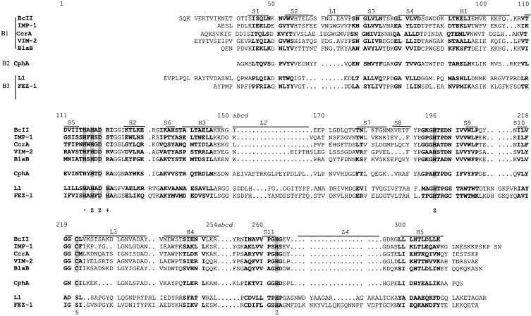
On the basis of the sequences, three subclasses of class B β-lactamases (B1 to B3) were identified, and a standard numbering scheme (BBL numbering) was proposed (13) by analogy to the ABL numbering scheme which has been widely used for class A β-lactamases. Due to the general low degree of identity between subclass sequences (<20%), classical alignment programs produce unreliable results. The proposed alignment (13) was facilitated by the availability of X-ray structures for B1 and B3 enzymes. Crystallographic structures have been described for several B1 enzymes: Bacillus cereus BcII (4, 11), Bacteroides fragilis CcrA (5, 8), Pseudomonas aeruginosa IMP-1 (7) and VIM-2 (unpublished data), and Chryseobacterium meningosepticum BlaB (14). Structural data are also available for two B3 enzymes: Stenotroptromonas maltophilia L1 (28) and Legionella gormanii FEZ-1 (15). Recently, we solved the first X-ray structure of a subclass B2 enzyme (CphA) produced by various species of Aeromonas (G. Garau, C. Bebrone, C. Anne, M. Galleni, J.-M. Frère, and O. Dideberg, unpublished data). Using all available three-dimensional structures, it is now possible to propose a bonafide structural alignment of the class B β-lactamases, and accordingly, to update the first proposed BBL scheme (Fig. 1).
FIG. 1.
Structural alignment of eight class B β-lactamases with known X-ray structures. The sequences are referred to by their familiar names. BCII, B. cereus 569H (16); IMP-1, P. aeruginosa 101/477 (17); CcrA, B. fragilis TAL3636 (25); VIM-2, P. aeruginosa species (24); BlaB, C. meningosepticum NCTC10585 (26); CphA, Aeromonas hydrophila AE036 (20); L1, S. maltophilia IID1275 (29); FEZ-1, L. gormanii ATCC33297T (1). The BBL numbering is defined. Conserved secondary-structure elements for the three subclasses are indicated above the sequences: S, β-strand; H, helix; L, loop. Amino acid insertions in newly sequenced enzymes are represented by lowercase letters. The zinc ligands in at least one subclass are shaded and labeled as follows: Z, residues conserved in the three subclasses; •, residues conserved in subclass B1 and some enzymes of subclass B3; +, residue conserved in subclass B3; §, residues conserved in subclasses B1 and B2. L2 and L4 represent largely variable regions. The 14 sequence fragments of structurally conserved positions, which cover the entirety of all sequences, are shown in boldface.
For the three-dimensional structure comparison of the eight available structures, we used the program TOP (18) with the new option MAPS, allowing multiple alignments of protein structures. In addition, the program produces two ranking scores: the sequence identities of aligned residues and the structural diversity. The structural-diversity score was defined as RMS/(Nmatch/N0)3/2, where RMS is the root mean square deviation of the distances between matched Cα atoms, Nmatch is the number of matching residues, and (Nmatch/N0) is the matching fraction of two compared structures. N0 = (N1 + N2/2), where N1 and N2 are the numbers of amino acids in the two compared proteins. This score estimates the evolutionary distance between proteins. These two scores are shown in Table 1 for all known X-ray structures.
TABLE 1.
Sequence identities of aligned residues and structure diversity among proteins
| Protein | Sequence identity/structural diversitya
|
|||||||
|---|---|---|---|---|---|---|---|---|
| CphA | BCII | CcrA | IMP-1 | VIM-2 | BlaB | L1 | FEZ-1 | |
| CphA | 0.32 | 0.30 | 0.24 | 0.28 | 0.29 | 0.20 | 0.15 | |
| BCII | 1.19 | 0.37 | 0.39 | 0.39 | 0.37 | 0.22 | 0.15 | |
| CcrA | 1.17 | 1.02 | 0.36 | 0.33 | 0.30 | 0.16 | 0.12 | |
| IMP-1 | 1.20 | 1.20 | 1.13 | 0.35 | 0.33 | 0.17 | 0.15 | |
| VIM-2 | 1.11 | 1.08 | 1.13 | 1.23 | 0.28 | 0.18 | 0.13 | |
| BlaB | 1.26 | 1.19 | 1.17 | 1.38 | 1.19 | 0.18 | 0.18 | |
| L1 | 1.72 | 1.60 | 1.58 | 1.79 | 1.62 | 1.68 | 0.33 | |
| FEZ-1 | 1.78 | 1.61 | 1.57 | 1.67 | 1.72 | 1.70 | 1.31 | |
In the upper right triangle (sequence diversity), the largest values characterize the most similar proteins; the opposite is true for the lower left triangle (structural diversity).
Figure 1 displays the proposed alignment and numbering. Interestingly, the numbering of the important class B residues is conserved between old and new alignments. Improvements in the alignment concern mainly N and C termini and small shifts along the sequences. The main result of the new alignment is the identification of 14 sequence fragments of structurally conserved positions, which cover the entirety of all sequences (Fig. 1); they belong mainly to secondary-structure elements (α helices or β sheets). Notably, all Zn ligands are structurally aligned.
The following comments can be made. (i) Only sequences of proteins of known structures are shown. (ii) For residues in lightface, the fact that they have the same number does not imply that they are structurally equivalent. (iii) For newly discovered enzymes, any insertion departing from the present numbering can be characterized by lowercase letters following the number of the last residue of the consensus sequence.
Table 2 shows the numbering of the putative zinc ligands. Not all proteins of known sequence are shown. Only enzymes with <50% sequence identity compared to the first reported sequence are included in the table.
TABLE 2.
Numbering of important class B residues
| Protein | Zn1 ligand | Zn2 ligand | ||||
|---|---|---|---|---|---|---|
| Consensus BBL B1 | His116 | His118 | His196 | Asp120 | Cys221 | His263 |
| BcII | His86 | His88 | His149 | Asp90 | Cys168 | His210 |
| IMP-1 | His77 | His79 | His139 | Asp81 | Cys158 | His197 |
| CcrA | His99 | His101 | His162 | Asp103 | Cys181 | His223 |
| VIM-2 | His88 | His90 | His153 | Asp92 | Cys172 | His214 |
| BlaB | His76 | His78 | His139 | Asp80 | Cys158 | His200 |
| IND-1 | His96 | His98 | His159 | Asp100 | Cys178 | His220 |
| SPM-1a | His76 | His78 | His165 | Asp80 | Cys184 | His221 |
| JOHN-1a | His76 | His78 | His159 | Asp80 | Cys178 | His220 |
| TUS-1a | His94 | His96 | His157 | Asp98 | Cys176 | His217 |
| Consensus BBL B2 | Asn116 | His118 | His196 | Asp120 | Cys221 | His263 |
| CphA | Asn69 | His71 | His148 | Asp73 | Cys167 | His205 |
| Sfh-I | Asn72 | His74 | His151 | Asp76 | Cys170 | His212 |
| Consensus BBL B3 | His/Gln116 | His118 | His196 | Asp120 | His121 | His263 |
| L1 | His84 | His86 | His160 | Asp88 | His89 | His225 |
| FEZ-1 | His71 | His73 | His149 | Asp75 | His76 | His215 |
| GOB-1a | Gln80 | His82 | His157 | Asp84 | His85 | His213 |
| THIN-Ba | His105 | His107 | His185 | Asp109 | His110 | His253 |
| CAU-1a | His96 | His98 | His172 | Asp100 | His101 | His237 |
In 1997, Neuwald et al. (23) detected a few proteins that have sequence similarities to (and may have given rise to) Zn-β-lactamases. They include enzymes with large variations in function (sulfatase; DNA cross-link repair enzyme) and which are encoded by yeast, plant, or bacterial open reading frames. Human glyoxalase II was also shown to belong to the superfamily. More recently, 17 groups with known functions were identified (9). In order to evaluate the structural diversity of the Zn-β-lactamase superfamily, human glyoxalase II (3) and rubredoxin oxygen-oxidoreductase from Desulfovibrio gigas (12) were also aligned using TOP, along with one member of each subclass. Table 3 shows the sequence identities and structural diversity of the two proteins and BCII, CphA, and FEZ-1. As expected, low sequence identity corresponds to a high structural-diversity score. The structural-diversity scores for proteins belonging to a superfamily range from 1.4 to 2, in contrast to 3.5 to 4 for proteins with different folds (18). Interestingly and surprisingly, FEZ-1 is closer to glyoxalase II and rubredoxin oxygen-oxidoreductase than to BcII or CphA.
TABLE 3.
Sequence identities of aligned residues and structural diversity among proteins
| Protein | Sequence identy/structural diversitya
|
||||
|---|---|---|---|---|---|
| CphA | BCII | FEZ-1 | ROO | GOX | |
| CphA | 0.32 | 0.15 | 0.20 | 0.23 | |
| BCII | 1.19 | 0.15 | 0.17 | 0.24 | |
| FEZ-1 | 1.78 | 1.61 | 0.16 | 0.21 | |
| ROO | 1.78 | 1.64 | 1.47 | 0.21 | |
| GOX | 1.61 | 1.62 | 1.40 | 1.47 | |
Sequency identities, upper right triangle; structural diversity, lower left triangle. ROO, rubredoxin oxygen-oxidoruductase; GOX, human glyoxylase II.
In the structural alignment, a large number of amino acid changes and insertions-deletions are observed. One hypothesis is that an ancient protein gave rise to the different subclasses of Zn-β-lactamases. A few candidates for the ancient protein are those related to essential biological functions within the cell, such as DNA or RNA processing or DNA repair (9). Nature used a limited number of scaffolds to generate a large variety of biological functions. Zn-β-lactamases are good examples of such a selection.
Acknowledgments
This work was supported by a grant from the European Union (HPRN-CT-2002-00264) and PAI P5/33 from the Belgian government.
The views expressed in this Commentary do not necessarily reflect the views of the journal or of ASM.
REFERENCES
- 1.Boschi, L., P. S. Mercuri, M. L. Riccio, G. Amicosante, M. Galleni, J. M. Frère, and G. M. Rossolini. 2000. The Legionella (Fluoribacter) gormanii metallo-beta-lactamase: a new member of the highly divergent lineage of molecular-subclass B3 beta-lactamases. Antimicrob. Agents Chemother. 44:1538-1543. [DOI] [PMC free article] [PubMed] [Google Scholar]
- 2.Bush, K. 1998. Metallo-beta-lactamases: a class apart. Clin. Infect. Dis. 27:S48-S53. [DOI] [PubMed] [Google Scholar]
- 3.Cameron, A. D., M. Ridderstrom, B. Olin, and B. Mannervik. 1999. Crystal structure of human glyoxalase II and its complex with a glutathione thiolester substrate analogue. Struct. Fold Des. 7:1067-1078. [DOI] [PubMed] [Google Scholar]
- 4.Carfi, A., E. Duée, M. Galleni, J. M. Frère, and O. Dideberg. 1998. 1.85 Å resolution structure of the zinc(II) beta-lactamase from Bacillus cereus. Acta Crystallogr. D 54:313-323. [DOI] [PubMed] [Google Scholar]
- 5.Carfi, A., E. Duée, R. Paul-Soto, M. Galleni, J.-M. Frère, and O. Dideberg. 1998. X-ray structure of the ZnII beta-lactamase from Bacteroides fragilis in an orthorhombic crystal form. Acta Crystallogr. D 54:45-57. [DOI] [PubMed] [Google Scholar]
- 6.Carfi, A., S. Parès, E. Duée, M. Galleni, C. Duez, J. M. Frère, and O. Dideberg. 1995. The 3-D structure of a zinc metallo-beta-lactamase from Bacillus cereus reveals a new type of protein fold. EMBO J. 14:4914-4921. [DOI] [PMC free article] [PubMed] [Google Scholar]
- 7.Concha, N. O., C. A. Janson, P. Rowling, S. Pearson, C. A. Cheever, B. P. Clarke, C. Lewis, M. Galleni, J. M. Frère, D. J. Payne, J. H. Bateson, and S. S. Abdel-Meguid. 2000. Crystal structure of the IMP-1 metallo beta-lactamase from Pseudomonas aeruginosa and its complex with a mercaptocarboxylate inhibitor: binding determinants of a potent, broad-spectrum inhibitor. Biochemistry 39:4288-4298. [DOI] [PubMed] [Google Scholar]
- 8.Concha, N. O., B. A. Rasmussen, K. Bush, and O. Herzberg. 1996. Crystal structure of the wide-spectrum binuclear zinc β-lactamase from Bacteroides fragilis. Structure 4:823-836. [DOI] [PubMed] [Google Scholar]
- 9.Daiyasu, H., K. Osaka, Y. Ishino, and H. Toh. 2001. Expansion of the zinc metallo-hydrolase family of the beta-lactamase fold. FEBS Lett. 503:1-6. [DOI] [PubMed] [Google Scholar]
- 10.Docquier, J. D., F. Pantanella, F. Giuliani, M. C. Thaller, G. Amicosante, M. Galleni, J.-M. Frère, K. Bush, and G. M. Rossolini. 2002. CAU-1, a subclass B3 metallo-beta-lactamase of low substrate affinity encoded by an ortholog present in the Caulobacter crescentus chromosome. Antimicrob. Agents Chemother. 46:1823-1830. [DOI] [PMC free article] [PubMed] [Google Scholar]
- 11.Fabiane, S. M., M. K. Sohi, T. Wan, D. J. Payne, J. H. Bateson, T. Mitchell, and B. J. Sutton. 1998. Crystal structure of the zinc-dependent beta-lactamase from Bacillus cereus at 1.9 Å resolution: binuclear active site with features of a mononuclear enzyme. Biochemistry 37:12404-12411. [DOI] [PubMed] [Google Scholar]
- 12.Frazao, C., G. Silva, C. M. Gomes, P. Matias, R. Coelho, L. Sieker, S. Macedo, M. Y. Liu, S. Oliveira, M. Teixeira, A. V. Xavier, C. Rodrigues-Pousada, M. A. Carrondo, and J. Le Gall. 2000. Structure of a dioxygen reduction enzyme from Desulfovibrio gigas. Nat. Struct. Biol. 7:1041-1045. [DOI] [PubMed] [Google Scholar]
- 13.Galleni, M., J. Lamotte-Brasseur, G. M. Rossolini, J. Spencer, O. Dideberg, and J. M. Frère. 2001. Standard numbering scheme for class B beta-lactamases. Antimicrob. Agents Chemother. 45:660-663. [DOI] [PMC free article] [PubMed] [Google Scholar]
- 14.Garcia-Saez, I., J. Hopkins, C. Papamicael, N. Franceschini, G. Amicosante, G. M. Rossolini, M. Galleni, J.-M. Frère, and O. Dideberg. 2003. The 1.5-A structure of Chryseobacterium meningosepticum zinc β-lactamase in complex with the inhibitor, d-captopril. J. Biol. Chem. 278:23868-23873. [DOI] [PubMed] [Google Scholar]
- 15.Garcia-Sáez, I., P. S. Mercuri, C. Papamicael, R. Kahn, J.-M. Frère, M. Galleni, G. M. Rossolini, and O. Dideberg. 2003. Three-dimensional structure of FEZ-1, a monomeric subclass B3 metallo-beta-lactamase from Fluoribacter gormanii, in native form and in complex with d-captopril. J. Mol. Biol. 325:651-660. [DOI] [PubMed] [Google Scholar]
- 16.Hussain, M., A. Carlino, M. J. Madonna, and J. O. Lampen. 1985. Cloning and sequencing of the metallothioprotein β-lactamase II gene of Bacillus cereus 569/H in Escherichia coli. J. Bacteriol. 164:223-229. [DOI] [PMC free article] [PubMed] [Google Scholar]
- 17.Laraki, N., M. Galleni, I. Thamm, M. L. Riccio, G. Amicosante, J. M. Frère, and G. M. Rossolini. 1999. Structure of In31, a blaIMP-containing Pseudomonas aeruginosa integron phyletically related to In5, which carries an unusual array of gene cassettes. Antimicrob. Agents Chemother. 43:890-901. [DOI] [PMC free article] [PubMed] [Google Scholar]
- 18.Lu, G. 2000. TOP: a new method for protein structure comparisons and similarity searches. J. Appl. Crystallogr. 33:176-183. [Google Scholar]
- 19.Mammeri, H., S. Bellais, and P. Nordmann. 2002. Chromosome-encoded beta-lactamases TUS-1 and MUS-1 from Myroides odoratus and Myroides odoratimimus (formerly Flavobacterium odoratum), new members of the lineage of molecular subclass B1 metalloenzymes. Antimicrob. Agents Chemother. 46:3561-3567. [DOI] [PMC free article] [PubMed] [Google Scholar]
- 20.Massidda, O., G. M. Rossolini, and G. Satta. 1991. The Aeromonas hydrophila cphA gene: molecular heterogeneity among class B metallo-β-lactamases. J. Bacteriol. 173:4611-4617. [DOI] [PMC free article] [PubMed] [Google Scholar]
- 21.Matagne, A., A. Dubus, M. Galleni, and J. M. Frere. 1999. The beta-lactamase cycle: a tale of selective pressure and bacterial ingenuity. Nat. Prod. Rep. 16:1-19. [DOI] [PubMed] [Google Scholar]
- 22.Naas, T., S. Bellais, and P. Nordmann. 2003. Molecular and biochemical characterization of a carbapenem-hydrolysing beta-lactamase from Flavobacterium johnsoniae. J. Antimicrob. Chemother. 51:267-273. [DOI] [PubMed] [Google Scholar]
- 23.Neuwald, A. F., J. S. Liu, D. J. Lipman, and C. E. Lawrence. 1997. Extracting protein alignment models from the sequence database. Nucleic Acids Res. 25:1665-1677. [DOI] [PMC free article] [PubMed] [Google Scholar]
- 24.Poirel, L., T. Naas, D. Nicolas, L. Collet, S. Bellais, J. D. Cavallo, and P. Nordmann. 2000. Characterization of VIM-2, a carbapenem-hydrolyzing metallo-beta-lactamase and its plasmid- and integron-borne gene from a Pseudomonas aeruginosa clinical isolate in France. Antimicrob. Agents Chemother. 44:891-897. [DOI] [PMC free article] [PubMed] [Google Scholar]
- 25.Rasmussen, B. A., Y. Gluzman, and F. P. Tally. 1990. Cloning and sequencing of the class B β-lactamase gene (ccrA) from Bacteroides fragilis TAL3636. Antimicrob. Agents Chemother. 34:1590-1592. [DOI] [PMC free article] [PubMed] [Google Scholar]
- 26.Rossolini, G. M., N. Franceschini, M. L. Riccio, P. S. Mercuri, M. Perilli, M. Galleni, J. M. Frère, and G. Amicosante. 1998. Characterization and sequence of the Chryseobacterium (Flavobacterium) meningosepticum carbapenemase: a new molecular class B beta-lactamase showing a broad substrate profile. Biochem. J. 332:145-152. [DOI] [PMC free article] [PubMed] [Google Scholar]
- 27.Toleman, M. A., A. M. Simm, T. A. Murphy, A. C. Gales, D. J. Biedenbach, R. N. Jones, and T. R. Walsh. 2002. Molecular characterization of SPM-1, a novel metallo-beta-lactamase isolated in Latin America: report from the SENTRY antimicrobial surveillance programme. J. Antimicrob. Chemother. 50:673-679. [DOI] [PubMed] [Google Scholar]
- 28.Ullah, J. H., T. R. Walsh, I. A. Taylor, D. C. Emery, C. S. Verma, S. J. Gamblin, and J. Spencer. 1998. The crystal structure of the L1 metallo-beta-lactamase from Stenotrophomonas maltophilia at 1.7 Å resolution. J. Mol. Biol. 284:125-136. [DOI] [PubMed] [Google Scholar]
- 29.Walsh, T. R., L. Hall, S. J. Assinder, W. W. Nichols, S. J. Cartwright, A. P. MacGowan, and P. M. Bennett. 1994. Sequence analysis of the L1 metallo-β-lactamase from Xanthomonas maltophilia. Biochim. Biophys. Acta 1218:199-201. [DOI] [PubMed] [Google Scholar]


