Significance
How cells specifically express only a single gene among numerous equivalent copies within their genomes is one of the unsolved mysteries in the field of eukaryotic gene expression. The molecular mechanisms that underlie mutually exclusive gene expression are the key for understanding the virulence of Plasmodium falciparum, the parasite responsible for the deadliest form of human malaria. P. falciparum expresses its primary virulence determinants in a mutually exclusive manner and evades human immune attack through switches in expression between different variants of a large gene family named var. We found that var-specific antisense long noncoding RNA molecules incorporate into chromatin and determine how parasites select a single gene for expression while the rest of the family is maintained silenced.
Keywords: malaria, Plasmodium falciparum, var genes, noncoding RNA, exclusive expression
Abstract
The virulence of Plasmodium falciparum, the causative agent of the deadliest form of human malaria, is attributed to its ability to evade human immunity through antigenic variation. These parasites alternate between expression of variable antigens, encoded by members of a multicopy gene family named var. Immune evasion through antigenic variation depends on tight regulation of var gene expression, ensuring that only a single var gene is expressed at a time while the rest of the family is maintained transcriptionally silent. Understanding how a single gene is chosen for activation is critical for understanding mutually exclusive expression but remains a mystery. Here, we show that antisense long noncoding RNAs (lncRNAs) initiating from var introns are associated with the single active var gene at the time in the cell cycle when the single var upstream promoter is active. We demonstrate that these antisense transcripts are incorporated into chromatin, and that expression of these antisense lncRNAs in trans triggers activation of a silent var gene in a sequence- and dose-dependent manner. On the other hand, interference with these lncRNAs using complement peptide nucleic acid molecules down-regulated the active var gene, erased the epigenetic memory, and induced expression switching. Altogether, our data provide evidence that these antisense lncRNAs play a key role in regulating var gene activation and mutually exclusive expression.
The most devastating form of human malaria is caused by Plasmodium falciparum. This protozoan parasite is estimated to be responsible for the death of up to 1 million people each year, primarily pregnant woman and young children (1). This intracellular parasite replicates within the circulating blood of an infected individual, and its virulence is attributed to its ability to modify the surface of the infected RBCs (iRBCs) and to undergo antigenic variation (2). The main antigenic ligands responsible for the cytoadhesive properties of the iRBCs are variable surface proteins named P. falciparum erythrocyte membrane protein-1 (PfEMP1) (3). These highly polymorphic proteins bind several endothelial receptors, leading to sequestration of iRBCs, which then avoid clearance by the spleen. To avoid the antibody-mediated response against these antigens, the parasites have evolved a tight mechanism of antigenic switches between expression of different variants of PfEMP1 (4). PfEMP1s are encoded by the multicopy gene family named var, and each of the ∼60 var genes in the parasite’s genome encodes a different variant of PfEMP1 (5). The var genes are expressed in a mutually exclusive manner (i.e., the parasites express only a single var gene at a time, whereas the rest of the var gene family remains transcriptionally silent) (6, 7). Thus, switches in expression to different var genes, each expressed in a mutually exclusive manner, enable the parasite to evade immune attack and maintain long-term chronic infections.
Changes in var gene expression and the resulting antigenic variation appear to be controlled at the level of transcription (6) and do not require DNA rearrangements. Therefore, silencing and activation of these genes are believed to be epigenetically regulated and were shown to involve processes of chromatin modification, subnuclear localization, promoter/promoter interactions, and sterile RNAs (8).
Each individual var gene contains the regulatory elements that enable it to maintain a transcriptionally silent state, even while an adjacent gene may be active, and to be “counted” by the mutually exclusive expression mechanism. Apparently, functional antigen production is not required for mutually exclusive expression (9, 10), which, in turn, was found to be dependent on the activity of two promoters found in each var gene, one upstream of the coding region and responsible for production of the mRNA and the second within a conserved intron found within each gene (11–13). Transcriptional activity of both var upstream and intron promoters is mediated by PolII (14), and whereas the var upstream promoter gives rise to the mRNA coding for PfEMP1 production, the bidirectional intron promoter gives rise to rather large sterile transcripts: about 2–2.5 kb in the forward orientation and 1.7 kb in the reverse orientation (15). These long noncoding RNAs (lncRNAs) do not have ORFs, and although they are capped, they are not polyadenylated, indicating that these sterile transcripts might be retained and function in the nucleus. Indeed, var-intron sense lncRNAs have been shown to be associated with chromatin (15). It has been postulated that the sense sterile transcripts of var introns might have a role in regulating epigenetic memory by imprinting var genes for silencing similar to chromosome X inactivation (Xist) in mammals and dosage compensation (Rox1 and Rox2) in Drosophila (15). There are several indications to support this hypothesis: First, the intron promoter is transcriptionally active in the forward orientation at the time in the cell cycle when var upstream promoters are silent (16); second, the sequence of the transcripts in this orientation is similar among most of the var gene family because it is encoded by the highly conserved second exon of var genes; and, third, these transcripts appear to be present in all var genes that were examined, whereas var upstream promoters are subjected to mutually exclusive expression (5).
Very little is known about the var-intron antisense lncRNAs, and although some studies associated these transcripts with silent var genes, others provided evidence associating them with active genes (14, 15, 17–19). Nonetheless, their possible role in var gene regulation remains unknown. Given the fact that the intronic antisense lncRNA complements the variable region of var genes, we hypothesized that the antisense lncRNAs might be involved in activation of a specific var gene. Here, we show that these antisense lncRNAs are associated specifically with the single active var gene at the time of the cell cycle when var genes are active. Moreover, we demonstrate that expression of these transcripts in trans induces var gene activation, and that interference with these antisense transcripts leads to repression of the active gene, erases epigenetic memory, and induces expression switching.
Results
Antisense Noncoding RNAs Expressed by var Introns Are Associated with the Active var Gene.
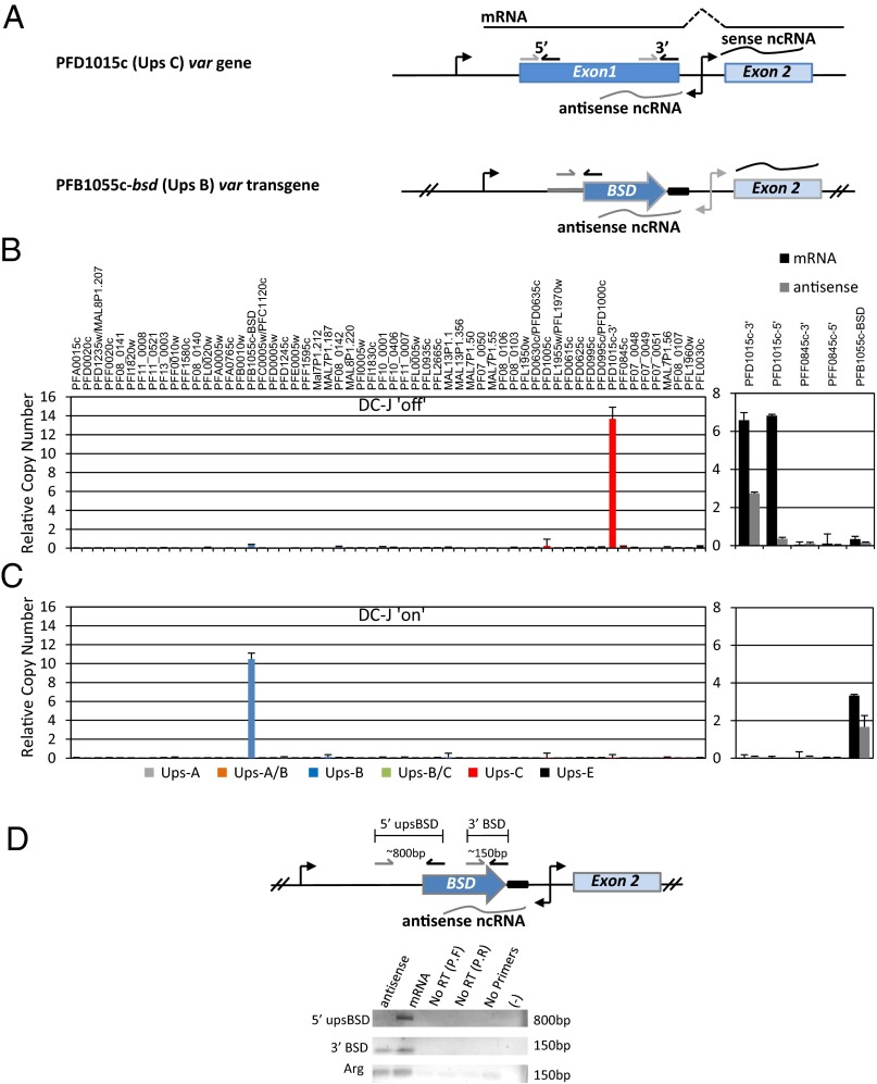
We initially observed that the levels of var transcripts measured by RT-quantitative PCR (qPCR) on cDNA produced with random primers, using a primer set designed to the 3′ of exon 1, are approximately double the levels measured by a primer set designed to the 5′ of the same exon (Fig. S1 A and B), although both primer sets had identical amplification efficiency. We reasoned that because the promoter within the intron is bidirectional, the 3′ primer set could potentially detect both the var mRNA and the antisense lncRNA of the intron. We therefore hypothesized that the antisense transcripts could be associated with the active var gene. To test if var-antisense transcripts are associated with the particular transcriptional status of an individual var gene, we initially used three clonal populations of the NF54 line (C3, C350, and chondroitin sulfate A-selected), each predominantly expressing a different var upstream promoter (PFD1005c, PFB1055c, and PFL0030c, respectively) (Fig. S1C, Left). To differentiate between the mRNA and the antisense lncRNA, we synthesized strand-specific cDNAs from each clonal population (Materials and Methods), and the levels of mRNA and intron antisense lncRNAs were measured from the predominantly active var gene and two silent genes in each of these populations. In the active genes, both mRNA and antisense lncRNA were detected using the primer set designed to amplify from the 3′ of exon 1. However, only mRNA, and not the antisense transcripts, was detected when using the primer set designed to amplify at the 5′ of exon 1, a region that the antisense transcript does not reach. In contrast, no mRNA or antisense lncRNA was detected from the silent var genes in each of the clonal populations (Fig. S1C, Right). To confirm these observations, we used the DC-J transgenic parasite line (9), which allows selecting for activation of a specific var gene (PFB1055c-bsd), thereby ensuring that the rest of the family is completely silent (Fig. 1A). We initially analyzed a clonal population of this line in which the transgene was silent and another var gene (PFD1015c) was active (Fig. 1B, Left Upper). We performed the same strand-specific cDNA synthesis as described above to differentiate between mRNA and the intronic antisense lncRNA. As in the NF54 WT clones, we could detect the antisense lncRNA only from the active gene, whereas no transcript was detected from the silent var genes (PFB1055c-bsd and PFF0845c) (Fig. 1B, Right Upper). As expected, the antisense transcripts were only detected by primers designated to the 3′ of exon 1. This parasite population was then grown under drug selection (2 μg/mL blasticidin S), thus forcing it to switch and exclusively express another var gene (PFB1055c-bsd) (Fig. 1C, Left Lower). In these parasites as well, the intronic antisense lncRNAs were detected exclusively from the active var gene PFB1055c-bsd, and not from the silent var genes (Fig. 1C, Right Lower). In the DC-J transgenic line, the long first exon of PFB1055c was replaced with the short bsd cassette (1,350 bp). Therefore, to be able to distinguish between detection of mRNA and antisense, we used RT-PCR with a forward primer located at the 5′ UTR of PFB1055c and a reverse primer located in the bsd coding region. Both mRNA and antisense from the active var gene were detected using the 3′BSD primer set, whereas the 5′upsBSD primer set could detect only the mRNA (Fig. 1D). In addition, these intronic antisense transcripts were only detected from the active gene at the time in the cell cycle when the var upstream promoter is active. In both DC-J “on” and “off” drug selection, as well as in the NF54 clones C3 and G6, the antisense transcripts were only detected in tightly synchronized parasites 18 h postinvasion (hpi) and not in late stages (36 hpi) (Fig. S1D). These observations are consistent with recent RNA-sequencing data from clonal parasite populations (17). Altogether, these data shows that var antisense lncRNAs are associated with the single active var gene and that these transcripts are detected in late ring stages at the time when the single var upstream promoter is transcriptionally active.
Fig. 1.
Antisense lncRNA is associated with the active var gene. The antisense lncRNA of var genes is transcribed simultaneously with var mRNA. (A) Schematic of the active var genes in the DC-J–off and DC-J–on lines (Upper and Lower, respectively), indicating the locations of the specific primers used for strand-specific cDNA production and RT-qPCR. Steady-state mRNA levels of the entire var gene family measured by RT-qPCR from DC-J–off (B, Left) and DC-J–on (C, Left). (B and C, Right) Strand-specific cDNA levels of mRNA (black) and antisense transcripts (gray) measured by RT-qPCR. RNA was extracted from tightly synchronized ring-stage parasites (∼18 h postinvasion). (D) Specificity of the strand-specific antisense detection. Schematic of the PFB1055c-bsd transgene indicating primer location (Upper) and RT-PCR (Lower), showing the specific detection of the antisense transcript only with the 3′BSD primer set. Amplification in the absence of reverse transcriptase with each of the forward or reverse primers [No RT (P.F) and No RT (P.R) respectively], no primers, and no template (−) were used as negative controls, whereas arginyl-tRNA synthetase was used as the positive control. Error bars represent SEs. The PbDT 3′ UTR is marked with a black rectangle, and the DC-J integration site at the 5′ UTR is marked with a bold gray line.
Antisense lncRNA Is Incorporated into Chromatin.
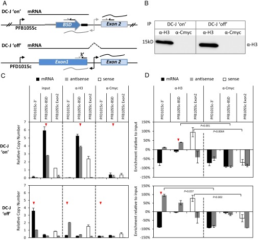
The var antisense lncRNA is transcribed by PolII (14), and although it is capped, it appears not to be polyadenylated, and thus is believed to remain in the nucleus (15). In recent years, it has been observed that transcriptional enhancers can recruit PolII to specific loci and mediate transcription of lncRNAs, which were postulated to be involved in gene regulation, possibly through incorporation into chromatin structures and recruitment of transcription factors and epigenetic modulators (20). Therefore, as a first step to investigate the possibility that var antisense transcripts have a regulatory role, we were interested to check if they are associated with chromatin. We performed ChIP using antibodies against the core histone H3, and α–C-myc antibodies were used as negative controls. The precipitated RNA was used as a template for strand-specific cDNA synthesis that enabled us to compare the level of enrichment of these antisense transcripts in active and silent var genes. We used the same clones of the DC-J transgenic line mentioned above (Fig. 1) after drug selection, where the transgene (PFB1055c-bsd) is active and all the remaining var genes are silent (DC-J–on), and compared it with the clone that grew without drug and predominantly expressed PFD1015c (DC-J–off). An additional advantage of using this line is that it enables us to distinguish between the PFB1055c-bsd mRNA, antisense lncRNA, and sense lncRNA by using different sets of primers (Fig. 2A). Following ChIP and RNA recovery of synchronized ring-stage parasites, the transcript levels were measured by RT-qPCR and the level of enrichment was calculated relative to the values from the same parasite cultures before performing ChIP (input). We found that in DC-J–on, the antisense of the active gene (PFB1055c-bsd) was significantly enriched in the α-H3 ChIP fraction compared with the input, whereas the antisense transcript of the silent gene could not be detected (Fig. 2 C and D, Upper). Similarly, in the DC-J–off line, where PFD1015c was predominantly expressed, its antisense transcript was significantly enriched in the ChIP fraction, whereas only low levels of antisense transcripts could be detected from PFB1055c-bsd, which is mostly silenced in this line (Fig. 2 C and D, Lower). In both lines, the sense noncoding transcripts of the intron, which are known to be incorporated into chromatin (15) and are expressed from numerous silent var genes, were significantly enriched in the α-H3 fraction as expected, indicating successful IP of these sterile transcripts. Similar to previous studies, we could detect low levels of sense transcripts in early stages even though these sense transcript are known to be highly transcribed in late stages (15, 16, 21). No transcripts were enriched when α–C-myc antibodies were used as negative controls for the IPs (Fig. 2 C and D), as expected, because chromatin was selectively pulled down by α-H3 and not α–C-myc antibodies (Fig. 2B). Interestingly, detectable levels of mRNA of the selected transgene (PFB1055c-bsd) were precipitated with H3 antibody, whereas no levels of mRNA transcripts were detected when the endogenous PFD1015c was active in the DC-J–off line. However, only the lncRNAs were enriched by ChIP, whereas mRNAs were not. These results suggest that the var antisense lncRNAs, which are associated with the single active var gene, are incorporated into chromatin.
Fig. 2.
Antisense lncRNA is associated with chromatin. RNA immunoprecipitation of var transcripts was performed on tightly synchronized ring-stage parasites (∼18 h postinvasion) using antibodies against the core histone H3 or with antibodies against C-myc as a negative control. (A) Schematic of the active var genes in each clone indicating the location of the specific primer sets that were used for strand-specific cDNA synthesis and RT-qPCR. The PbDT 3′ UTR is marked with a black rectangle. (B) Western blot of the fractions that were immunoprecipitated with α-H3 and α–C-myc antibodies from DC-J–on and DC-J–off populations and detected by the α-H3 antibody. (C) Levels of sense and antisense var transcripts detected by RT-qPCR from the IP samples (α-H3 or α–C-myc) and non-IP samples (input). Different primer sets presented on top of each column were used differentially to detect mRNA (black), antisense (gray), and sense ncRNA (white). (D) Enrichment levels of var sense and antisense transcripts detected by RT-qPCR and calculated as the relative percentile of the levels detected in the IP samples (α-H3 or α–C-myc) relative to the input. The levels of strand-specific cDNA were measured by RT-qPCR relative to arginyl-tRNA synthetase (PFL0900c). The active gene in each parasite line is marked with a red arrowhead. Each value is the average of two biological replicates and three technical triplicates. Error bars represent SEs. Samples differences were tested using the Student’s t test at P < 0.05.
Silent var Gene Is Activated by Expression of Its Specific Antisense Transcript.
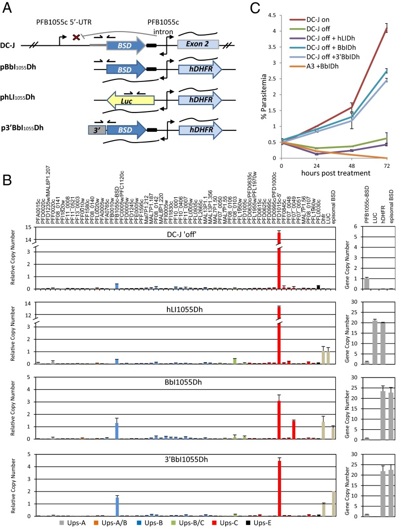
The association of the var-intron–derived antisense lncRNA with the single active var gene and their interaction with chromatin led us to hypothesize that these particular sterile transcripts might play a role in var gene activation. To address this possibility, we transfected the DC-J–off line, where the transgene PFB1055c-bsd is silent (and does not express antisense noncoding RNA) and the endogenous var gene PFD1015c is predominantly active, with a plasmid that expresses the specific antisense transcript of the silent var PFB1055c-bsd (Fig. 3). We monitored the expression of the entire var gene family using RT-qPCR to determine if the expression of the antisense lncRNAs in trans could influence var gene expression. To exclude the possible effect of spontaneous switching events, these patterns were compared with the expression of the entire var gene family in DC-J–off parasites that were not transfected and grown in parallel. In addition, we compared var expression with parasites that were transfected with a mock plasmid expressing luciferase instead of the sterile transcripts, as well as a plasmid in which the antisense transcript is fused to an unrelated 3′ UTR (Fig. 3A). Both parasite populations that were either not transfected or transfected with the mock plasmid had similar var expression patterns, and both predominantly expressed PFD1015c (Fig. 3B, Upper and Middle Upper, respectively). However, this expression pattern was altered in the DC-J–off parasites transfected with the plasmid expressing the PFB1055c intronic antisense transcripts. In this population, the transcript levels of PFD1015c were down-regulated, whereas the transcript level of PFB1055-bsd was up-regulated more than threefold without any blasticidin selection (Fig. 3B, Middle Lower). These data suggest that expression of particular var antisense lncRNAs in trans can alter their mRNA expression patterns and increase the activation rate of a specific var gene. Interestingly, parasites that were transfected with episomes in which the antisense transcripts were fused to an unrelated 3′ UTR showed similar activation levels of the PFB1055-bsd and down-regulation of PFD1015c (Fig. 3B, Lower). We noted that the level of steady-state antisense lncRNAs was approximately doubled when they were fused to the 3′ UTR. All transfected parasite populations carried similar copy numbers of the expression vectors (Fig. 3B, Right).
Fig. 3.
Silent var gene is activated by expression of its specific antisense transcript. (A) Schematics of the PFB1055-bsd recombinant locus in the DC-J parasite line (Upper); the plasmid pBbI1055Dh, which was used to express the specific antisense lncRNA of PFB1055-bsd (Middle Upper); the plasmid phLI1055Dh, which expresses an unrelated sequence (luciferase) that was used as a negative control (Middle Lower); and the plasmid p3′BbIDh, which expresses the antisense lncRNA of PFB1055-bsd fused to an unrelated 3′ UTR (Lower). The locations of the differential primers used for RT-qPCR are indicated by arrows. The PbDT 3′ UTR is marked with a black rectangle. (B, Left) Steady-state mRNA levels of the entire var gene family measured by RT-qPCR from DC-J–off (Upper), DC-J–off transfected with the control plasmid phLI1055Dh expressing an unrelated sequence (Middle Upper), DC-J–off transfected with the plasmid pBbI1055Dh expressing the var-specific antisense transcript (Middle Lower), and DC-J–off transfected with the plasmid p3′BbI1055Dh expressing the var-specific antisense transcript terminated by an unrelated 3′ UTR (Lower). RNA was extracted from tightly synchronized ring-stage parasites (∼18 h postinvasion). (B, Right) Plasmid copy number measured by qPCR of genomic DNA is presented. (C) Growth curves of parasite populations grown under selection with 2 μg/mL blasticidin. Growth curves were calculated for untransfected DC-J–on parasites that were preselected using blasticidin (brown); untransfected DC-J–off, where the bsd is predominantly silent (green); DC-J–off transfected with the mock plasmid phLI1055Dh (purple); DC-J–off transfected with the plasmid expressing the var-specific antisense pBbI1055Dh (light blue); DC-J–off transfected with the plasmid expressing the var-specific antisense fused with the 3′ UTR p3′BbI1055Dh (gray); and WT NF54 clone (A3) transfected with the plasmid expressing the var-specific antisense pBbI1055Dh (orange). Error bars represent SEs.
To verify that the up-regulation effect of the episomal antisense is sequence-specific, the antisense expression vector was transfected into a clonal NF54 population (A3) that predominantly expresses PFF0845c. This WT line does not contain the sequence found in the transgene PFB1055c-bsd, and thus does not share any sequence similarity with the episomal antisense transcripts. As we hypothesized, the antisense of the transgene PFB1055c-bsd had no effect on var transcription in the WT NF54 population, indicating that the mechanism of activation is sequence-specific (Fig. S2).
We tested whether this transcriptional alteration is indeed a consequence of a switch in var gene expression by putting these parasites under blasticidin pressure and measured their growth rates. We assumed that the growth rate of parasites in which the PFB1055c-bsd transgene is activated, and therefore express bsd, will be faster than in parasites in which this gene is initially silent. As additional controls, we used DC-J–on parasites that had been previously selected on 2 μg/mL blasticidin, activated PFB1055c-bsd, and therefore are blasticidin-resistant. In addition, we tested the growth of blasticidin-sensitive NF54 parasites (clone A3) transfected with the vector expressing the PFB1055c-bsd antisense lncRNAs. We found that the growth rate of the DC-J–off parasites expressing the PFB1055c-bsd antisense lncRNAS from either the BI1055Dh or 3′BI1055Dh expression vector was higher than in parasites that were not transfected (DC-J–off) or in parasites that were transfected with the mock plasmid (DC-J–off + hLI1055Dh) (Fig. 3C). The WT NF54 parasites transfected with the antisense expression vector (A3 + BI1055Dh) died after blasticidin selection and never recovered (Fig. 3C). Similar up-regulation of a silent var gene was obtained when expressing these antisense noncoding RNAs (ncRNAs) in another transgenic line [B15C2 (9)] where var PFL0020w was integrated with bsd cassette that enables its activation in a mutually exclusive manner (Fig. S3). Without bsd selection, these parasites undergo normal in vitro var switching. We identified a parasite population where the PFL0020w-bsd transgene was silent (B15C2-G) and transfected it with vectors expressing either the specific antisense lncRNAs (BbIYh) or a mock plasmid expressing an unrelated sequence (hLIYh) (Fig. S3A). In these experiments as well, the expression of the specific antisense transcripts activated the endogenous PFL0020c-bsd, whereas the mock plasmid had no effect (Fig. S3 B and C). These results suggest that expression of particular var antisense ncRNAs in trans accelerates var gene activation, providing the first evidence, to our knowledge, of the involvement of these sterile transcripts in var gene regulation.
Activation of var Genes by the Antisense lncRNA Is Sequence-Specific and Is Independent of PolII Transcription.
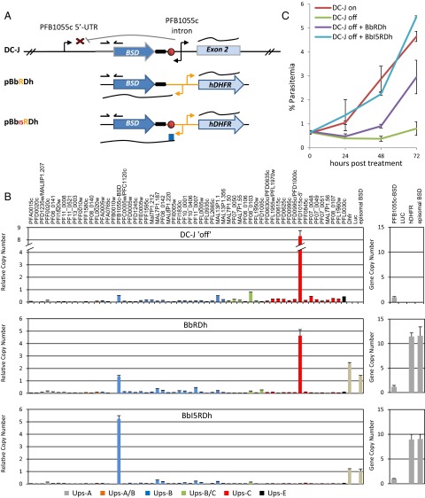
Both sense and antisense ncRNAs derived from the promoter found within the var intron are transcribed by PolII (14). We were interested to test if activation of var genes by the antisense transcripts is PolII-dependent. Therefore, the PolII-dependent promoter in our expression vectors was replaced with a PolI-dependent promoter (rRNA promoter) so that the antisense of the transgene PFB1055c-bsd is transcribed by PolI (pBbRDh). We have previously shown that var silencing is regulated by specific DNA elements [pairing elements (PEs)] found in each var upstream regulatory region and intron (22). As a consequence of their position within each intron, these PEs are found also in each of the var antisense transcripts. Therefore, in one of our expression vectors, we fused the sequence containing the var PE motif immediately after the PolI promoter (pBbI5RDh) (Fig. 4A). These plasmids were transfected into the DC-J–off clone, where the transgene PFB1055c-bsd is silent (Fig. 4B, Upper). We found that the var antisense transcripts activated the PFB1055c-bsd even when they are expressed by a PolI promoter. Moreover, when the PEs are added to these transcripts, the level of activation increased as reflected in the increase in bsd mRNA transcription level, as well as in the increase in the growth rate under blasticidin selection (Fig. 4 B and C). Transfection of the same parasite line with mock plasmids expressing unrelated sequences by a PolI promoter, with or without the PEs, did not alter var gene activation (Fig. S4). These results indicate that activation of var genes by the antisense transcripts is sequence-specific but is independent of PolII transcription per se.
Fig. 4.
Activation of var genes by the antisense lncRNA is sequence-specific and independent of PolII transcription. The var antisense lncRNA expressed by a PolI promoter activates a silent var gene. (A) Schematics of the PFB1055-bsd recombinant locus in the DC-J parasite line. (Upper) Location of the PEs in the var intron is indicated by a red circle. Two plasmids were used to express the antisense lncRNA of PFB1055-bsd by a PolI promoter: the plasmid pBbRDh (Middle) and the plasmid pBbI5RDh, where the PolI promoter was fused to the var PEs (Lower). The PbDT 3′ UTR is marked with a black rectangle. (B, Left) Steady-state mRNA levels of the entire var gene family measured by RT-qPCR from DC-J–off (Upper), DC-J–off transfected with the plasmid pBbRDh expressing the var-specific (PFB1055-bsd) antisense transcript (Middle), and DC-J–off transfected with the plasmid pBbI5RDh expressing the var-specific antisense transcript fused to the PEs (Lower). RNA was extracted from tightly synchronized ring-stage parasites (∼18 h postinvasion). (B, Right) Plasmid copy number measured by qPCR of genomic DNA is presented. (C) Growth curves of parasite populations growing under selection by 2 μg/mL blasticidin. Growth curves were calculated for untransfected DC-J–on parasites after preselection with blasticidin (brown), untransfected DC-J–off in which the bsd gene is predominantly silent (green), DC-J–off transfected with the plasmid pBbRDh (purple), and DC-J–off transfected with the plasmid pBbI5RDh with the PEs (light blue). Error bars represent SEs.
Dose-Dependent Transcriptional Activation of Endogenous var Genes by Gradual Overexpression of Their Specific var Antisense lncRNA.
To explore the possibility that the antisense ncRNAs can activate var genes in a dose-dependent manner, we took advantage of a parasite line that contains several copies of var-luciferase (var-luc) transgenes, which were integrated into the genome as four concatameric repeats [D10 (23)]. In each of these four var-luc transgenes, luciferase expression is controlled by the var upstream promoters, but because these var upstream promoters are paired with an intron, they are silent by default and express no luciferase. In addition to these paired var upstream promoters, this parasite line contains a var upstream promoter that is unpaired with an intron, and thus constitutively expresses low levels of luciferase (Fig. S5A, Upper). This parasite line was then transfected with a plasmid that expresses the specific antisense of the transgenes (pLhIBb) or a mock plasmid expressing an unrelated sequence (phGIBb) (Fig. S5A). The use of blasticidin as a selectable marker enables fine-tuned selection of parasites that carry increasing numbers of the expression vectors, and thus enables one to express increasing levels of any gene of interest as previously shown (21, 24). Following selection on 2 μg/mL blasticidin, the cultures were split and exposed to increasing concentrations of 6 and 10 μg/mL blasticidin. As expected, both new lines expressed increasing levels of either the episomal antisense (luc) or the unrelated sequence (GFP) (Fig. S5B) from the pLhIBb and phGIBb plasmids, respectively. In each of these transfected lines, the activity of the endogenous var-luc was monitored by RT-qPCR (Fig. S5C) and by quantifying protein activity (Fig. S5D). We found that as we increased expression of the var-luc antisense lncRNAs, the levels of luciferase from the endogenous var-luc had increased as well in a dose-dependent manner both at the level of steady-state mRNA and luciferase activity (Fig. S5 C and D, dark gray columns). However, increased expression of the unrelated GFP sequence had no effect on var-luc activation (Fig. S5 C and D, light gray columns). Overall, our data show that a cluster of multicopy var genes can be gradually activated by fine-tuned overexpression of their specific antisense lncRNAs, providing evidence for their role in var transcriptional activation.
Interference with the Antisense lncRNAs Down-Regulates the Active var Gene, Erases the Epigenetic Memory, and Induces var Gene Switching.
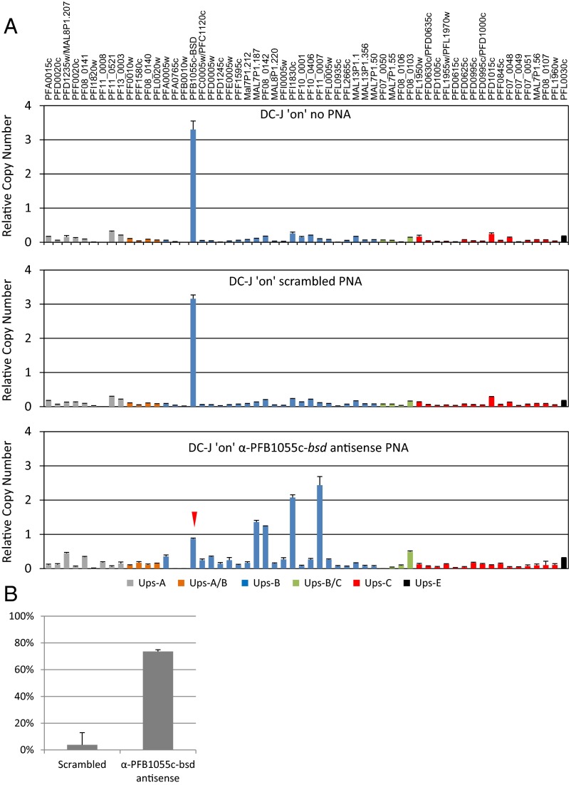
After demonstrating that silent var genes could be activated by expressing their specific antisense lncRNAs, we were interested to test if interference with the antisense lncRNAs of the active gene would down-regulate it. We recently showed that peptide nucleic acids (PNAs) could be used to down-regulate gene expression by hybridizing to mRNAs (25). We designed a PNA molecule that specifically complements a sequence of 17 bp found specifically in the antisense lncRNA of the active var gene (PFD1015c) in the DC-J–off parasite line. This parasite culture was split and incubated with 10 μM complementary PNA or scrambled PNA that contains the same nucleotides, which were shuffled and thus contained no sequence similarity to the P. falciparum genome. An additional culture was grown in parallel without any PNA treatment. We found that the population that was incubated with the scrambled PNA predominantly expressed PFD1015c similar to the population cultured in parallel that was not incubated with PNA (Fig. S6A, Upper and Middle). However, in the population that was incubated with the PNA molecules that complement the antisense of PFD1015c, this gene was significantly down-regulated (Fig. S6A, Lower) and expressed steady-state mRNA levels that were ∼70% lower than the parasites that were not incubated with PNA or those parasites incubated with the scrambled PNA (Fig. S6B).
It has been shown previously that the slow switch rate of internal var genes, such as PFD1015c (26), is maintained through tight regulation of epigenetic memory (27) and that this memory is maintained by active transcription of the active gene (24). We decided to test if down-regulation of a telomeric var gene that has a higher switch rate would erase the epigenetic memory and cause antigenic switching. We therefore designed PNAs that complement the antisense of the telomeric active var gene (PFD1055c-bsd) in the DC-J–on line and compared var transcription patterns in cultures that were incubated with the specific PNA with those cultures that were not incubated or were incubated with the scrambled PNA. We found that incubation with the scrambled PNA had no effect on var transcription and PFB1055c-bsd was predominantly expressed similar to the culture that was not incubated with PNA (Fig. 5A, Upper and Middle). However, the active gene (PFB1055c-bsd) was significantly down-regulated (∼70%) in the culture that was incubated with the specific PNAs that complement its antisense lncRNAs (Fig. 5 A, Lower and B). Furthermore, by down-regulating the transcription levels of this gene, we erased the epigenetic memory that imprinted this gene as the active gene and induced switching to activation of other var genes in the population, which became transcriptionally heterogeneous. Altogether, this set of experiments indicates that interference with the antisense lncRNAs down-regulates the active var gene, erases the epigenetic memory, and induces var gene switching.
Fig. 5.
Interference with the antisense lncRNAs down-regulates the active var gene, erases epigenetic memory, and induces var gene switching. PNAs complementary to the antisense transcript of the active var gene (PFB1055c-bsd) in the DC-J–on parasite population (growing without selection) were used to block its activity by hybridization. (A) Steady-state mRNA levels of the entire var gene family measured by RT-qPCR from DC-J–on with no PNA treatment (Upper), DC-J–on incubated with 10 μM scrambled PNA (Middle), and DC-J–on incubated with 10 μM specific α–PFB1055c-bsd antisense PNA (Lower). RNA was extracted from tightly synchronized ring-stage parasites (∼18 h postinvasion). (B) Levels of down-regulation of PFB1055c-bsd were calculated as the ratio between the levels of steady-state mRNA in PNA-treated parasites relative to nontreated parasites. These levels are presented as a percentage of down-regulation. All values presented are the average of three biological replicates and three technical triplicates. Error bars represent SEs.
Discussion
The lncRNAs were shown to play an important role in epigenetic regulation of gene transcription in several eukaryotic systems (28). However, although ncRNAs were predicted to be abundant in Plasmodium (29–34), their function remained unknown. Here, we provide evidence that antisense lncRNAs are involved in var gene activation. These transcripts are specifically associated with the active gene and are detected from late ring stages parallel to the time during the intraerythrocytic development when the mRNA-producing var upstream promoter is active. In contrast to the mRNA, which is translated into PfEMP1, we show that these antisense lncRNAs are associated with chromatin. We also show that expression of particular var antisense lncRNAs in trans activates the corresponding endogenous var gene in a sequence- and dose-dependent manner. Last, we present evidence that interference with these antisense lncRNAs leads to transcriptional repression of an endogenous active var gene, which triggers var gene switching. Thus far, the promoter found in var introns has been implicated in var silencing and considered to act as a silencer of upstream var promoter (11–13, 35). The data presented here suggest that the bidirectional promoter within var intron may act as a silencer when expressing the sense lncRNAs and as an enhancer when expressing the antisense transcripts. It is still unclear what determines the timing and orientation of the intron promoter activity, and perhaps it involves some alternative transcription start site (TSS) found in the intron promoter (15), which could be associated with different promoter orientation. Recent data provided evidence for the mechanism of var gene silencing and epigenetic memory linking PolII transcription of sense lncRNA to recruitment of a var-specific heterochromatin marker (17, 36). The mechanisms by which PolII transcription in the antisense orientation is involved in var gene activation are unknown; nonetheless, our results indicate that these antisense transcripts activate var genes independent of PolII transcription.
The best-studied examples for epigenetic regulation by ncRNAs include Xist in mammals (37–39) and Rox1 and Rox2 in Drosophila (40, 41), where ncRNAs incorporated into chromatin structures initiate a cascade of events that epigenetically mark these particular loci for either silent heterochromatin or active euchromatin. Recent studies have shown that lncRNAs are associated with enhancer regions and have important roles in activating transcription of neighboring genes (42–46). These lncRNAs were proposed to incorporate into chromatin and to play a role in recruiting transcription factors and chromatin modulators required for gene activation (20). In mouse cortical neurons, PolII transcription of enhancer ncRNAs was positively correlated with the level of RNA synthesis at nearby genes, suggesting that long-distance interactions between enhancers and promoters are necessary for activation (43). In addition, in HOXA activation, it was shown that HOTTIP (HOXA transcript at the distal tip) lncRNA functions in cis on its target genes by modifying chromatin structure and coordinates activation by recruiting specific chromatin-modifying factors to a specific genome location (45, 47). The incorporation of var antisense lncRNAs into chromatin and their function in a sequence-dependent manner could indicate that similar mechanisms are involved in var gene activation. These antisense lncRNAs may contribute to remodeling of chromatin conformation and recruitment of activation factors to the locus of the active var gene. In addition, var antisense transcripts may contribute to the maintenance of epigenetic memory by recruiting chromatin-modifying enzymes that drive specific histone marks, which epigenetically imprint the active locus for many cells’ cycles and ensure a very slow switch rate. Furthermore, var silencing by the intron promoter was shown to be mediated by insulator-like PEs that bind specific nuclear proteins (22). The intronic PEs are found upstream to the intronic TSS; therefore, the antisense transcripts contain the complementary sequence of the PEs. The data presented here indicate that the presence of the PE sequence increases the level of activation by the antisense transcripts. It is possible that when incorporating into chromatin, these transcripts could hybridize to the cDNA sequence and interfere with the binding of nuclear proteins to the PEs. Preventing binding of proteins associated with silencing might influence chromatin conformation and change it from compact heterochromatin into loose euchromatin that enables a single var gene to be accessible for the transcriptional machinery (Fig. S7).
Our findings that antisense lncRNAs are involved in var gene activation open new avenues to explore mutually exclusive expression of var genes, particularly how a single var gene is chosen to be active and what mechanisms are involved in the epigenetic memory.
Materials and Methods
Details of plasmid construction and methodologies that have been previously described are provided in SI Materials and Methods.
Extraction of DNA, RNA, cDNA Synthesis, and RT-qPCR.
Extraction of genomic DNA and RNA and cDNA synthesis were performed as described (9, 24). Strand-specific cDNAs from mRNA and sense transcripts were synthesized by using specific reverse (R) primers, whereas cDNA from antisense transcripts was synthesized using forward (F) primers for specific var genes as detailed in each figure and listed in Table S1. The cDNA of the control genes was synthesized with both F and R primers specific to the following: arginyl-tRNA synthetase (PFL0900c), seryl-tRNA synthetase (PF07_0073), actin (PFL2215), and glutaminyl-tRNA synthetase (PF13_0170). For each cDNA synthesis, we have included three negative controls: without reverse transcriptase (no reverse transcriptase-F and no reverse transcriptase-R, respectively) and without primers. All primers used are listed in Table S1. RT-qPCR was performed as described (48). Additional primers used in this study are listed in Table S1. All values presented are relative copy numbers to the housekeeping gene arginyl-tRNA synthetase (PFL0900c). Each value is the average of at least two biological replicates and three technical triplicates.
RNA Immunoprecipitation.
RNA immunoprecipitation was done as described (49) using α-H3 antibody (Ab1791; Abcam) bound to Protein-G Sepharose beads (Santa Cruz Biotechnology). Beads bound to α–C-myc antibody (Santa Cruz Biotechnology) or no antibody were used as negative controls. Bound RNA was collected in TRIzol LS Reagent (Life Technologies) and purified on a PureLink column (Invitrogen).
PNA Synthesis and Treatment.
PNA sequences, equipped with a C-terminal octalysine moiety, were synthesized as described (25). PNA sequences are detailed in Table S2. DC-J–on and DC-J–off parasite lines were tightly synchronized as described above. Late-stage parasites were recovered and placed back in culture. Twenty-two hours after synchronization, the drug selection from the DC-J–on parasite line was removed and each parasite line was divided into three cultures and incubated for an additional 48 h with 10 μM PNA designed to complement the antisense lncRNA of the active gene, 10 μM scrambled PNA, and no PNA treatment. Parasites from each culture were then collected for RT-qPCR analysis as described above.
Supplementary Material
Acknowledgments
We thank Dr. Yoav Smith for his help in performing the statistical analysis and Dr. Adina Heinberg for critical reading of the manuscript. This work was supported partially by Israeli Academy for Science Grant 141/13 and continued to be funded by European Research Council (erc.europa.eu) Consolidator Grant 615412 (to R.D.). I.A.-A. was supported by the Abisch–Frenkel Foundation.
Footnotes
The authors declare no conflict of interest.
This article is a PNAS Direct Submission.
This article contains supporting information online at www.pnas.org/lookup/suppl/doi:10.1073/pnas.1420855112/-/DCSupplemental.
References
- 1.WHO 2010. World Malaria Report 2010 (World Health Organization, Geneva)
- 2.Miller LH, Baruch DI, Marsh K, Doumbo OK. The pathogenic basis of malaria. Nature. 2002;415(6872):673–679. doi: 10.1038/415673a. [DOI] [PubMed] [Google Scholar]
- 3.Baruch DI, Gormely JA, Ma C, Howard RJ, Pasloske BL. Plasmodium falciparum erythrocyte membrane protein 1 is a parasitized erythrocyte receptor for adherence to CD36, thrombospondin, and intercellular adhesion molecule 1. Proc Natl Acad Sci USA. 1996;93(8):3497–3502. doi: 10.1073/pnas.93.8.3497. [DOI] [PMC free article] [PubMed] [Google Scholar]
- 4.Smith JD, et al. Switches in expression of Plasmodium falciparum var genes correlate with changes in antigenic and cytoadherent phenotypes of infected erythrocytes. Cell. 1995;82(1):101–110. doi: 10.1016/0092-8674(95)90056-x. [DOI] [PMC free article] [PubMed] [Google Scholar]
- 5.Su XZ, et al. The large diverse gene family var encodes proteins involved in cytoadherence and antigenic variation of Plasmodium falciparum-infected erythrocytes. Cell. 1995;82(1):89–100. doi: 10.1016/0092-8674(95)90055-1. [DOI] [PubMed] [Google Scholar]
- 6.Scherf A, et al. Antigenic variation in malaria: in situ switching, relaxed and mutually exclusive transcription of var genes during intra-erythrocytic development in Plasmodium falciparum. EMBO J. 1998;17(18):5418–5426. doi: 10.1093/emboj/17.18.5418. [DOI] [PMC free article] [PubMed] [Google Scholar]
- 7.Chen Q, et al. Developmental selection of var gene expression in Plasmodium falciparum. Nature. 1998;394(6691):392–395. doi: 10.1038/28660. [DOI] [PubMed] [Google Scholar]
- 8.Dzikowski R, Deitsch KW. Genetics of antigenic variation in Plasmodium falciparum. Curr Genet. 2009;55(2):103–110. doi: 10.1007/s00294-009-0233-2. [DOI] [PMC free article] [PubMed] [Google Scholar]
- 9.Dzikowski R, Frank M, Deitsch K. Mutually exclusive expression of virulence genes by malaria parasites is regulated independently of antigen production. PLoS Pathog. 2006;2(3):e22. doi: 10.1371/journal.ppat.0020022. [DOI] [PMC free article] [PubMed] [Google Scholar]
- 10.Voss TS, et al. A var gene promoter controls allelic exclusion of virulence genes in Plasmodium falciparum malaria. Nature. 2006;439(7079):1004–1008. doi: 10.1038/nature04407. [DOI] [PubMed] [Google Scholar]
- 11.Calderwood MS, Gannoun-Zaki L, Wellems TE, Deitsch KW. Plasmodium falciparum var genes are regulated by two regions with separate promoters, one upstream of the coding region and a second within the intron. J Biol Chem. 2003;278(36):34125–34132. doi: 10.1074/jbc.M213065200. [DOI] [PubMed] [Google Scholar]
- 12.Gannoun-Zaki L, Jost A, Mu J, Deitsch KW, Wellems TE. A silenced Plasmodium falciparum var promoter can be activated in vivo through spontaneous deletion of a silencing element in the intron. Eukaryot Cell. 2005;4(2):490–492. doi: 10.1128/EC.4.2.490-492.2005. [DOI] [PMC free article] [PubMed] [Google Scholar]
- 13.Dzikowski R, et al. Mechanisms underlying mutually exclusive expression of virulence genes by malaria parasites. EMBO Rep. 2007;8(10):959–965. doi: 10.1038/sj.embor.7401063. [DOI] [PMC free article] [PubMed] [Google Scholar]
- 14.Kyes S, et al. Plasmodium falciparum var gene expression is developmentally controlled at the level of RNA polymerase II-mediated transcription initiation. Mol Microbiol. 2007;63(4):1237–1247. doi: 10.1111/j.1365-2958.2007.05587.x. [DOI] [PubMed] [Google Scholar]
- 15.Epp C, Li F, Howitt CA, Chookajorn T, Deitsch KW. Chromatin associated sense and antisense noncoding RNAs are transcribed from the var gene family of virulence genes of the malaria parasite Plasmodium falciparum. RNA. 2009;15(1):116–127. doi: 10.1261/rna.1080109. [DOI] [PMC free article] [PubMed] [Google Scholar]
- 16.Kyes SA, et al. A well-conserved Plasmodium falciparum var gene shows an unusual stage-specific transcript pattern. Mol Microbiol. 2003;48(5):1339–1348. doi: 10.1046/j.1365-2958.2003.03505.x. [DOI] [PMC free article] [PubMed] [Google Scholar]
- 17.Jiang L, et al. PfSETvs methylation of histone H3K36 represses virulence genes in Plasmodium falciparum. Nature. 2013;499(7457):223–227. doi: 10.1038/nature12361. [DOI] [PMC free article] [PubMed] [Google Scholar]
- 18.Zhang Q, et al. Exonuclease-mediated degradation of nascent RNA silences genes linked to severe malaria. Nature. 2014;513(7518):431–435. doi: 10.1038/nature13468. [DOI] [PubMed] [Google Scholar]
- 19.Ralph SA, et al. Transcriptome analysis of antigenic variation in Plasmodium falciparum—var silencing is not dependent on antisense RNA. Genome Biol. 2005;6(11):R93. doi: 10.1186/gb-2005-6-11-r93. [DOI] [PMC free article] [PubMed] [Google Scholar]
- 20.Ong CT, Corces VG. Enhancer function: New insights into the regulation of tissue-specific gene expression. Nat Rev Genet. 2011;12(4):283–293. doi: 10.1038/nrg2957. [DOI] [PMC free article] [PubMed] [Google Scholar]
- 21.Epp C, Raskolnikov D, Deitsch KW. A regulatable transgene expression system for cultured Plasmodium falciparum parasites. Malar J. 2008;7:86. doi: 10.1186/1475-2875-7-86. [DOI] [PMC free article] [PubMed] [Google Scholar]
- 22.Avraham I, Schreier J, Dzikowski R. Insulator-like pairing elements regulate silencing and mutually exclusive expression in the malaria parasite Plasmodium falciparum. Proc Natl Acad Sci USA. 2012;109(52):E3678–E3686. doi: 10.1073/pnas.1214572109. [DOI] [PMC free article] [PubMed] [Google Scholar]
- 23.Frank M, et al. Strict pairing of var promoters and introns is required for var gene silencing in the malaria parasite Plasmodium falciparum. J Biol Chem. 2006;281(15):9942–9952. doi: 10.1074/jbc.M513067200. [DOI] [PMC free article] [PubMed] [Google Scholar]
- 24.Dzikowski R, Deitsch KW. Active transcription is required for maintenance of epigenetic memory in the malaria parasite Plasmodium falciparum. J Mol Biol. 2008;382(2):288–297. doi: 10.1016/j.jmb.2008.07.015. [DOI] [PMC free article] [PubMed] [Google Scholar]
- 25.Kolevzon N, Nasereddin A, Naik S, Yavin E, Dzikowski R. Use of peptide nucleic acids to manipulate gene expression in the malaria parasite Plasmodium falciparum. PLoS ONE. 2014;9(1):e86802. doi: 10.1371/journal.pone.0086802. [DOI] [PMC free article] [PubMed] [Google Scholar]
- 26.Frank M, Dzikowski R, Amulic B, Deitsch K. Variable switching rates of malaria virulence genes are associated with chromosomal position. Mol Microbiol. 2007;64(6):1486–1498. doi: 10.1111/j.1365-2958.2007.05736.x. [DOI] [PMC free article] [PubMed] [Google Scholar]
- 27.Chookajorn T, et al. Epigenetic memory at malaria virulence genes. Proc Natl Acad Sci USA. 2007;104(3):899–902. doi: 10.1073/pnas.0609084103. [DOI] [PMC free article] [PubMed] [Google Scholar]
- 28.Mercer TR, Dinger ME, Mattick JS. Long non-coding RNAs: Insights into functions. Nat Rev Genet. 2009;10(3):155–159. doi: 10.1038/nrg2521. [DOI] [PubMed] [Google Scholar]
- 29.Mourier T, et al. Genome-wide discovery and verification of novel structured RNAs in Plasmodium falciparum. Genome Res. 2008;18(2):281–292. doi: 10.1101/gr.6836108. [DOI] [PMC free article] [PubMed] [Google Scholar]
- 30.Gunasekera AM, et al. Widespread distribution of antisense transcripts in the Plasmodium falciparum genome. Mol Biochem Parasitol. 2004;136(1):35–42. doi: 10.1016/j.molbiopara.2004.02.007. [DOI] [PubMed] [Google Scholar]
- 31.Patankar S, Munasinghe A, Shoaibi A, Cummings LM, Wirth DF. Serial analysis of gene expression in Plasmodium falciparum reveals the global expression profile of erythrocytic stages and the presence of anti-sense transcripts in the malarial parasite. Mol Biol Cell. 2001;12(10):3114–3125. doi: 10.1091/mbc.12.10.3114. [DOI] [PMC free article] [PubMed] [Google Scholar]
- 32.Li F, Sonbuchner L, Kyes SA, Epp C, Deitsch KW. Nuclear non-coding RNAs are transcribed from the centromeres of Plasmodium falciparum and are associated with centromeric chromatin. J Biol Chem. 2008;283(9):5692–5698. doi: 10.1074/jbc.M707344200. [DOI] [PubMed] [Google Scholar]
- 33.Broadbent KM, et al. A global transcriptional analysis of Plasmodium falciparum malaria reveals a novel family of telomere-associated lncRNAs. Genome Biol. 2011;12(6):R56. doi: 10.1186/gb-2011-12-6-r56. [DOI] [PMC free article] [PubMed] [Google Scholar]
- 34.Sierra-Miranda M, et al. Two long non-coding RNAs generated from subtelomeric regions accumulate in a novel perinuclear compartment in Plasmodium falciparum. Mol Biochem Parasitol. 2012;185(1):36–47. doi: 10.1016/j.molbiopara.2012.06.005. [DOI] [PMC free article] [PubMed] [Google Scholar]
- 35.Deitsch KW, Calderwood MS, Wellems TE. Malaria. Cooperative silencing elements in var genes. Nature. 2001;412(6850):875–876. doi: 10.1038/35091146. [DOI] [PubMed] [Google Scholar]
- 36.Ukaegbu UE, et al. Recruitment of PfSET2 by RNA polymerase II to variant antigen encoding loci contributes to antigenic variation in P. falciparum. PLoS Pathog. 2014;10(1):e1003854. doi: 10.1371/journal.ppat.1003854. [DOI] [PMC free article] [PubMed] [Google Scholar]
- 37.Rasmussen TP, Wutz AP, Pehrson JR, Jaenisch RR. Expression of Xist RNA is sufficient to initiate macrochromatin body formation. Chromosoma. 2001;110(6):411–420. doi: 10.1007/s004120100158. [DOI] [PubMed] [Google Scholar]
- 38.Lee JT, Davidow LS, Warshawsky D. Tsix, a gene antisense to Xist at the X-inactivation centre. Nat Genet. 1999;21(4):400–404. doi: 10.1038/7734. [DOI] [PubMed] [Google Scholar]
- 39.Jeon Y, Sarma K, Lee JT. New and Xisting regulatory mechanisms of X chromosome inactivation. Curr Opin Genet Dev. 2012;22(2):62–71. doi: 10.1016/j.gde.2012.02.007. [DOI] [PMC free article] [PubMed] [Google Scholar]
- 40.Meller VH, et al. Ordered assembly of roX RNAs into MSL complexes on the dosage-compensated X chromosome in Drosophila. Curr Biol. 2000;10(3):136–143. doi: 10.1016/s0960-9822(00)00311-0. [DOI] [PubMed] [Google Scholar]
- 41.Park Y, Kelley RL, Oh H, Kuroda MI, Meller VH. Extent of chromatin spreading determined by roX RNA recruitment of MSL proteins. Science. 2002;298(5598):1620–1623. doi: 10.1126/science.1076686. [DOI] [PubMed] [Google Scholar]
- 42.Ørom UA, et al. Long noncoding RNAs with enhancer-like function in human cells. Cell. 2010;143(1):46–58. doi: 10.1016/j.cell.2010.09.001. [DOI] [PMC free article] [PubMed] [Google Scholar]
- 43.Kim TK, et al. Widespread transcription at neuronal activity-regulated enhancers. Nature. 2010;465(7295):182–187. doi: 10.1038/nature09033. [DOI] [PMC free article] [PubMed] [Google Scholar]
- 44.De Santa F, et al. A large fraction of extragenic RNA pol II transcription sites overlap enhancers. PLoS Biol. 2010;8(5):e1000384. doi: 10.1371/journal.pbio.1000384. [DOI] [PMC free article] [PubMed] [Google Scholar]
- 45.Wang KC, et al. A long noncoding RNA maintains active chromatin to coordinate homeotic gene expression. Nature. 2011;472(7341):120–124. doi: 10.1038/nature09819. [DOI] [PMC free article] [PubMed] [Google Scholar]
- 46.Ørom UA, Shiekhattar R. Long non-coding RNAs and enhancers. Curr Opin Genet Dev. 2011;21(2):194–198. doi: 10.1016/j.gde.2011.01.020. [DOI] [PMC free article] [PubMed] [Google Scholar]
- 47.Tsai MC, et al. Long noncoding RNA as modular scaffold of histone modification complexes. Science. 2010;329(5992):689–693. doi: 10.1126/science.1192002. [DOI] [PMC free article] [PubMed] [Google Scholar]
- 48.Fastman Y, Noble R, Recker M, Dzikowski R. Erasing the epigenetic memory and beginning to switch—The onset of antigenic switching of var genes in Plasmodium falciparum. PLoS ONE. 2012;7(3):e34168. doi: 10.1371/journal.pone.0034168. [DOI] [PMC free article] [PubMed] [Google Scholar]
- 49.Mair GR, et al. Regulation of sexual development of Plasmodium by translational repression. Science. 2006;313(5787):667–669. doi: 10.1126/science.1125129. [DOI] [PMC free article] [PubMed] [Google Scholar]
Associated Data
This section collects any data citations, data availability statements, or supplementary materials included in this article.


