Abstract
Organ formation requires a delicate balance of positive and negative regulators. In Drosophila eye development, wingless (wg) is expressed at the lateral margins of the eye disc and serves to block retinal development. The T-box gene optomotor-blind (omb) is expressed in a similar pattern and is regulated by Wg. Omb mediates part of Wg activity in blocking eye development. Omb exerts its function primarily by blocking cell proliferation. These effects occur predominantly in the ventral margin. Our results suggest that the primary effect of Omb is the blocking of Jak/STAT signaling by repressing transcription of upd which encodes the Jak receptor ligand Unpaired.
Introduction
The Drosophila compound eye originates from the eye-antenna anlage in the embryo. These cells proliferate and form the eye-antennal disc in the larva. In the mid-third instar eye disc, a wave of cell cycle coordination and apical cellular constriction, called the morphogenetic furrow (MF) forms at the posterior margin and progressively moves toward anterior. Posterior to the MF, retinal cell fates are specified by a series of cellular interactions [1,2,3,4]. The early steps of eye development involve at least three aspects: specification of eye fate, control of cell proliferation, and initiation and progression of the MF.
A large number of genes are involved in promoting eye development. Eye fate is specified by the retinal determination gene network which includes the transcription factors encoded by eyeless (ey), twin of eyeless (toy), sine oculis (so), eyes absent (eya), and dachshund (dac) [5,6]. Cell proliferation is highly regulated. Undifferentiated cells anterior to the MF undergo proliferation that is promoted by Notch signaling, the Pax protein Eyg, a combination of the transcription factors Eyeless, Homothorax (Hth), Teashirt (Tsh) and the transcriptional coactivator Yorkie (Yki), as well as Upd/Jak/STAT signaling [7,8,9,10,11,12,13,14]. MF initiation and progression are promoted by the Decapentaplegic (Dpp), Hedgehog (Hh) and Upd/Jak/STAT signaling pathways [11,15,16,17,18,19,20,21,22,23,24].
However, developmental processes rarely proceed by agonistic action alone but tend to be held in check by interaction between agonists and antagonists. The necessity to keep retinal development in bounds is obvious in the eye-antennal imaginal disc since this disc, in addition to the retina, gives rise to much of the exterior of the adult head [25,26,27]. Molecules with the ability to block eye development include Patched (Ptc) and other negative regulators of Hh signaling, Wingless (Wg) and the positive components of its signaling pathway, the transcription factors and cofactors encoded by homothorax (hth), teashirt (tsh), hairy (h), extramacrochaetae (emc), pannier (pnr), Chip, arrowhead (awh) and Lim1 [6,13,28,29,30,31,32,33,34].
Of these anti-retinal genes, Wg is the only signaling ligand and appears to be the most important anti-retinal factor. In the third instar eye disc, wg is expressed in the lateral margins and prevents inappropriate marginal morphogenetic furrow initiation [30,35]. Wg exerts its anti-retinal function by several routes. First, Wg blocks MF initiation [30,35]. A primary target is Dpp, which is essential for MF initiation [15,36]. Wg signaling represses dpp transcription and Dpp signaling at a step downstream of receptor activation [37,38]. Second, Wg also blocks MF progression [35] and neuronal differentiation through repression of Daughterless (Da) [38].
Which gene is induced by Wg to mediate its anti-retinal functions? One prime candidate is optomotor-blind (omb, FlyBase bifid, bi) which is expressed in the lateral margins in a pattern similar to the wg expression domain [39]. Ectopic expression of either wg or its downstream effector armadillo (arm) induces the expression of omb near the lateral margins [40]. Omb encodes a T-domain transcription factor and is required for the development of the optic lobes, wing, abdomen, and terminalia [41,42,43,44,45,46,47,48,49,50,51]. The polar eye disc expression and the fact that ectopic omb can completely block eye development [52] led us to investigate the role of omb in this process, and its relationship with Wg.
We show that Omb antagonizes eye development primarily at the level of cell proliferation. We further identified a molecular pathway downregulated by Omb. Our results suggest that the main effects of Omb are a block of Jak/STAT signaling by suppressing transcription of upd encoding the Jak/STAT ligand Unpaired. The block of Jak/STAT signaling accounts for the effect of Omb on cell proliferation. Our results also show that Omb mediates part of the Wg anti-retinal effects.
Materials and Methods
Fly stocks
Fly culture and crosses were performed according to standard procedure at 25°C unless noted otherwise. Transgenic expression lines: UAS-omb and hsp70-omb [47], UAS-arm [40], UAS-dpp [53], UAS-upd, UAS-hop [54]. STAT92E 397[55], STAT92E 06346 (STAT P1681-lacZ, in [56,57]). dpp C40.6-GAL4 [53], omb P3-GAL4 (GAL4-bi md 653 in [58]; cf. [59], ey-GAL4 [37] and omb P7-GAL4 (omb3 in [60]; cf. [59] were used as GAL4 drivers. Alleles used are: omb bi (regulatory hypomorph, [41]), l(1)omb D4, l(1)omb 3198, and l(1)omb 15 (molecularly defined null mutants, [61,62]), omb For (gain-of-function mutant caused by a large downstream insertion [51]) and omb P7-GAL4 is hemi- and homozygous lethal and was used both as an omb allele and as GAL4-driver in the omb expression domain [60]. lacZ reporter lines are: omb-lacZ (omb P 1 in [63]), dpp-lacZ (BS3.0)[3], mirr-lacZ [63], fng-lacZ [64], and wg-lacZ [65]. w omb P 1 l(1)omb 3198 was obtained by intragenic recombination. dpp-lacZ (BS3.0), dpp-GAL4c 40.6 and wg IL114 were kindly provided by Jessica Treisman, omb For by Marc Fortini, Act5C>CD2>GAL4 [23], tub>CD2>omb and tub>CD2>GAL4 by Christian Dahmann. Other fly stocks were obtained from the Bloomington Drosophila Stock Center and the Mid-America Drosophila Stock Center (Bowling Green, Ohio).
Construction of 10X STAT-GFP-nls
GFP-nls sequence was amplified from pH-Stinger [66] by PCR primers (GGTTCAGGGGGAGGTGTGGG; ACTCGAGGCAGCCAAGCTGATCCTCTAGGG) and then cloned into 10XSTAT-luciferase [66] by Xho I and Xba I to generate the10X-STAT-GFP-nls construct. Germline transformants were generated as described previously [67].
Clonal induction
Positively labeled flp-out expression clones were generated by crossing UAS-lines to hs-FLP 122; Act5C>y + >GAL4 UAS-GFP S65T [68]. Heat shock induction of hs-FLP 122 was at 37°C for 30 min at 24–48 hr after egg laying. l(1)omb D4 and control clones were generated by incubating hs-flp 122 hs-GFP FRT19/FRT19 or hs-flp 122 hs-GFP FRT19/l(1)omb D4 FRT19 larvae at 48–60 h AEL at 38°C for 30 min. Larvae were raised at 25°C for 48 h. Before dissection, larvae were subjected to 37°C for 1h and then shifted back to 25°C for 1h to allow GFP expression. omb gain-of-function and control clones were generated by incubating hs-flp 122; tub>CD2>GAL4; UAS-GFP or hs-flp 122; tub>CD2>omb; UAS-GFP larvae at 36–48h AEL for 30 min at 37°C. Larvae were dissected after 72 h at 25°C.
Immunohistochemistry
Late third instar larval imaginal discs were dissected and stained. Primary antibodies were rat anti-Elav 7E8A10 (1:500, Developmental Studies Hybridoma Bank, U. of Iowa (DSHB, Iowa), rabbit anti-ß-galactosidase (1:1000, Cappel), mouse anti-Eya 10H6 (1:200, DSHB, Iowa), rabbit anti-BarH1(S12) (1:1000, gift from Tetsuya Kojima), rabbit anti-Omb (1:1000, [47,49]), rabbit anti-phospho-histone H3 (anti-PH3) (1:200–1:1000, Upstate Biotechnology), rabbit anti-Caspase-3 (cleaved) (1:200, Upstate Biotechnology), mouse anti-CD2 (rat) (1: 2000, Serotec), and mouse anti-Wg 4D4 (1:200, DSHB, Iowa), mouse anti-BrdU (1:50, Roche). Secondary antibodies (Jackson ImmunoResearch) were FITC-, Cy3- or Cy5-conjugated anti-rabbit, anti-rat and anti-mouse. Confocal microscopy was performed on a Zeiss LSM 310 or LSM 510. X-Gal staining of lacZ expression was done as described [63]. Anti-BrdU staining was performed as described [9].
RNA in situ hybridization
upd RNA in situ hybridization is executed as described [9].
Tissue sections
Semi-thin plastic eye sections were performed according to [69].
Scanning electron microscopy and determination of ommatidial number
Scanning electron micrographs of adult eyes were obtained as described [52]. Due to the curvature of the eye, the ommatidial number N cannot be obtained from a single micrograph. A given eye was photographed from different angles. Dust particles or aberrations in the bristle pattern allowed the alignment of the otherwise repetitive structures such that error-free counting was possible.
Results
omb negatively regulates eye size
In the eye imaginal disc, Omb is expressed in two cell types. Within the main epithelium, Omb is expressed at the dorsal and ventral margins (S1A Fig., arrow). Omb is also expressed in the retinal basal glial cells that lie at the basal level of the eye disc and in the optic stalk [39,70] (S1A Fig.). Only the epithelial expression will be considered here. We found that loss-of-function and gain-of-function omb mutations caused changes in eye size.
In omb hypomorphic allele combinations and omb knock-down, the Omb level was reduced in both margins and the eye disc was enlarged (S1B Fig.). This was observed for the omb hypomorph omb bi in combination with any of three molecularly characterized omb null alleles, l(1)omb D4, l(1)omb 282, and l(1)omb 3198 [61,62]. In these adults, the eye was enlarged with an increase in ommatidial number (N) by up to 25%, from 750–800 in wild type to 850–1000 in the mutant (wild type: N = 782, SD = 11.5, n = 6; omb bi /l(1)omb 282: N = 952, SD = 61, n = 9).
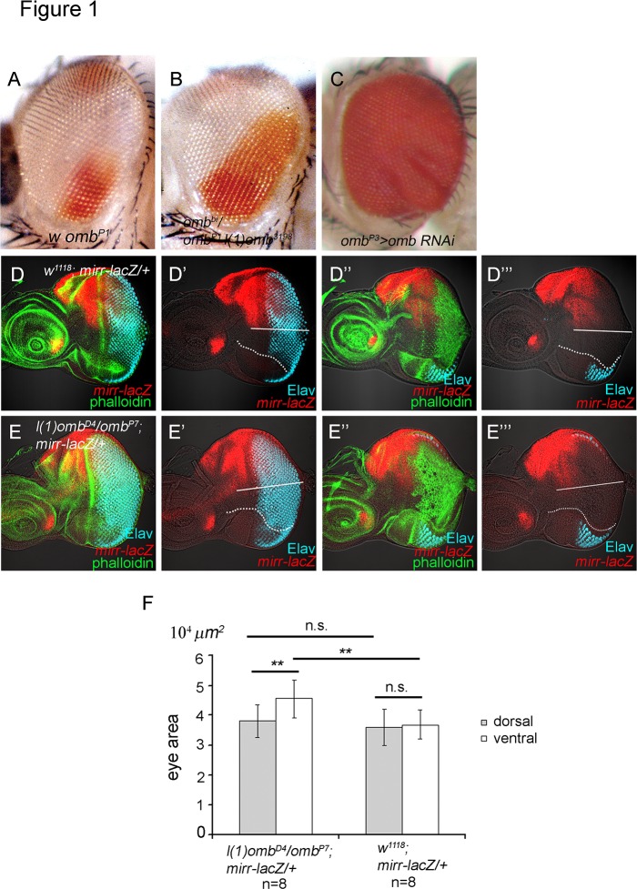
The expansion of the eye occurred primarily ventrally. The dorsal-ventral distinction was based on several criteria. Using the enhancer trap insertion omb P1 [63] as marker for dorsal and ventral ommatidia (Fig. 1A), only an increase in the ventral expression domain could be observed in the adult eye of omb hypomorphs (Fig. 1B). In the larval eye disc, the location of the dorsoventral (DV) midline was defined by the location of the optic stalk (S2A-D, Fig., arrowhead), the inversion of ommatidial chirality based on anti-Bar staining [71] (S2A-B Fig.), the ventral-specific fng-lacZ expression [64] (S2C-D Fig.) and the dorsal-specific mirr-lacZ expression (Fig. 1D, E). There was an obvious enlargement of the ventral eye disc in l(1)omb D4/omb P7 hypomorphic larvae (Fig. 1D-F; S2B, D Fig.). We followed the developmental progress of omb P7 eye discs, based on the number of ommatidial rows, and found that the number of ommatidia in the ventral region was consistently higher relative to that in the dorsal region, which was not different from wild type (S2E Fig.). By all these criteria, an overgrowth of the ventral relative to the dorsal part was evident in the omb hypomorphic mutant eye.
Fig 1. omb expression level influences eye size.
(A) w omb P1 (an enhancer trap insertion that does not affect omb expression and function, Sun et al., 1995), (B) w omb P 1 l(1)omb 3198 /w omb bi. The expanded territory of ventral eye fate is clearly evident. Because of the increased size, the eye surface is more convex. Therefore, the unaffected dorsal pigmentation is not fully visible under this angle. (C) omb P3 >omb-RNAi showed strong overgrowth in the eye. The overgrowth is stronger in the ventral than in the dorsal part of the eye. The eye is convoluted. (D-E) mirr-lacZ (anti-beta-galactosidase, red). Phalloidin staining (green). Elav (blue). (D-D”’) mirr-lacZ/+ eye disc showing the dorsal-specific expression of mirr-lacZ. D, D’ and D”, D”’ are two focal planes. The D”, D”’ focal plane shows the ventral flap. (E-E”’) l(1) omb D4 / omb P7 eye disc. E, E’ and E”, E”’ are two focal planes. The E”, E”’ focal plane shows the ventral flap. The dorsal and ventral eye regions were distinguished (separated by a white line) based on mirr-lacZ and the position of the optic stalk. Two different focal planes are acquired in each eye disc. The area of eye disc including the ventral flap, based on two focal planes, were measured by the software, Zeiss Zen 2009. The results are summarized in (F). The ventral area of l(1)omb D4 /omb P7; mirr-lacZ/+ are significantly enlarged compared to that of mirr-lacZ/+. The dorsal area of l(1)omb D4 /omb P7; mirr-lacZ/+ are not significant increased compared to that of mirr-lacZ/+. Differences (*) presented in (E) and (J) are significant (Student′s t-test, **, p<0.05; n.s., non-significant). In all panels anterior is left and dorsal up.
These loss-of-function effects were also observed when omb was knocked down by RNAi. Expressing omb-RNAi [48] in its own expression domain using omb P3-GAL4 caused a strong reduction of Omb level in the margins of eye disc, the retinal basal glia and in the antenna disc This resulted in a strong overgrowth of eye disc (S1B Fig.) and adult eye (Fig. 1B). When omb-RNAi expression was driven by GMR-GAL4, the size of eye disc and adult eye was normal (not shown). This is consistent with omb expression not overlapping with the activity of the GMR-GAL4 driver, which is restricted to cells posterior to the MF [72].
In contrast to the loss-of-function effects, gain-of-function of omb caused reduction or elimination of the eye. In the regulatory dominant gain-of-function allele omb For [51], Omb was overexpressed in the lateral margins and in the retinal basal glia (S1C Fig.). omb For larvae had smaller eye discs (S1C Fig.) and the adults had a reduced number of ommatidia and a posterior indentation in the eye (wild type: N = 782, SD = 11.5, n = 9; omb For: N = 507, SD = 57.6, n = 6). Targeted mis-expression of omb in the lateral and posterior margins by dpp-GAL4 (dpp>omb) causes a strong reduction or total absence of the adult eye [52]. Specific overexpression of omb at the lateral margins using 30A-GAL4 [73] caused a decrease in ommatidial number N that depended on the strength of the UAS-omb line (UAS-omb 4–15: N = 601.9, SD = 52.3, n = 10; UAS-omb 2–17: N = 670, SD = 32.3, n = 9).
In summary, the loss and gain-of-function phenotypes of omb indicate that omb is a negative regulator of eye development. The effect is stronger on the ventral side of the eye.
omb blocks cell proliferation during eye development
Deviations from normal eye size can arise by several mechanisms. Changes in proliferation, cell death, morphogenetic furrow progression or retinal differentiation can all affect eye size. We tested the effect of loss and gain of omb on proliferation and cell death.
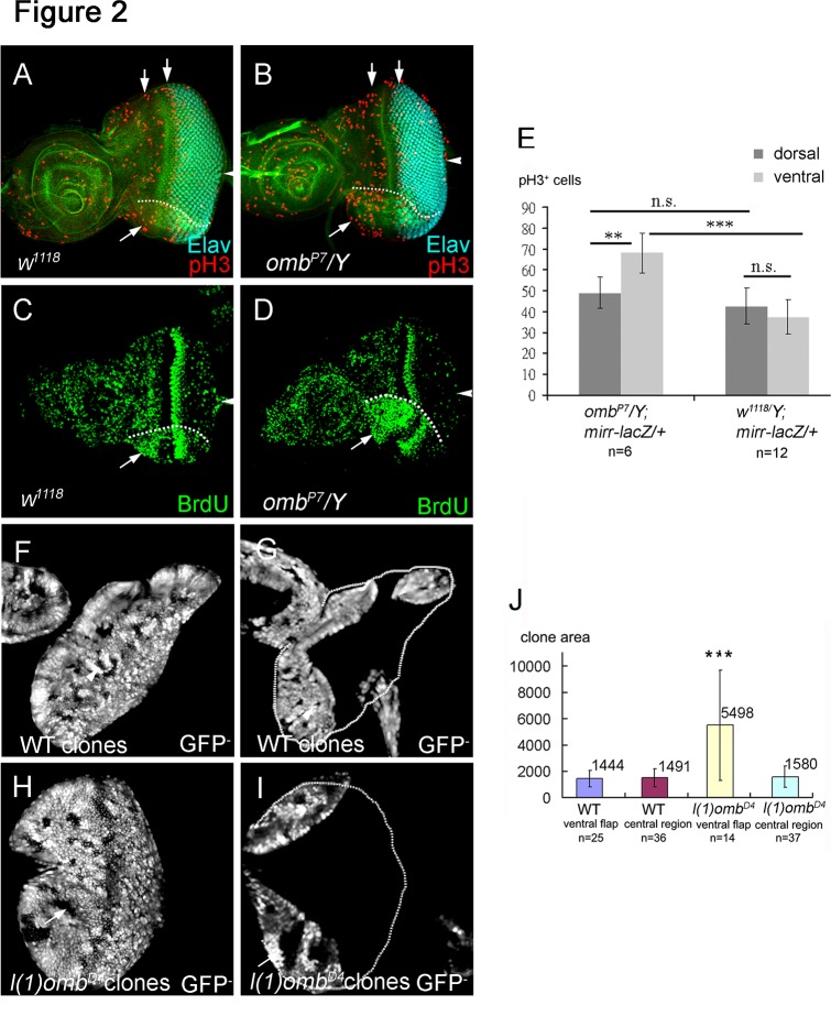
In omb hypomorphs, cell cycle activity was increased in the ventral eye disc, as monitored by histone H3 phosphorylation (pH3) (omb P7 /Y, Fig. 2B, compare with wild type eye disc in 2A; quantified comparison in 2E) or BrdU incorporation (omb P7 /Y, Fig. 2D, compare with wild type eye disc in 2C) as markers for cell proliferation. There are two mitotic waves in the eye disc. The first mitotic wave occurs anterior to the MF and affects the cell population which will be recruited to form ommatidial clusters. Changing cell proliferation in the first mitotic wave will change the number of ommatidia [9,10]. omb hypomorphic eye discs showed an increase of mitosis in the first mitotic wave (Fig. 2B, D), as expected by their increase in ommatidial numbers. In contrast, the second mitotic wave occurs behind the MF and affects the number of cellular components assembled into the ommatidia [74]. Since omb is expressed in the anterior lateral margins, it is not expected to affect the second mitotic wave. This was confirmed in omb hypomorphic mutants (Fig. 2B, D) and by knocking down omb by GMR>omb-RNAi, which resulted in normally sized adult eyes (not shown). Therefore, omb appears to affect cell divisions in the undifferentiated region anterior to the MF.
Fig 2. Omb blocks cell proliferation in eye disc.
Cell proliferation was monitored by staining against the mitotic marker phospho-histone 3 (pH3), BrdU incorporation, and by comparing clone size in late third instar eye discs. (A-D) Arrowhead points to the position of the optic stalk. (A) Wild type eye disc showing the two mitotic waves (arrows) labeled by anti-pH3 (red). (B) The omb P7 mutant showed an increased number of pH3-positive nuclei in the ventral eye compared to wild type. (C, D) BrdU incorporation showed an increase of proliferating cells in the ventral flap (arrow) of the omb P7 mutant eye disc (D) compared to the wild type eye disc (C). (E) mirr-lacZ was used to mark the dorsal region. pH3 positive cells were scored in omb P7 /Y; mirr-lacZ/+ and mirr-lacZ dorsal and ventral eyes. In order to include the ventral flap area, the images of several optical sections were merged. The quantification results are summarized in (E). The mitotic cells in ventral eye of omb P7 is significant increased compared to ventral eye in wild type (p<0.05). (F, G) A wild type eye disc with clones (marked by the absence of GFP) at two focal planes to show the central region (F) and the ventral and dorsal flap regions (G). The clones were of similar size in all regions (summarized in J). (H, I) An eye disc with l(1)omb D4 clones (marked by the absence of GFP) at two focal planes to show the central (H) and ventral and dorsal flap regions (I). The wild type clones and l(1)omb D4 mutant clones were induced at the same time. The l(1)omb D4 clones in the ventral flap were on average about 3.5 times larger than omb clones in the central region of the disc or than wild type clones (summarized in J). Differences (*) presented in (E) and (J) are significant (Student′s t-test, *** p<0.001; **, p<0.05).
We also analyzed the effect of omb loss of function mutant clones on cell proliferation. The l(1)omb D4 null allele was used because it yielded stronger effects than the hypomorphic alleles In wild type eye discs, control clones (marked by lack of GFP) were of similar size relative to their twin spots, irrespective of location (Fig. 2F, G shows two different focal planes to allow clone size visualization and measurement in the infolded margins, in particular the "ventral flap"; data are summarized in Fig. 2J). As expected from the restricted omb expression pattern and from the phenotype of omb loss-of-function mutants, omb null mutant clones had a proliferative advantage relative to their twin spots (omb + /omb +) only in the ventral margin (Fig. 2I, arrow) but not in the center of the disc (Fig. 2H, arrow) or in the dorsal margin (Fig. 2I). omb clones in the ventral regions were about 3.5 times larger than omb clones in the central region of the disc or than wild type clones (summarized in Fig. 2J).
We next analyzed whether apoptosis plays a role in the omb over-expression phenotype. There is little cell death in wild type larval eye discs [75]. Before onset of retinal differentiation, dpp-GAL4 c 40.6 is expressed in the lateral and posterior margins; later it is restricted to the lateral eye disc margins. The expression in the lateral margins partially overlaps with the omb expression domain in the progenitor region (cf. S6 Fig.). Expression of omb driven by dpp-GAL4 (dpp>omb) caused a strong reduction to total absence of the adult eye [52] and lack of retinal differentiation in the eye disc Enhanced apoptosis could be detected in the posterior margin of the dpp>omb+GFP eye disc (S3A. Fig.). Coexpressing the anti-apoptotic factor p35 (dpp>omb+p35) did not rescue adult eye size (data not shown) nor retinal differentiation in eye disc, although apoptosis was strongly reduced (S3B Fig.). These results suggest that apoptosis is not primarily responsible for eye size reduction at the larval and adult stages and that Omb mainly affects eye size by blocking cell proliferation.
Omb can block retinal differentiation
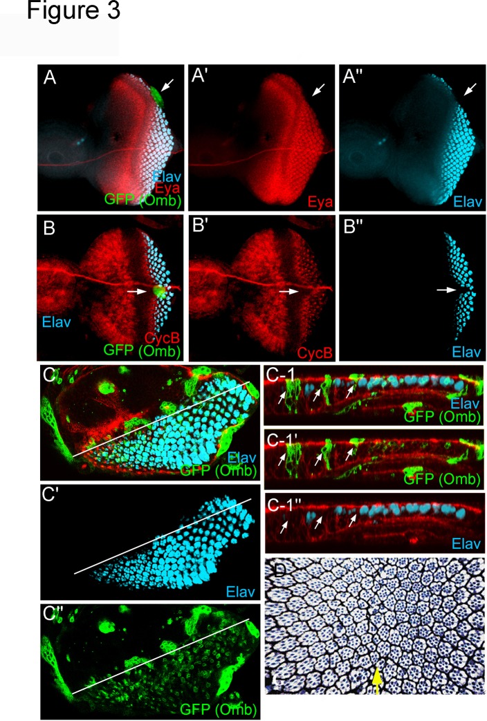
In addition to the effect on cell proliferation, Omb ectopic expression can block retinal differentiation. Ectopic clonal omb expression at the posterior margin prevented MF initiation (Fig. 3A). Ectopic clonal omb expression in the path of the MF blocked its progression (Fig. 3B) and neural differentiation (Fig. 3C-C-1”, arrow). Transient overexpression of omb by heat-induced expression of hs-omb in the entire eye field caused a dorso-ventral scar in the adult eye (Fig. 3D, arrow) characteristic of furrow-stop mutations [19]. Anterior and posterior to the scar, retinal differentiation proceeded normally, indicating that Omb does not irreversibly arrest MF progression and retinal differentiation. Previously we have shown that sustained omb expression posterior to the MF severely disturbs ommatidial development [52].
Fig 3. Ectopic omb expression can block morphogenetic furrow initiation, progression, and differentiation.
Flip-out induced omb expression clones (Act5C>omb) marked by GFP coexpression repressed Elav (cyan) and Eya (red) expression. (A-A”) A clone at the posterior margin (arrow) inhibited MF initiation. (B-B”) A clone at the MF (arrow) inhibited MF progression (as indicated by CycB pattern, red) and neuronal differentiation (Elav, cyan). (C-C”) Omb expression level in Act5C>omb clones varied. Omb expression in a single ommatidial clusters (arrows) could autonomously block neuronal differentiation (Elav, cyan). The Z-section along the white line is shown in C-1 to C-1”. The relative level of Omb induction correlates to the signal of coexpressed GFP. (D) Tangential semi-thin sections through an adult eye of an hs-omb transgenic fly exposed to a single 1hr 37°C heat shock during mid-L3. Ommaditial patterning resumed normally beyond the dorso-ventral scar (arrow).
Increase in ommatidial number in omb hypomorphs apparently did not occur at the expense of gena tissue (the rim of naked tissue between retina and vibrissae) (Fig. 1B, C). This indicates that Omb in the lateral margins does not act to prevent spreading of eye fate into adjacent tissue domains. Rather, the increase in eye size in omb hypomorphs appears caused by overproliferation of retinal precursors in the ventral eye field (s. above).
Omb inhibits cell proliferation by blocking Jak/STAT signaling through repression of upd transcription
To understand the mechanism by which Omb impedes proliferation in the ventral anterior eye disc, we tested the effect of Omb on the Upd/Jak/STAT signaling pathway, which promotes cell proliferation [7,9,10,18,24], as well as MF initiation [15,17,18,22,24].
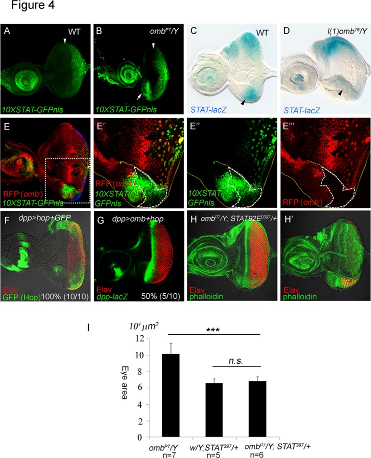
The ligand Unpaired (Upd, FlyBase: outstretched, os) of the Jak/STAT pathway is expressed in the ventral eye disc at first instar and in the posterior center of the eye disc at second and early third instar [9,10,57]. Upd, acting through the Jak/STAT signaling pathway, promotes cell proliferation and represses wg transcription to promote MF initiation [7,9,10,18,24]. STAT signaling is induced by Upd and can be detected using STAT reporters containing STAT binding sites [9,10,18,24,76]. Grh-STAT-lacZ and 10X-STAT-GFP reporter expression is high in the posterior region, consistent with STAT activity being induced by the Upd ligand [24]. 10X-STAT-GFP is expressed in the posterior part of the second instar eye disc, before MF initiation. In the third instar eye disc, the 10XSTAT-GFP signal is much reduced and represents perdurance from earlier expression [76]. In the omb hypomorph omb P7, STAT activity was ectopically activated in the ventral margin (Fig. 4B and S4 Fig.), as monitored by expression of 10XSTAT-GFP-nls (constructed in this study) which is normally expressed only in the posterior region of the eye disc (Fig. 4A). omb P3 >omb-RNAi yielded similar results (not shown). A STAT-lacZ enhancer trap reporter, although not fully recapitulating the STAT92E mRNA pattern, is known to be negatively regulated by Jak/STAT activity [43,57] and, therefore, can be used as a reporter for Jak/STAT activity. In the wild type L3 eye disc, its expression was higher in the lateral poles and lower around the DV midline (Fig. 4C, see also [57]). In the l(1)omb 15 eye disc, STAT-lacZ expression was lost in the ventral region (Fig. 4D), suggesting an elevated STAT activity in this region. To determine whether omb regulates Jak/STAT activity cell-autonomously, we generated l(1)omb D4 mutant clones. We found that 10XSTAT-GFP-nls was nonautonomously induced in ventral clones (Fig. 4E-E”’). These results suggest that Omb normally acts to repress Jak/STAT activity in the ventral region of the eye disc.
Fig 4. Omb blocks Jak/STAT signaling.
10XSTAT-GFP is a reporter of Jak/STAT signaling [76]. We added a nuclear localizing signal (nls) to obtain 10XSTAT-GFP-nls. (A) 10XSTAT-GFP-nls expression pattern (GFP, green) in wild type third instar eye disc. (B) The 10XSTAT-GFP-nls was ectopically expressed in the ventral eye margin (arrow) in an omb P7 hypomorphic mutant eye disc. (A, B) The position of the MF, based on the DIC image, is marked by an arrowhead. (C) STAT-lacZ is repressed by Jak/STAT signaling. In wild type late third instar eye disc, its expression was strong in the lateral poles and weaker around the DV midline, as reported [57]. (D) In l(1)omb 15 /Y eye discs, STAT-lacZ expression was attenuated in the ventral region. (E-E”’) 10XSTAT-GFP-nls (green) was ectopically induced in l(1)omb D4 mutant clones (clone marked by loss of RFP (red) expression and by dashed line). (E’-E”’) Higher magnification of the square marked in (E). 10XSTAT-GFP-nls was non-autonomously induced by loss of omb in the ventral margin. (F) dpp>hop+GFP caused an enlargement of the eye disc (Elav, red; GFP, green). (G) Coexpression of hop with omb (dpp>omb+hop) could largely rescue the dpp>omb phenotype (dpp-lacZ, green; Elav, red). (H-H’) Reducing STAT dosage in omb P7 /Y; STAT92E 397 /+ larvae reduced the size of the ventral retinal field compared to that in omb P7 /Y (Fig. 2B). Different focal planes of omb P7 /Y; STAT92E 397 /+ were shown in H and H’. The quantified eye areas are summarized in (I).
In order to determine, at which level of the Jak/STAT signaling cascade Omb inhibits this pathway, we coexpressed the Janus kinase gene hopscotch (hop) with omb (dpp>omb+hop) (Fig. 4G). Hop largely rescued retinal development indicating that Omb acts upstream of hop. dpp>hop (not shown) and dpp>hop+GFP (Fig. 4F) caused an enlargement of the eye disc, consistent with the role of Jak/STAT signaling in promoting cell proliferation. These results suggest that repression of Jak/STAT activity is a major mechanism by which ectopic Omb blocks eye development. We then asked whether increased Jak/STAT signaling can account for the omb loss-of-function phenotype. We reduced the STAT92E dose in the omb P7 mutant background, (omb P7 /Y; STAT 06346 /+ and omb P7; STAT92E 397 /+), which caused attenuation of the ventral outgrowth of eye disc (Fig. 4H, H’, two focal planes; quantified comparison in 4I). These results suggest that the overgrowth phenotype elicited by reduced omb expression requires Jak/STAT activity. Therefore, in its normal function Omb appears to suppress inappropriate Jak/STAT signaling at the ventral margin.
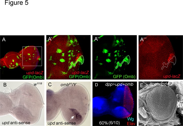
Upd is the ligand of Jak/STAT pathway and expressed in the center of the posterior margin of the eye disc in L2 and L3 larvae (S5A-D, F Fig.; [9,57]). We examined whether omb could suppress Jak/STAT activity by downregulating upd expression. Expression of omb along the posterior margin (in dpp>omb+GFP) suppressed upd-lacZ expression (S5E, E’ Fig.). Clonal expression of omb repressed upd-lacZ cell autonomously (Fig. 5A-A”’). We further tested whether upd expression is affected in omb mutant eye discs. We performed RNA in situ hybridization on wild type and omb P7 eye discs and found that upd was derepressed in the ventral margin in omb P7 (Fig. 5C) compared wild type (Fig. 5B). Thus, ectopically expressed Omb can suppress upd transcription, and Omb in its normal expression domain restricts upd transcription in the ventral eye margin. Moreover, coexpression of omb with upd (in dpp>omb+upd) largely rescued retinal development in the eye disc (Fig. 5D) and the adult eye (Fig. 5E). These results suggest that Omb, in its endogenous expression domain, acts by repressing upd, thus limiting cell proliferation in the ventral eye. Our results further suggest that repression of Jak/STAT signaling occurs at the transcriptional level of upd and is the major mechanism by which Omb blocks cell proliferation in eye development.
Fig 5. omb can repress upd transcription.
(A-A”’) omb expression clone induced by Act5C-GAL4 suppressed upd-lacZ expression (Act5C>omb+GFP in upd-lacZ). (A’-A”’) are a higher magnification of the area marked in (A). GFP (green) marks the Omb expressing cells. Omb expressing cells suppressed upd-lacZ (red). (B-C) RNA in situ hybridization of third instar eye discs. (B) In late third instar, no signal was detected by upd anti-sense probe in wild type eye disc. (C) upd mRNA was ectopically expressed in the ventral eye margin of omb P7 /Y. (D, E) Coexpression of omb with upd (in dpp>omb+upd) partially or fully rescued retinal development in the eye disc (D) and adult eye (E). (Elav, red; Wg, blue)
Omb is a mediator of Wg anti-retinal function
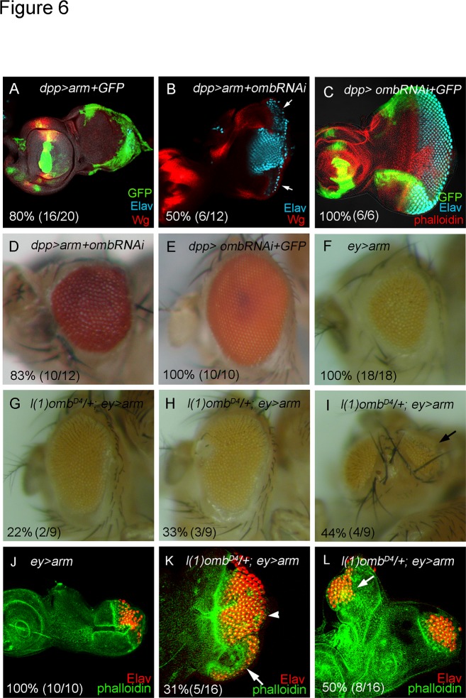
Since Wg and Omb are expressed in a similar pattern and both block retinal development, we asked whether the anti-retinal activity of the Wg signal is mediated by Omb. The Wg effector Arm was expressed to mimic Wg signaling. In dpp>arm+GFP, eye disc size was reduced and no neuronal differentiation was detected (Fig. 6A), consistent with a block of eye development by Wg signaling. dpp expression along the posterior and lateral margins has little overlap with endogenous wg expression (S6A’-E’, A”-E” Fig.). Therefore, this experiment tests the effect of ectopic Wg signaling. When omb was knocked down in the background of dpp>arm (in dpp>arm+omb-RNAi), disc size and neuronal differentiation were partly recovered (Fig. 6B). Adult eye size also was largely restored (Fig. 6D). Knock down of Omb in the dpp expression domain (dpp>omb-RNAi+GFP) caused no significant effect on retinal development (Fig. 6C, E). Misexpression of arm by ey-GAL4 (ey>arm) caused a reduction of adult eye size and retinal differentiation in eye disc (Fig. 6F, J). When omb dosage was reduced (l(1)omb D4/+), the ey>arm phenotype was partially rescued with full penetrance (Fig. 6G-I, K-L). These results suggest that Omb is one of the mediators of Wg activity in blocking eye development.
Fig 6. Functional relationship between wg and omb.
(A) dpp>arm+GFP eye discs were reduced in size and showed no neuronal differentiation (Wg, red; Elav, cyan; GFP, Green). (B, D) dpp>arm+omb-RNAi caused partial rescue of eye disc size and neuronal differentiation (B) and adult eye (D). (C, E) dpp>omb-RNAi+GFP did not affect retinal development in eye disc (C) and adult eye (E). Misexpression of arm by ey-GAL4 (ey>arm) caused eye size reduction in adult eye (F) and in eye disc (J). Reduction of omb genetic dosage (l(1)omb D4 /+) in the background of ey>arm partially rescued the eye size with full penetrance in adult (G-I) and in eye disc (K, L). 31% of these eye discs showed ventral expansion of retinal differentiation. Interestingly, 44~50% of l(1)ombD4/+; ey>arm flies have dorsal ectopic eyes in adult (I) and in eye discs (6L).
Discussion
omb represses retinal development
In this study, we demonstrate that omb is a negative regulator of retinal development. Omb can block eye development at several levels: cell proliferation, MF initiation, and progression. In omb loss-of-function mutant or RNAi-knockdown animals, the most prominent phenotype was an enlargement of the ventral eye (Fig. 1) due to extra cell proliferation (Fig. 2). omb mutant clones in its expression domain at the ventral margin were 3.5 times larger than control clones (Fig. 2). These loss-of-function results were supported by the opposite effect in gain-of-function experiments. In omb gain-of-function animals, the size of eye disc (S1C Fig.) and adult eye was reduced (data not shown). In addition, omb-expressing clones blocked MF initiation (Fig. 3A) and progression (Fig. 3B). Expression of omb along the margins (dpp>omb+GFP) could completely block retinal development (S3B-C Fig.).
Act5C>omb clones were not detected in late third instar eye discs. Rare clones were observed only when larvae were raised at 17°C and examined at early to mid-third instar (Fig. 4). These clones were often round and sorted out from the neuroepithelial layer. This behavior has previously been observed for omb gain-of-function clones in the wing imaginal disc [49].
We identified the Jak/STAT signaling pathway asdownregulated by Omb. In omb loss-of-function animals, the 10XSTAT-GFP-nls and upd-lacZ expression were elevated in the ventral region (Figs. 4B, 5C). Conversely, when omb was ectopically expressed in the posterior and lateral margins, upd-lacZ expression at the margins was reduced (S5E, E’ Fig.). These results show that upd transcription is repressed by Omb in the lateral margins, especially the ventral margin.
The repression of the Upd signaling cascades by Omb is of developmental relevance. Ectopic expression of omb in the margins (dpp>omb+GFP) blocked retinal development (S3C,5E Figs.). Coexpression with of hop (dpp>omb+hop) could nearly fully restore retinal development in the eye disc (Fig. 4G). Reducing the dosage of STAT (in omb P7 /Y; STAT92E 397 /+) partially suppressed the omb P7 enlarged eye phenotype (Fig. 4H), suggesting that STAT signaling is downstream of omb and involved in causing its mutant phenotype. These results indicate that the repression of the Upd/Jak/STAT pathways is responsible for the block of retinal development by Omb.
upd is transcriptionally repressed by Omb. Is it a direct transcriptional target of Omb? Omb is a transcription factor of the T-box family all members of which bind to a common consensus element, the T-box binding element (TBE, [77]). Based on transcript microarray data and cell culture studies, Omb acts predominantly as a transcriptional repressor ([52]; A. Klebes and G. O. Pflugfelder, unpublished data). A bioinformatic search using a position weight matrix constructed from bona fide TBEs identified a well conserved high-affinity potential TBE in the upstream region of upd (G. O. Pflugfelder, unpublished data). This finding and the cell-autonomous repression of upd by Omb (Fig. 5A) as well as the derepression of upd in the ventral omb domain in omb mutant eye discs (Fig. 5C) suggest that repression of upd by Omb may be direct. However, Omb cannot be the only factor that prevents upd expression from the lateral margins, because dorsally upd is not derepressed in omb mutant eye discs.
Omb partially mediates the anti-retinal function of Wg signaling
Since Wg and Omb are expressed in similar patterns and since both can block retinal development at multiple steps, the question arises whether Omb mediates Wg signaling. We found that the anti-retinal effects of ectopic Wg signaling (ey>arm and dpp>arm) were attenuated when the omb dosage was reduced. The partial rescue suggests that Omb mediates part of the Wg effect and that additional factor(s) are likely to be involved. Our analyses show that the effect of Omb is partly similar to that of Wg and partly different (Fig. 6).
A clear difference was the opposite effect on cell proliferation. Omb repressed cell proliferation, through a block of Upd/Jak/STAT signaling. In contrast, enhanced Wg signaling (in axin mutant) causes overgrowth [78], whereas loss of Wg results in reduction of eye disc size [30,35]. Therefore, Wg can promote cell proliferation.
Loss of omb affected primarily development of the ventral eye margin. The ventral bias included effects on STAT activity. Therefore, the effect of wg in the dorsal side may be mediated by another factor, either independent of Omb or functionally redundant with Omb. Dorsal eye fate is governed by the expression of members of the Iroquois gene complex (Iro-C)[79]. Its activity modulates the function of genes that are symmetrically expressed at the poles of the eye disc, like teashirt, for instance [79,80]. The specification of dorsal rim ommatidia late in eye development provides an example of how Omb function is modulated at the lateral eye margins.
During pupal eye development, a dorsal Wg gradient specifies three cell fates at the dorsal eye boundary: pigment rim, polarization-sensitive dorsal rim (DR) ommatidia, and bald ommatidia (lacking the mechanosensory hairs). Omb which is induced by Wg, is sufficient to induce the DR fate in the dorsal eye. Ectopic omb induces the dorsally restricted expression of homothorax (hth) which together with ubiquitous extradenticle (exd) allows the formation of Hth/Exd complexes which specify DR development [81]. Dorsalisation of the ventral eye by ubiquitous expression of Iro-C genes causes DR fate also along the ventral margin. The monopolar Iro-C expression thus determines the different functional outcome of symmetrical omb expression at the two margins. Like in early eye development, loss of omb has little consequence for dorsal eye development during specification of DR ommatidia. The discrepancy between strong effect in omb gain-of-function and little effect in omb loss-of-function dorsal eye phenotypes has been attributed to a redundant function exerted by the related T-box genes Dorsocross (Doc) [82].
Intriguingly, the closely omb-related vertebrate T-box genes Tbx2/3/5 are all expressed in a polar pattern, first in the dorsal eye cup and later in the dorsal retina [83,84,85,86]. A role of human TBX5 in eye development is apparent from the frequent ophtalmological symptoms of patients suffering from Holt Oram Syndrome (TBX5 haploinsufficiency) [87,88]. The related expression patterns of Tbx2/3/5 and omb in eye development suggest conservation of ancient functions which were already present in an evolutionary precursor before the split into the protostome and deuterostome lineages [89], in spite of the widely differing mechanisms of eye ontogenesis in metazoans [90,91].
Supporting Information
Eye-antennal discs stained with anti-Omb (green) and anti-Elav (red). Marginal expression is indicated by arrows, expression in retinal basal glial cells by arrowheads. (A) wild type, (B) omb P3 >omb-RNAi, (C) omb For, (D) omb P3 >GFP and (E) omb P7 >GFP. In omb For, Omb is increased in the dorsal and ventral margin and in the retinal basal glia. The disc size is reduced (C). The marginal expression of omb P7-GAL4 was broader than that of omb P3-GAL4. In (C) and (D) the retinal basal glial cells were below the focal plane.
(TIF)
(A-D) The boundary of dorsal/ventral fields in third instar eye discs was monitored by the position of the optic stalk (white arrowhead), anti-Bar antibody staining (A, B) and the ventrally expressed fng-lacZ (C, D). Dotted lines mark the MF and the projection from the optic stalk entry point onto the MF. In (A, B) the dotted line also visualises the line of mirror symmetry in the Bar expression pattern. The BarH1 and BarH2 expression in photoreceptor cells R1 and R6 [71] is mirror-symmetrical with regard to the equator. The solid line in (C, D) marks the dorsal boundary of the ventral fng-lacZ expression domain. (A) w 1118 /Y, (B) omb P7 /Y, (C) fng-lacZ and (D) omb P7 /Y; fng-lacZ. The D/V eye field was symmetrical in the third instar eye disc of wild type, but the ventral field was expanded in omb P7 /Y. (E) The number of rows of ommatidia in each eye disc, and the numbers of ommatidia in the dorsal and ventral eye fields were counted at different stages of eye disc development. In wild type eye disc, the dorsal and ventral eye fields were always of equal size. In omb P7 /Y, the ventral eye field was consistently larger than the dorsal field.
(TIF)
(A) dpp-GAL4 driven GFP (dpp>GFP) expression (GFP, green) overlapped with the omb-lacZ expression (red) domain in the lateral margins. In early eye disc, dpp-GAL4 expression is similar to that of dpp-lacZ (S6 Fig.) in the posterior and lateral margins. Unlike dpp-lacZ, dpp-GAL4 is not expressed in the progressing MF in mid to late third instar eye disc. (B) dpp>omb+GFP, as dpp>omb, completely blocked eye development in adult (B) and in late third instar eye disc (C). The eye disc has no neuronal differentiation (Elav, blue) but has elevated activated caspase 3 (red). (D) Blocking apoptosis by coexpression of p35 (dpp>omb+p35) significantly reduced the caspase 3 signal but did not rescue eye size or retinal differentiation. Scale bar: 50um.
(TIF)
10XSTAT-GFPnls is a Jak/STAT reporter. (A-C) Jak/STAT activity in omb P7 eye discs. (A-A”) 10XSTAT-GFPnls was found in the posterior eye field in the early third instar eye disc of omb P7. (B-B”) Jak/STAT activity was activated in posterior eye field and ventral margin (arrow) of mid-third instar larvae. (C, C”) Jak/STAT activity was detected in the posterior eye field as well as ventral margin (arrow) in the late third eye field. Elav (red), GFP (green).
(TIF)
(A-C’) The expression pattern of omb P1-lacZ (red) and upd>GFP (green) did not overlap in late second (A), early third (B) and late-third instar eye discs (C). omb P1-lacZ is also expressed in the retinal basal glia which lies at the basal surface and does not overlap with the upd expressing cells in the neuroepithelial layer (not shown). (D) The expression pattern of upd-lacZ (red) in wild type. Elav (cyan). (E, E’) dpp>omb+GFP (GFP, green) suppressed upd-lacZ expression (red) at the center of the posterior margin (arrow)
(TIF)
(A-E) The expression patterns of omb (visualized by omb P3 >GFP, green), (A’-E’) Wg (anti-Wg, red), and (A”-E”) dpp (represented by dpp-lacZ, blue) were followed during eye-antennal disc development from early second instar to late third instar. (A”’-E”’) shows the merge images of omb P3 >GFP, dpp-lacZ and Wg immunostaining.
(TIF)
Acknowledgments
We thank Jessica Treisman, Mark Fortini, Christian Dahmann, the Bloomington Stock Center, the Bowling Green Mid-America Drosophila Stock Center and the VDRC Stock Center for providing fly stocks. We are grateful to Cheng-Ting Chien for valuable suggestions. We are grateful to Chen-shu Hsieh, Chun-lan Hsu, Yu-Chi Yang, and Erika Jost for preparing fly food and maintaining fly stocks, to Chiou-Yang Tang for making transgenic flies, to Su-Ping Lee and the IMB Imaging Core for help in confocal microscopy, and to Ying-Rou Lee and Hsin-Yi Huang for help in SEM.
Data Availability
All relevant data are within the paper and its Supporting Information files.
Funding Statement
This study was supported by grants to Y.H.S. (NSC 88-2312-B-001-016, NSC 90-2312-B-001-006, NSC 91-2312-B-001-006) to Y.C.T. (NSC 99-2311-B-029-002-MY3, NSC-102-2633-B-029-002) from the National Science Council (http://www.most.gov.tw/mp.aspx?mp=7) of the Republic of China, to G.O.P. (Pf 163/11-1) from Deutsche Forschungsgemeinschaft (http://www.dfg.de/en/index.jsp) and a ROC-Germany exchange program (PPP 87-I-2911-01). The funders had no role in study design, data collection and analysis, decision to publish, or preparation of the manuscript.
References
- 1. Freeman M (1997) Cell determination strategies in the Drosophila eye. Development 124: 261–270. [DOI] [PubMed] [Google Scholar]
- 2. Wolff T, Ready DF (1991) The beginning of pattern formation in the Drosophila compound eye: the morphogenetic furrow and the second mitotic wave. Development 113: 841–850. [DOI] [PubMed] [Google Scholar]
- 3. Blackman RK, Sanicola M, Raftery LA, Gillevet T, Gelbart WM (1991) An extensive 3' cis-regulatory region directs the imaginal disk expression of decapentaplegic, a member of the TGF-beta family in Drosophila . Development 111: 657–666. [DOI] [PubMed] [Google Scholar]
- 4. Wolff TaR DN, editor (1993) The development of Drosophila melanogaster NY. pp1277–1325 p. [Google Scholar]
- 5. Silver SJ, Rebay I (2005) Signaling circuitries in development: insights from the retinal determination gene network. Development 132: 3–13. [DOI] [PubMed] [Google Scholar]
- 6. Kumar JP (2010) Retinal determination the beginning of eye development. Curr Top Dev Biol 93: 1–28. 10.1016/B978-0-12-385044-7.00001-1 [DOI] [PMC free article] [PubMed] [Google Scholar]
- 7. Chao JL, Tsai YC, Chiu SJ, Sun YH (2004) Localized Notch signal acts through eyg and upd to promote global growth in Drosophila eye. Development 131: 3839–3847. [DOI] [PubMed] [Google Scholar]
- 8. Dominguez M, de Celis JF (1998) A dorsal/ventral boundary established by Notch controls growth and polarity in the Drosophila eye. Nature 396: 276–278. [DOI] [PubMed] [Google Scholar]
- 9. Tsai YC, Sun YH (2004) Long-range effect of upd, a ligand for Jak/STAT pathway, on cell cycle in Drosophila eye development. Genesis 39: 141–153. [DOI] [PubMed] [Google Scholar]
- 10. Bach EA, Vincent S, Zeidler MP, Perrimon N (2003) A sensitized genetic screen to identify novel regulators and components of the Drosophila janus kinase/signal transducer and activator of transcription pathway. Genetics 165: 1149–1166. [DOI] [PMC free article] [PubMed] [Google Scholar]
- 11. Treisman JE (2013) Retinal differentiation in Drosophila . Wiley Interdiscip Rev Dev Biol 2: 545–557. 10.1002/wdev.100 [DOI] [PMC free article] [PubMed] [Google Scholar]
- 12. Peng HW, Slattery M, Mann RS (2009) Transcription factor choice in the Hippo signaling pathway: homothorax and yorkie regulation of the microRNA bantam in the progenitor domain of the Drosophila eye imaginal disc. Genes Dev 23: 2307–2319. 10.1101/gad.1820009 [DOI] [PMC free article] [PubMed] [Google Scholar]
- 13. Bessa J, Gebelein B, Pichaud F, Casares F, Mann RS (2002) Combinatorial control of Drosophila eye development by eyeless, homothorax, and teashirt . Genes Dev 16: 2415–2427. [DOI] [PMC free article] [PubMed] [Google Scholar]
- 14. Lopes CS, Casares F (2010) hth maintains the pool of eye progenitors and its downregulation by Dpp and Hh couples retinal fate acquisition with cell cycle exit. Dev Biol 339: 78–88. 10.1016/j.ydbio.2009.12.020 [DOI] [PubMed] [Google Scholar]
- 15. Wiersdorff V, Lecuit T, Cohen SM, Mlodzik M (1996) Mad acts downstream of Dpp receptors, revealing a differential requirement for dpp signaling in initiation and propagation of morphogenesis in the Drosophila eye. Development 122: 2153–2162. [DOI] [PubMed] [Google Scholar]
- 16. Chanut F, Heberlein U (1997) Retinal morphogenesis in Drosophila: hints from an eye-specific decapentaplegic allele. Dev Genet 20: 197–207. [DOI] [PubMed] [Google Scholar]
- 17. Borod ER, Heberlein U (1998) Mutual regulation of decapentaplegic and hedgehog during the initiation of differentiation in the Drosophila retina. Dev Biol 197: 187–197. [DOI] [PubMed] [Google Scholar]
- 18. Tsai YC, Yao JG, Chen PH, Posakony JW, Barolo S, et al. (2007) Upd/Jak/STAT signaling represses wg transcription to allow initiation of morphogenetic furrow in Drosophila eye development. Dev Biol 306: 760–771. [DOI] [PubMed] [Google Scholar]
- 19. Heberlein U, Wolff T, Rubin GM (1993) The TGF beta homolog dpp and the segment polarity gene hedgehog are required for propagation of a morphogenetic wave in the Drosophila retina. Cell 75: 913–926. [DOI] [PubMed] [Google Scholar]
- 20. Pan D, Rubin GM (1995) cAMP-dependent protein kinase and hedgehog act antagonistically in regulating decapentaplegic transcription in Drosophila imaginal discs. Cell 80: 543–552. [DOI] [PubMed] [Google Scholar]
- 21. Strutt DI, Mlodzik M (1995) Ommatidial polarity in the Drosophila eye is determined by the direction of furrow progression and local interactions. Development 121: 4247–4256. [DOI] [PubMed] [Google Scholar]
- 22. Chanut F, Heberlein U (1997) Role of decapentaplegic in initiation and progression of the morphogenetic furrow in the developing Drosophila retina. Development 124: 559–567. [DOI] [PubMed] [Google Scholar]
- 23. Pignoni F, Zipursky SL (1997) Induction of Drosophila eye development by decapentaplegic . Development 124: 271–278. [DOI] [PubMed] [Google Scholar]
- 24. Ekas LA, Baeg GH, Flaherty MS, Ayala-Camargo A, Bach EA (2006) JAK/STAT signaling promotes regional specification by negatively regulating wingless expression in Drosophila . Development 133: 4721–4729. [DOI] [PubMed] [Google Scholar]
- 25. Haynie JL, Bryant PJ (1986) Development of the eye-antenna imaginal disc and morphogenesis of the adult head in Drosophila melanogaster . J Exp Zool 237: 293–308. [DOI] [PubMed] [Google Scholar]
- 26. Dominguez M, Casares F (2005) Organ specification-growth control connection: new in-sights from the Drosophila eye-antennal disc. Dev Dyn 232: 673–684. [DOI] [PubMed] [Google Scholar]
- 27. Kumar JP (2011) My what big eyes you have: how the Drosophila retina grows. Dev Neurobiol 71: 1133–1152. 10.1002/dneu.20921 [DOI] [PMC free article] [PubMed] [Google Scholar]
- 28. Brown NL, Sattler CA, Paddock SW, Carroll SB (1995) Hairy and emc negatively regulate morphogenetic furrow progression in the Drosophila eye. Cell 80: 879–887. [DOI] [PubMed] [Google Scholar]
- 29. Cadigan KM, Jou AD, Nusse R (2002) Wingless blocks bristle formation and morphogenetic furrow progression in the eye through repression of Daughterless. Development 129: 3393–3402. [DOI] [PubMed] [Google Scholar]
- 30. Ma C, Moses K (1995) Wingless and patched are negative regulators of the morphogenetic furrow and can affect tissue polarity in the developing Drosophila compound eye. Development 121: 2279–2289. [DOI] [PubMed] [Google Scholar]
- 31. Pai CY, Kuo TS, Jaw TJ, Kurant E, Chen CT, et al. (1998) The Homothorax homeoprotein activates the nuclear localization of another homeoprotein, extradenticle, and suppresses eye development in Drosophila . Genes Dev 12: 435–446. [DOI] [PMC free article] [PubMed] [Google Scholar]
- 32. Singh A, Choi KW (2003) Initial state of the Drosophila eye before dorsoventral specification is equivalent to ventral. Development 130: 6351–6360. [DOI] [PubMed] [Google Scholar]
- 33. Roignant JY, Legent K, Janody F, Treisman JE (2010) The transcriptional co-factor Chip acts with LIM-homeodomain proteins to set the boundary of the eye field in Drosophila . Development 137: 273–281. 10.1242/dev.041244 [DOI] [PMC free article] [PubMed] [Google Scholar]
- 34. Oros SM, Tare M, Kango-Singh M, Singh A (2010) Dorsal eye selector pannier (pnr) suppresses the eye fate to define dorsal margin of the Drosophila eye. Dev Biol 346: 258–271. 10.1016/j.ydbio.2010.07.030 [DOI] [PMC free article] [PubMed] [Google Scholar]
- 35. Treisman JE, Rubin GM (1995) wingless inhibits morphogenetic furrow movement in the Drosophila eye disc. Development 121: 3519–3527. [DOI] [PubMed] [Google Scholar]
- 36. Burke R, Basler K (1996) Hedgehog-dependent patterning in the Drosophila eye can occur in the absence of Dpp signaling. Dev Biol 179: 360–368. [DOI] [PubMed] [Google Scholar]
- 37. Hazelett DJ, Bourouis M, Walldorf U, Treisman JE (1998) decapentaplegic and wingless are regulated by eyes absent and eyegone and interact to direct the pattern of retinal differentiation in the eye disc. Development 125: 3741–3751. [DOI] [PubMed] [Google Scholar]
- 38. Cadigan KM (2002) Regulating morphogen gradients in the Drosophila wing. Semin Cell Dev Biol 13: 83–90. [DOI] [PubMed] [Google Scholar]
- 39. Poeck B, Hofbauer A, Pflugfelder GO (1993) Expression of the Drosophila optomotor-blind gene transcript in neuronal and glial cells of the developing nervous system. Development 117: 1017–1029. [DOI] [PubMed] [Google Scholar]
- 40. Zecca M, Basler K, Struhl G (1996) Direct and long-range action of a wingless morphogen gradient. Cell 87: 833–844. [DOI] [PubMed] [Google Scholar]
- 41. Pflugfelder GO, Heisenberg M (1995) Optomotor-blind of Drosophila melanogaster: a neurogenetic approach to optic lobe development and optomotor behaviour. Comp Biochem Physiol A Physiol 110: 185–202. [DOI] [PubMed] [Google Scholar]
- 42. Cook O, Biehs B, Bier E (2004) brinker and optomotor-blind act coordinately to initiate development of the L5 wing vein primordium in Drosophila . Development 131: 2113–2124. [DOI] [PubMed] [Google Scholar]
- 43. del Alamo Rodriguez D, Terriente Felix J, Diaz-Benjumea FJ (2004) The role of the T-box gene optomotor-blind in patterning the Drosophila wing. Dev Biol 268: 481–492. [DOI] [PubMed] [Google Scholar]
- 44. Gorfinkiel N, Sanchez L, Guerrero I (1999) Drosophila terminalia as an appendage-like structure. Mech Dev 86: 113–123. [DOI] [PubMed] [Google Scholar]
- 45. Kopp A, Duncan I (1997) Control of cell fate and polarity in the adult abdominal segments of Drosophila by optomotor-blind . Development 124: 3715–3726. [DOI] [PubMed] [Google Scholar]
- 46. Shen J, Dahmann C (2005) The role of Dpp signaling in maintaining the Drosophila anteroposterior compartment boundary. Dev Biol 279: 31–43. [DOI] [PubMed] [Google Scholar]
- 47. Grimm S, Pflugfelder GO (1996) Control of the gene optomotor-blind in Drosophila wing development by decapentaplegic and wingless . Science 271: 1601–1604. [DOI] [PubMed] [Google Scholar]
- 48. Shen J, Dorner C, Bahlo A, Pflugfelder GO (2008) optomotor-blind suppresses instability at the A/P compartment boundary of the Drosophila wing. Mech Dev 125: 233–246. 10.1016/j.mod.2007.11.006 [DOI] [PubMed] [Google Scholar]
- 49. Shen J, Dahmann C, Pflugfelder GO (2010) Spatial discontinuity of optomotor-blind expression in the Drosophila wing imaginal disc disrupts epithelial architecture and promotes cell sorting. BMC Dev Biol 10: 23 10.1186/1471-213X-10-23 [DOI] [PMC free article] [PubMed] [Google Scholar]
- 50. Zhang X, Luo D, Pflugfelder GO, Shen J (2013) Dpp signaling inhibits proliferation in the Drosophila wing by Omb-dependent regional control of bantam . Development 140: 2917–2922. 10.1242/dev.094300 [DOI] [PubMed] [Google Scholar]
- 51. Pflugfelder GO (2009) omb and circumstance. J Neurogenet 23: 15–33. 10.1080/01677060802471619 [DOI] [PubMed] [Google Scholar]
- 52. Porsch M, Sauer M, Schulze S, Bahlo A, Roth M, et al. (2005) The relative role of the T-domain and flanking sequences for developmental control and transcriptional regulation in protein chimeras of Drosophila OMB and ORG-1. Mech Dev 122: 81–96. [DOI] [PubMed] [Google Scholar]
- 53. Staehling-Hampton K, Jackson PD, Clark MJ, Brand AH, Hoffmann FM (1994) Specificity of bone morphogenetic protein-related factors: cell fate and gene expression changes in Drosophila embryo induced by decapentaplegic but not 60A . Cell Growth & Differentiation 5: 585–593. [PubMed] [Google Scholar]
- 54. Luo H, Asha H, Kockel L, Parke T, Mlodzik M, et al. (1999) The Drosophila Jak kinase hopscotch is required for multiple developmental processes in the eye. Dev Biol 213: 432–441. [DOI] [PubMed] [Google Scholar]
- 55. Ekas LA, Cardozo TJ, Flaherty MS, McMillan EA, Gonsalves FC, et al. (2010) Characterization of a dominant-active STAT that promotes tumorigenesis in Drosophila . Dev Biol 344: 621–636. 10.1016/j.ydbio.2010.05.497 [DOI] [PMC free article] [PubMed] [Google Scholar]
- 56. Hou XS, Melnick MB, Perrimon N (1996) Marelle acts downstream of the Drosophila HOP/JAK kinase and encodes a protein similar to the mammalian STATs. Cell 84: 411–419. [DOI] [PubMed] [Google Scholar]
- 57. Zeidler MP, Perrimon N, Strutt DI (1999) Polarity determination in the Drosophila eye: a novel role for unpaired and JAK/STAT signaling. Genes Dev 13: 1342–1353. [DOI] [PMC free article] [PubMed] [Google Scholar]
- 58. Calleja M, Moreno E, Pelaz S, Morata G (1996) Visualization of gene expression in living adult Drosophila . Science 274: 252–255. [DOI] [PubMed] [Google Scholar]
- 59. Mayer LR, Diegelmann S, Abassi Y, Eichinger F, Pflugfelder GO (2013) Enhancer trap infidelity in Drosophila optomotor-blind . Fly (Austin) 7: 118–128. 10.4161/fly.23657 [DOI] [PMC free article] [PubMed] [Google Scholar]
- 60. Tang CY, Sun YH (2002) Use of mini-white as a reporter gene to screen for GAL4 insertions with spatially restricted expression pattern in the developing eye in Drosophila . Genesis 34: 39–45. [DOI] [PubMed] [Google Scholar]
- 61. Poeck B, Balles J, Pflugfelder GO (1993a) Transcript identification in the optomotor-blind locus of Drosophila melanogaster by intragenic recombination mapping and PCR-aided sequence analysis of lethal point mutations. Mol Gen Genetics 238: 325–332. [DOI] [PubMed] [Google Scholar]
- 62. Sen A, Gadomski C, Balles J, Abassi Y, Dorner C, et al. (2010) Null mutations in Drosophila Optomotor-blind affect T-domain residues conserved in all Tbx proteins. Mol Genet Genomics 283: 147–156. 10.1007/s00438-009-0505-z [DOI] [PubMed] [Google Scholar]
- 63. Sun YH, Tsai CJ, Green MM, Chao JL, Yu CT, et al. (1995) white as a reporter gene to detect transcriptional silencers specifying position-specific gene expression during Drosophila melanogaster eye development. Genetics 141: 1075–1086. [DOI] [PMC free article] [PubMed] [Google Scholar]
- 64. Cho KO, Choi KW (1998) Fringe is essential for mirror symmetry and morphogenesis in the Drosophila eye. Nature 396: 272–276. [DOI] [PubMed] [Google Scholar]
- 65. Kassis JA, Noll E, VanSickle EP, Odenwald WF, Perrimon N (1992) Altering the insertional specificity of a Drosophila transposable element. Proc Natl Acad Sci U S A 89: 1919–1923. [DOI] [PMC free article] [PubMed] [Google Scholar]
- 66. Barolo S, Carver LA, Posakony JW (2000) GFP and beta-galactosidase transformation vectors for promoter/enhancer analysis in Drosophila . Biotechniques 29: 726, 728, 730, 732. [DOI] [PubMed] [Google Scholar]
- 67. Jang CC, Chao JL, Jones N, Yao LC, Bessarab DA, et al. (2003) Two Pax genes, eye gone and eyeless, act cooperatively in promoting Drosophila eye development. Development 130: 2939–2951. [DOI] [PubMed] [Google Scholar]
- 68. Ito K, Awano W, Suzuki K, Hiromi Y, Yamamoto D (1997) The Drosophila mushroom body is a quadruple structure of clonal units each of which contains a virtually identical set of neurones and glial cells. Development 124: 761–771. [DOI] [PubMed] [Google Scholar]
- 69. Basler K, Hafen E (1988) Control of photoreceptor cell fate by the sevenless protein requires a functional tyrosine kinase domain. Cell 54: 299–311. [DOI] [PubMed] [Google Scholar]
- 70. Rangarajan R, Gong Q, Gaul U (1999) Migration and function of glia in the developing Drosophila eye. Development 126: 3285–3292. [DOI] [PubMed] [Google Scholar]
- 71. Higashijima S, Kojima T, Michiue T, Ishimaru S, Emori Y, et al. (1992) Dual Bar homeo box genes of Drosophila required in two photoreceptor cells, R1 and R6, and primary pigment cells for normal eye development. Genes Dev 6: 50–60. [DOI] [PubMed] [Google Scholar]
- 72. Freeman M (1996) Reiterative use of the EGF receptor triggers differentiation of all cell types in the Drosophila eye. Cell 87: 651–660. [DOI] [PubMed] [Google Scholar]
- 73. Brand AH, Perrimon N (1993) Targeted gene expression as a means of altering cell fates and generating dominant phenotypes. Development 118: 401–415. [DOI] [PubMed] [Google Scholar]
- 74. Ready DF, Hanson TE, Benzer S (1976) Development of the Drosophila retina, a neurocrystalline lattice. Dev Biol 53: 217–240. [DOI] [PubMed] [Google Scholar]
- 75. Bonini NM, Fortini ME (1999) Surviving Drosophila eye development: integrating cell death with differentiation during formatio of a neural structure. BioEssays 21: 991–1003. [DOI] [PubMed] [Google Scholar]
- 76. Bach EA, Ekas LA, Ayala-Camargo A, Flaherty MS, Lee H, et al. (2007) GFP reporters detect the activation of the Drosophila JAK/STAT pathway in vivo . Gene Expr Patterns 7: 323–331. [DOI] [PubMed] [Google Scholar]
- 77. Sen A, Grimm S, Hofmeyer K, Pflugfelder GO (2014) Optomotor-blind in the development of the Drosophila HS and VS lobula plate tangential cells. J Neurogenet: 1–49. 10.3109/01677063.2014.908875 [DOI] [PubMed] [Google Scholar]
- 78. Lee JD, Treisman JE (2001) The role of Wingless signaling in establishing the anteroposterior and dorsoventral axes of the eye disc. Development 128: 1519–1529. [DOI] [PubMed] [Google Scholar]
- 79. Singh A, Tare M, Puli OR, Kango-Singh M (2012) A glimpse into dorso-ventral patterning of the Drosophila eye. Dev Dyn 241: 69–84. 10.1002/dvdy.22764 [DOI] [PMC free article] [PubMed] [Google Scholar]
- 80. Gutierrez-Avino FJ, Ferres-Marco D, Dominguez M (2009) The position and function of the Notch-mediated eye growth organizer: the roles of JAK/STAT and four-jointed . EMBO Rep 10: 1051–1058. 10.1038/embor.2009.140 [DOI] [PMC free article] [PubMed] [Google Scholar]
- 81. Wernet MF, Desplan C (2014) Homothorax and Extradenticle alter the transcription factor network in Drosophila ommatidia at the dorsal rim of the retina. Development 141: 918–928. 10.1242/dev.103127 [DOI] [PMC free article] [PubMed] [Google Scholar]
- 82. Tomlinson A (2003) Patterning the peripheral retina of the fly: decoding a gradient. Dev Cell 5: 799–809. [DOI] [PubMed] [Google Scholar]
- 83. Behesti H, Papaioannou VE, Sowden JC (2009) Loss of Tbx2 delays optic vesicle invagination leading to small optic cups. Dev Biol 333: 360–372. 10.1016/j.ydbio.2009.06.026 [DOI] [PMC free article] [PubMed] [Google Scholar]
- 84. Gross JM, Dowling JE (2005) Tbx2b is essential for neuronal differentiation along the dorsal/ventral axis of the zebrafish retina. Proc Natl Acad Sci U S A 102: 4371–4376. [DOI] [PMC free article] [PubMed] [Google Scholar]
- 85. Leconte L, Lecoin L, Martin P, Saule S (2004) Pax6 interacts with cVax and Tbx5 to establish the dorsoventral boundary of the developing eye. J Biol Chem 279: 47272–47277. [DOI] [PubMed] [Google Scholar]
- 86. Sowden JC, Holt JK, Meins M, Smith HK, Bhattacharya SS (2001) Expression of Drosophila omb-related T-box genes in the developing human and mouse neural retina. Invest Ophthalmol Vis Sci 42: 3095–3102. [PubMed] [Google Scholar]
- 87. Gruenauer-Kloevekorn C, Reichel MB, Duncker GI, Froster UG (2005) Molecular genetic and ocular findings in patients with holt-oram syndrome. Ophthalmic Genet 26: 1–8. [DOI] [PubMed] [Google Scholar]
- 88. Newbury-Ecob RA, Leanage R, Raeburn JA, Young ID (1996) Holt-Oram syndrome: a clinical genetic study. J Med Genet 33: 300–307. [DOI] [PMC free article] [PubMed] [Google Scholar]
- 89. Chapman DL, Garvey N, Hancock S, Alexiou M, Agulnik SI, et al. (1996) Expression of the T-box family genes, Tbx1-Tbx5, during early mouse development. Dev Dyn 206: 379–390. [DOI] [PubMed] [Google Scholar]
- 90. Gehring WJ, Ikeo K (1999) Pax 6: mastering eye morphogenesis and eye evolution. Trends Genet 15: 371–377. [DOI] [PubMed] [Google Scholar]
- 91. Treisman JE (1999) A conserved blueprint for the eye? BioEssays 21: 843–850. [DOI] [PubMed] [Google Scholar]
Associated Data
This section collects any data citations, data availability statements, or supplementary materials included in this article.
Supplementary Materials
Eye-antennal discs stained with anti-Omb (green) and anti-Elav (red). Marginal expression is indicated by arrows, expression in retinal basal glial cells by arrowheads. (A) wild type, (B) omb P3 >omb-RNAi, (C) omb For, (D) omb P3 >GFP and (E) omb P7 >GFP. In omb For, Omb is increased in the dorsal and ventral margin and in the retinal basal glia. The disc size is reduced (C). The marginal expression of omb P7-GAL4 was broader than that of omb P3-GAL4. In (C) and (D) the retinal basal glial cells were below the focal plane.
(TIF)
(A-D) The boundary of dorsal/ventral fields in third instar eye discs was monitored by the position of the optic stalk (white arrowhead), anti-Bar antibody staining (A, B) and the ventrally expressed fng-lacZ (C, D). Dotted lines mark the MF and the projection from the optic stalk entry point onto the MF. In (A, B) the dotted line also visualises the line of mirror symmetry in the Bar expression pattern. The BarH1 and BarH2 expression in photoreceptor cells R1 and R6 [71] is mirror-symmetrical with regard to the equator. The solid line in (C, D) marks the dorsal boundary of the ventral fng-lacZ expression domain. (A) w 1118 /Y, (B) omb P7 /Y, (C) fng-lacZ and (D) omb P7 /Y; fng-lacZ. The D/V eye field was symmetrical in the third instar eye disc of wild type, but the ventral field was expanded in omb P7 /Y. (E) The number of rows of ommatidia in each eye disc, and the numbers of ommatidia in the dorsal and ventral eye fields were counted at different stages of eye disc development. In wild type eye disc, the dorsal and ventral eye fields were always of equal size. In omb P7 /Y, the ventral eye field was consistently larger than the dorsal field.
(TIF)
(A) dpp-GAL4 driven GFP (dpp>GFP) expression (GFP, green) overlapped with the omb-lacZ expression (red) domain in the lateral margins. In early eye disc, dpp-GAL4 expression is similar to that of dpp-lacZ (S6 Fig.) in the posterior and lateral margins. Unlike dpp-lacZ, dpp-GAL4 is not expressed in the progressing MF in mid to late third instar eye disc. (B) dpp>omb+GFP, as dpp>omb, completely blocked eye development in adult (B) and in late third instar eye disc (C). The eye disc has no neuronal differentiation (Elav, blue) but has elevated activated caspase 3 (red). (D) Blocking apoptosis by coexpression of p35 (dpp>omb+p35) significantly reduced the caspase 3 signal but did not rescue eye size or retinal differentiation. Scale bar: 50um.
(TIF)
10XSTAT-GFPnls is a Jak/STAT reporter. (A-C) Jak/STAT activity in omb P7 eye discs. (A-A”) 10XSTAT-GFPnls was found in the posterior eye field in the early third instar eye disc of omb P7. (B-B”) Jak/STAT activity was activated in posterior eye field and ventral margin (arrow) of mid-third instar larvae. (C, C”) Jak/STAT activity was detected in the posterior eye field as well as ventral margin (arrow) in the late third eye field. Elav (red), GFP (green).
(TIF)
(A-C’) The expression pattern of omb P1-lacZ (red) and upd>GFP (green) did not overlap in late second (A), early third (B) and late-third instar eye discs (C). omb P1-lacZ is also expressed in the retinal basal glia which lies at the basal surface and does not overlap with the upd expressing cells in the neuroepithelial layer (not shown). (D) The expression pattern of upd-lacZ (red) in wild type. Elav (cyan). (E, E’) dpp>omb+GFP (GFP, green) suppressed upd-lacZ expression (red) at the center of the posterior margin (arrow)
(TIF)
(A-E) The expression patterns of omb (visualized by omb P3 >GFP, green), (A’-E’) Wg (anti-Wg, red), and (A”-E”) dpp (represented by dpp-lacZ, blue) were followed during eye-antennal disc development from early second instar to late third instar. (A”’-E”’) shows the merge images of omb P3 >GFP, dpp-lacZ and Wg immunostaining.
(TIF)
Data Availability Statement
All relevant data are within the paper and its Supporting Information files.


