



Abstract
Immunosuppressive CD11b+Gr-1+ myeloid-derived suppressor cells (MDSC) accumulate in the livers of tumor-bearing mice. We studied hepatic MDSC in two murine models of immune mediated hepatitis. Unexpectedly, treatment of tumor bearing mice with Concanavalin A or α-Galactosylceramide resulted in increased ALT and AST serum levels in comparison to tumor free mice. Adoptive transfer of hepatic MDSC into naïve mice exacerbated Concanavalin A induced liver damage. Hepatic CD11b+Gr-1+ cells revealed a polarized pro-inflammatory gene signature after Concanavalin A treatment. An interferon gamma- dependent up-regulation of CD40 on hepatic CD11b+Gr-1+ cells along with an up-regulation of CD80, CD86, and CD1d after Concanavalin A treatment was observed. Concanavalin A treatment resulted in a loss of suppressor function by tumor-induced CD11b+Gr-1+ MDSC as well as enhanced reactive oxygen species-mediated hepatotoxicity. CD40 knockdown in hepatic MDSC led to increased arginase activity upon Concanavalin A treatment and lower ALT/AST serum levels. Finally, blockade of arginase activity in Cd40−/− tumor-induced myeloid cells resulted in exacerbation of hepatitis and increased reactive oxygen species production in vivo. Our findings indicate that in a setting of acute hepatitis, tumor-induced hepatic MDSC act as pro-inflammatory immune effector cells capable of killing hepatocytes in a CD40-dependent manner.
Keywords: immune mediated hepatitis, myeloid derived suppressor cells, CD40, Reactive Oxygen Species, Concanavalin A, α-Galactosylceramide
Introduction
Myeloid derived suppressor cells (MDSC) comprise immature myeloid progenitors at different stages of development, such as precursors of granulocytes, macrophages and dendritic cells (DC) [1],[2]. MDSC have immune regulatory properties and accumulate in tumors, blood, bone marrow, spleen and liver in tumor-bearing mice and cancer patients [2]–[10]. In mice, MDSC can be identified by their co-expression of CD11b and Gr-1. The hallmark of MDSC is their ability to suppress both adaptive and innate immune responses, which is achieved through multiple mechanisms [11]–[13].
Expansion of MDSC has been described in patients and murine models of hepatocellular carcinoma [7],[14]–[17], a tumor type typically associated with chronic inflammation. However, the role of MDSC in promoting inflammation-induced carcinogenesis is unclear. We have previously described the ability of MDSC to suppress T cell-mediated inflammatory responses in a murine colitis model [18],[19]. Here, we studied tumor-induced hepatic MDSC in two different models of immune mediated hepatitis. Based on our own findings and those of others suggesting an immune suppressor function of MDSC [1],[12],[20] we hypothesized that tumor-induced MDSC will also suppress inflammatory responses in immune-mediated hepatitis. Unexpectedly, we observed the opposite.
Different murine models of immune-mediated hepatitis have been described. Concanavalin A (Con A)-induced hepatitis is a well-established model for a T cell -mediated inflammatory condition similar to human autoimmune hepatitis [3],[21]–[24]. While the exact mechanism of Con A-mediated hepatitis is not understood in complete detail, studies from multiple investigators suggest that Con A activates Kupffer cells, T helper cells and NKT cells followed by cytokine release, which in turn induces liver damage [4],[6],[14],[19],[25]. Similarly, NKT cell activation also plays a crucial role in α-Galactosylceramide induced immune-mediated hepatitis [26]–[28].
Here, we report the unexpected finding that in contrast to their hallmark suppressive function, tumor-induced hepatic MDSC fail to ameliorate immune-mediated hepatitis and instead exacerbate ROS-mediated liver damage in vivo in a CD40-dependent manner.
Results
Presence of subcutaneous tumors exacerbates liver damage in two murine models of immune mediated hepatitis
CD11b+Gr-1+ MDSC accumulate in the liver of tumor-bearing (TB) mice (Supplementary Figure S1A and B). To study the immunomodulatory function of hepatic CD11b+Gr-1+ cells, naïve C57BL/6 tumor-free (TF) mice and mice bearing subcutaneous EL4 tumors were challenged with Con A. Sixteen hours later ALT/AST serum levels were measured. Unexpectedly, serum transaminase levels (ALT and AST) were significantly higher in EL4 TB mice (Figure 1A), suggesting more severe liver damage. Analysis of Ly6G+Ly6Clow and Ly6GnegLy6Chigh CD11b+ MDSC subsets did not reveal specific changes in distribution upon Con A challenge (data not shown). Then, Con A was injected into mice subcutaneously challenged with B16 GM-CSF tumor cells, since GM-CSF expressing tumors support accumulation of high numbers of MDSC ([7],[29],[30] and Supplementary Figure S1B). B16 GM-CSF TB mice succumbed following Con A challenge within a few hours in contrast to TF mice (Figure 1B). Higher ALT levels were also observed in CT26 GM-CSF BALB/c TB than in TF mice (Supplementary Figure S1C) and only 80% of CT26 GM-CSF TB mice survived Con A challenge in contrast to 100% TF mice (data not shown). Next, we challenged EL4 TB mice with α-GalCer, a glycolypid known to induce hepatitis in mice [5],[27],[31],[32]. Again, higher transaminase levels were observed in TB mice (Figure 1C).
Figure 1. Tumor-bearing mice develop more severe immune-mediated hepatitis than tumor-free littermates.
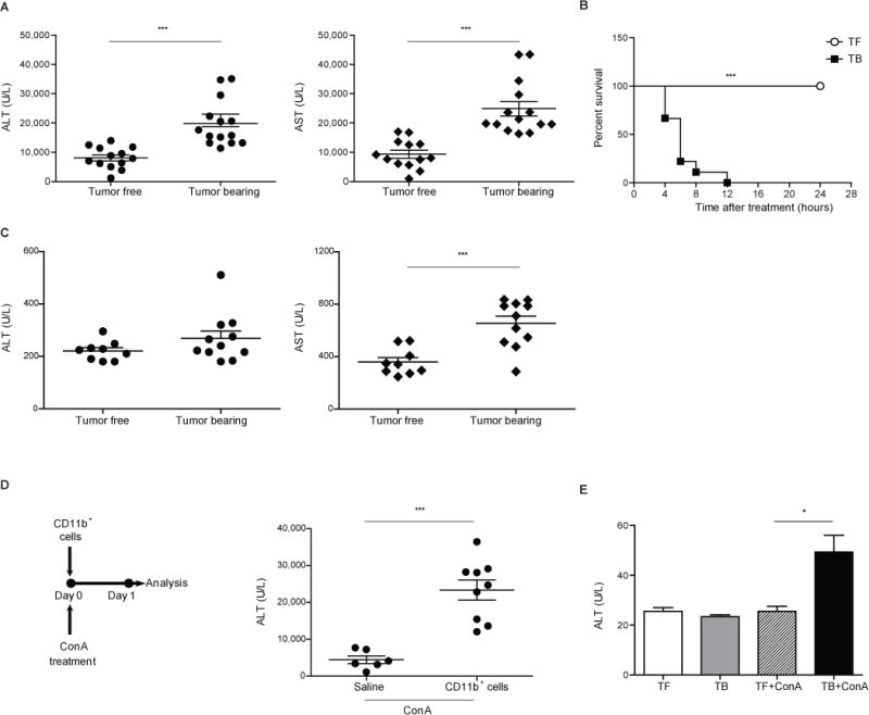
(A) Serum ALT/AST levels in TF (n=13) and EL4 TB (n=14) mice 16 hours after Con A treatment. (B) Kaplan-Meyer survival curve for cohorts of TF (n=5) and B16 GM-CSF TB mice (n=9) after Con A injection. (C) Serum ALT/AST levels in TF (n=7) and EL4 TB mice (n=8) 16 hours after α-GalCer injection. (D) Serum ALT values 16 hours after adoptive cell transfer of 5×10⁷ hepatic CD11b+ cells from B16 GM-CSF TB mice and Con A challenge (saline n=6; CD11b+ cells n=9). (E) TF (n=6) and EL4 TB (n=8) Rag-1−/− mice were injected either with saline or Con A. Serum ALT levels were determined as indicated above. Data are expressed as a mean ± SEM and are a cumulative of 6 (A), 2 (B), 3 (C–E) independent experiments. *P<0.05, ** P<0.01, ***P<0.001: Student’s t test (A,C, D and E) and Mantel-Cox log-rank test (B). TF = tumor-free; TB = tumor-bearing.
In order to link the presence of tumor-induced hepatic MDSC and exacerbation of liver damage, we purified hepatic CD11b+ cells from B16 GM-CSF TB mice (Supplementary Figure S1E) and adoptively transferred them into naïve mice prior to Con A challenge. Donor cells accumulated in the liver 1 hour after transfer (Supplementary Figure S1F). Mice receiving CD11b+ cells demonstrated higher ALT serum levels upon Con A challenge compared to control mice without adoptive transfer (Figure 1D), suggesting a direct link between the presence of tumor-induced myeloid cells in the liver and exacerbation of hepatitis. In control experiments we did not find an exacerbation of ALT/AST serum levels compared to naïve mice upon Con A challenge after transfer of bone marrow CD11b+ cells from naïve mice (data not shown). Next we decided to study the effects of MDSC depletion prior to Con A challenge. We have previously shown that anti Gr-1 fails to deplete tumor-induced MDSC in the liver [33],[34]. However, it has been reported that low dose 5-fluorouracil (5-FU) selectively kills tumor-induced MDSC [35]–[37]. Therefore, we treated tumor-bearing mice with 5-FU prior to Con A challenge which led in a reduction of hepatic MDSC (Supplementary Figure S1D). In parallel and as expected, ALT levels also dropped upon 5-FU treatment (Supplementary Figure S1D), suggesting that depletion of hepatic MDSC alleviated liver damage in subcutaneous TB mice. Finally, we also studied the effect of hepatic MDSC in the absence of T and NKT cells, which are known to be important effector cells in these models, using Rag-1−/− mice. Tumor growth led to recruitment of CD11b+Gr-1+ cells in the liver of Rag-1−/− mice (Supplementary Figure S1E). While Con A completely failed to induce inflammation in TF Rag-1−/−, hepatic MDSC accumulated in TB mice (Supplementary Figure S1G) and these mice showed modest ALT elevation upon Con A injection (Figure 1E), supporting our observation that hepatic MDSC are sufficient to cause liver damage.
Hepatic MDSC change into a mature and inflammatory phenotype upon Con A injection
In order to better understand how hepatic CD11b+Gr-1+ cells promoted liver damage we first studied their suppressor function upon Con A challenge in vitro. As expected CD11b+Gr1+ cells from TF mice did not demonstrate any suppressor activity after Con A injection. In contrast to hepatic MDSC obtained from TB mice without Con A treatment tumor-induced hepatic CD11b+Gr-1+ cells derived from mice after Con A injection lost their suppressor activity (Figure 2A and Supplementary Figure S2B). Next, to study molecular and phenotypic changes in hepatic MDSC upon Con A challenge we adoptively transferred CD45.1+ EL4-induced hepatic CD11b+Gr-1+ cells into naïve CD45.2+ mice and performed gene expression analysis, comparing mRNA profiles of donor cells sorted from livers of mice with and without Con A treatment. Results revealed an enriched inflammatory gene signature in transferred cells upon Con A challenge (Figure 2B). A significant increase in mRNA for IL-1α, IL-12, and TNF-α in hepatic CD11b+Gr-1+ cells three hours following Con A challenge was confirmed by qPCR (Figure 2C). Interestingly, flow cytometry analysis showed that tumor-induced hepatic CD11b+Gr-1high cells (gated as in Supplementary Figure S3A) substantially down-regulated CD244 surface expression upon Con A injection (Figure 2D), a molecule recently related to the granulocytic MDSC lineage [35],[38]. In a different set of experiments a striking increase in the expression of CD40, as well as CD80, CD86 and CD1d occurred by three hours after Con A injection in EL4 –induced hepatic CD11b+Gr-1low and CD11b+Gr-1high cells (Figures 2E, 2F and Supplementary Figures S3B and C). Similar results have been observed in two other tumor models (data not shown). In summary, acute liver inflammation resulted in the following effects: maturation of tumor-induced hepatic CD11b+Gr-1+ cells; induced the expression of pro-inflammatory cytokines and up regulation of costimulatory molecules including CD40; promoted loss of suppressor function.
Figure 2. Immune-mediated hepatitis shapes MDSC into inflammatory myeloid cells.
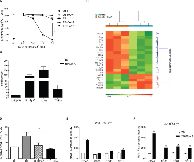
(A) Hepatic CD11b+Gr-1+ cells sorted from the livers of TF and EL4 TB mice, before or 3 hours after Con A challenge (n=4 mice/group) were cultured with 105 CFSE-labeled OT-I splenocytes at different ratios in the presence of OVA257–264 peptide (0.1 μg/ml). Proliferation of CFSE+CD8+ cells was evaluated after 48 hours. Data shown are mean ± SEM representative of 2 independent experiments. (B) Analysis of inflammation–related gene expression in EL4-induced CD45.1+ hepatic CD11b+Gr-1+ cells isolated from congenic mice 3 hours after adoptive transfer and induction of immune-mediated hepatitis. Dendrogram showing significant inflammation –related genes differentially expressed with fold change ≥ 2 Gene ontology score (n=7/group, pool of 3 independent experiments). Sample processing is described in Materials and Methods. (C) Increase in mRNA expression of several pro inflammatory genes in sorted liver CD11b+Gr-1+ cells from EL4 TB mice 3 hours after acute hepatitis induction (n=8 mice/ group; mRNA expression in sorted hepatic CD11b+Gr-1+ MDSC from TB mice without Con A injection was set to 1). Data shown are mean ± SEM representative of 2 independent experiments. (D) CD244 expression on hepatic CD11b+Gr-1high cells from either TF or TB mice before and 3 h after Con A challenge (n=2–4 mice/group). Cumulative data expressed as mean ± SEM are shown. (E–F) Mean Fluorescence Intensity (MFI) of CD40, CD80, CD86 and CD1d was determined either on CD11b+Gr-1high (E) or CD11b+Gr-1low (F) cells derived from EL4 TB mice, before or 3 hours after Con A treatment (n=3 mice/group). Data shown are mean ± SEM representative of 2 independent experiments *P<0.05, ** P<0.01, *** P<0.001: Student’s t test.
CD11b+Gr-1+ mediated liver damage is ROS-dependent
Our gene expression analysis on adoptively transferred CD45.1+ tumor-induced hepatic CD11b+Gr-1+ cells isolated from CD45.2+ recipient mice livers after acute liver injury showed significant enrichment (enrichment score=10.7 based on gene ontology analysis) of metabolic function, including genes responsible for respiratory burst, nitrogen and oxidation-reduction processes (Figures 3A and 3B). Reactive Oxygen Species (ROS) are known as a different mechanism by which MDSC suppress T cells [35],[39] but have also been reported to cause liver injury [40]–[43]. Although hepatic MDSC produced ROS ex vivo, increased ROS levels were found in hepatic CD11b+Gr-1+ cells from TB mice upon liver damage (Figure 3C). Since the increased ROS production by tumor-induced myeloid cells has been shown to be regulated by NADPH oxidase 2 (Nox2) [44], we isolated hepatic CD11b+Gr1+ cells from EL4 TB mice 3 hours after either saline or Con A injection and analyzed Nox2 expression. Nox2 mRNA levels were increased in tumor-induced hepatic CD11b+Gr1+ cells after Con A challenge (Figure 3D). Furthermore, congenic TB hepatic CD11b+ adoptive transfer experiments showed that donor-derived CD11b+Gr-1+ cells increased ROS production upon Con A challenge (Figure 3E).
Figure 3. Liver CD11b+Gr-1+ MDSC produce hepatocyte damage via ROS release.
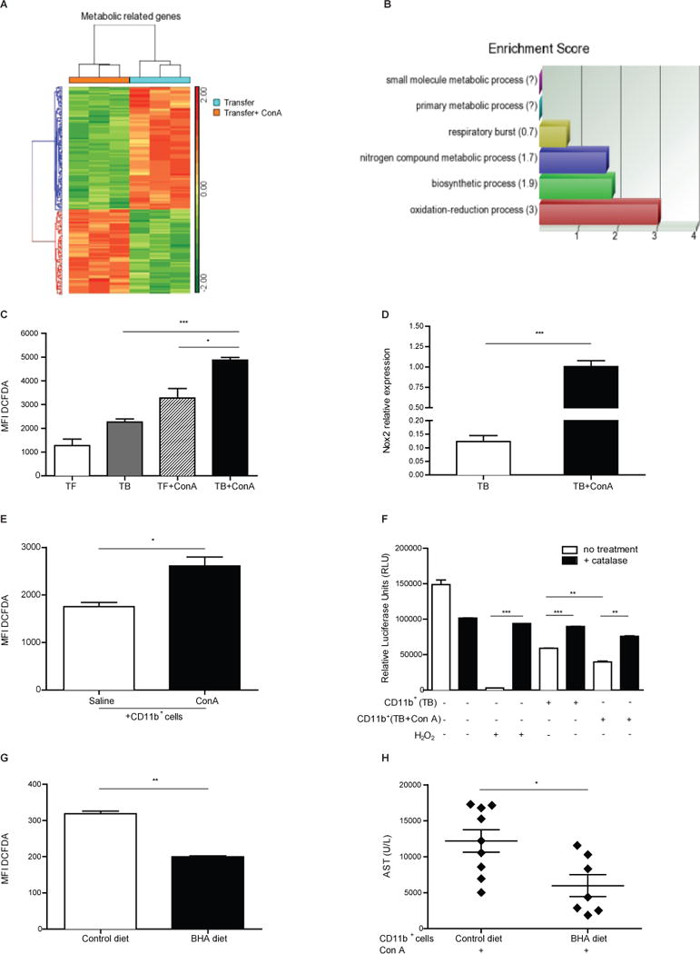
(A) Dendrogram showing significant metabolism –related genes differentially expressed with fold change ≥ 2 Gene ontology score in metabolic processes (n=7/group, pool of 3 independent experiments). (B) Enrichment score bar chart grouping metabolism-related genes in metabolic processes. (C) ROS production was evaluated by flow cytometry gating on hepatic CD11b+Gr-1+ cells from TF and EL4 TB mice, before and 3 hours after Con A treatment (n=2–4 mice/group). (D) NADPH Oxidase 2 (Nox2) mRNA expression on EL4-induced hepatic CD11b+Gr-1+ cells before and 3 hours after Con A treatment (n=2–4 mice/group). (E) ROS production by flow cytometry gating on hepatic CD11b+Gr-1+ in adoptively transferred clonotypic B16 GM-CSF-induced hepatic CD11b+ cells with or without Con A co-injection. (F) Luminescence intensity in luciferase-expressing RIL-175 cells cultured with or without hepatic CD11b+ cells derived from EL4 TB mice before and 3 hours after Con A challenge (n=3 mice/group). 1000U/ml catalase was used to block ROS production and 2 mM H2O2 was set as positive control. (G–H) B16 GM-CSF TB mice were subjected for 3 days to a butylated hydroxyanisole (BHA) diet (7 mg/g bodyweight) or control diet (n=3 mice/group). ROS production gated on hepatic CD11b+Gr-1+ cells was evaluated by flow cytometry (G). Hepatic myeloid cells were MACS isolated (>70% are CD11b+Gr-1+ cells) and adoptively transferred followed by Con A challenge. AST levels were measured after 16 hours (H). Data are shown as mean ± SEM derived from representative (A–D, F–G) and cumulative (E,H) of 3 (A–C) and 2 (D–H) independent experiments. *P<0.05, ** P<0.01, *** P<0.001: Student’s t test.
To better understand whether ROS production by hepatic CD11b+Gr-1+ cells may exacerbate acute liver damage, we next studied the cytotoxic activity of tumor-induced hepatic myeloid cells in vitro after Con A challenge. Interestingly, hepatic CD11b+ cells derived from TB mice injected with Con A significantly enhanced the killing of hepatoma cells, suggesting that Con A treatment exacerbates ROS-mediated liver cell killing by hepatic myeloid cells (Figure 3F). To further confirm this mechanism, we kept TB mice on a butylated hydroxyanisole (BHA) diet to block ROS production [45]. As expected, MDSC from BHA-fed mice produced less ROS in vitro than MDSC derived from mice on a normal diet (Figure 3G). Hepatic CD11b+ cells from B16 GM-CSF TB littermates kept on a BHA or control diet were transferred into naïve mice followed by Con A challenge. AST levels were lower in mice adoptively transferred with hepatic myeloid cells from BHA fed mice (Figure 3H).
CD40 dependent control of arginase function, ROS expression and suppressor function in hepatic MDSC
We first studied CD40 up regulation on tumor-induced hepatic myeloid suppressive cells upon Con A challenge and then performed studies using Cd40−/− TB mice. High serum TNF-α and IFN-γ levels have been previously described in response to Con A injection [46]. Similarly, we found elevated IFN-γ serum levels in TB mice after Con A injection (Supplementary Figure S4A). CD40 expression on hepatic myeloid cells from TB mice was elevated after incubation with IFN-γ but not with TNF-α (Supplementary Figure S4B). Likewise in vivo IFN-γ blockade prior to Con A treatment inhibited CD40 up regulation gated on transferred tumor-induced hepatic CD45.1+CD11b+Gr-1+ cells (Figure 4A and Supplementary Figure S4C), suggesting that IFN-γ modulates CD40 in vivo. Finally, we evaluated the effect of IFN-γ on arginase activity in tumor-induced hepatic myeloid cells upon Con A treatment. In vitro IFN-γ treated hepatic CD11b+ cells showed reduced activity of arginase, an enzyme related with myeloid suppressive activity (Figure 4B).
Figure 4. Immune-mediated hepatitis changes hepatic MDSC into inflammatory myeloid cells by CD40 ligation.
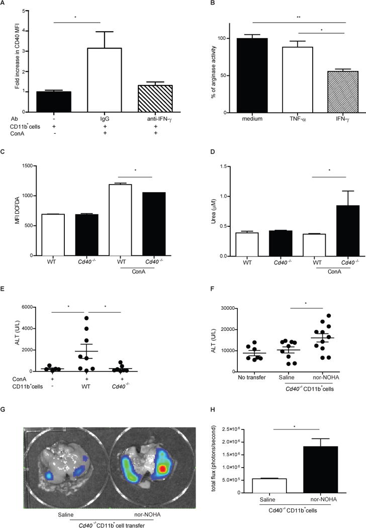
(A) CD40 MFI gated on CD45.1+CD11b+Gr-1+ cells 3 hours after congenic transfer of B16 GM-CSF-induced hepatic CD45.1+CD11b+ cells into CD45.2+ mice and Con A challenge. Data shown are mean ± SEM of 2 independent experiments. (B) Liver CD11b+ cells from B16 GM-CSF TB mice (n=3) were incubated for 16 hours in the presence of 0.1 μg/ml TNF-α or IFN-γ or were left untreated. Arginase activity was evaluated based on amount of urea release by the cell lysates. Data from untreated cells were set as 100%. Data shown are mean ± SEM representative of 2 independent experiments. (C–D) Function and phenotype of hepatic MDSC from either WT or Cd40−/− B16 GM-CSF TB mice before or 3 hours after Con A challenge (n=3/group): (C) ROS production ex vivo gated on hepatic CD11b+Gr-1+cells was evaluated by flow cytometry using Carboxy-H2-DCFDA. (D) Arginase activity was evaluated after overnight incubation of B16 GM-CSF TB liver CD11b+ cells. Data shown in C–D are mean ± SEM representative of at least 2 independent experiments. (E) 5×10⁷ liver CD11b+ cells from either WT or Cd40−/− B16 GM-CSF TB mice (n=3/group) were injected into naïve WT recipients and Con A was administered immediately. ALT (E) levels 16 hours after Con A are shown (n=5–8 mice/group). Cumulative data expressed as mean ± SEM, representative of 2 independent experiments. (F) liver CD11b+ cells from Cd40−/− B16 GM-CSF TB mice (n=3) were isolated and incubated either with PBS or arginase inhibitor nor-NOHA for 1 hour. Then 5×10⁷ cells were injected into naïve recipients followed by Con A injection. ALT levels 16 hours after Con A are shown (n=5–8 mice/group). Cumulative data expressed as mean ± SEM, representative of 2 independent experiments; (G–H) Mice treated as described in (F), were inoculated with 5 μg L-012, which shows luminescence after chemical reaction with ROS. Data show a representative in vivo image (G) and quantification of bioluminescence (H) (n=3 mice/group). Cumulative data expressed as mean ± SEM, representative 2 independent experiments. *P<0.05, *** P<0.001: Student’s t test.
Using wild type and Cd40−/− B16 GM-CSF TB mice before and 3 hours after Con A treatment, hepatic CD11b+Gr-1+ cells were tested for production of ROS and arginase activity was measured in tumor-induced hepatic CD11b+ cells. ROS production was decreased in hepatic CD11b+Gr-1+ cells derived from Cd40−/− mice compared to wild type controls, while arginase activity was higher upon Con A challenge (Figures 4C and 4D). As metabolic functions diverged upon acute inflammation in wild type and Cd40−/− hepatic CD11b+Gr-1+ cells, we tested whether hepatic Cd40−/− MDSC were able to ameliorate Con A-induced hepatitis in vivo. Hepatic CD11b+ cells were isolated from B16 GM-CSF TB wild type or Cd40−/− mice and transferred into naïve wild type mice followed by Con A injection. Serum ALT levels were consistently lower in mice after transfer of cells derived from Cd40−/− mice (Figures 4E). In contrast higher serum ALT levels were observed after transfer of B16 GM-CSF TB Cd40−/− myeloid cells, in which arginase activity was blocked by nor-NOHA (Figures 4F). Finally, adoptively transferred nor-NOHA treated Cd40−/− CD11b+ cells showed more efficient ROS production upon Con A treatment of recipient mice in contrast to saline treated cells (Figures 4G and 4H). In summary, acute inflammation-induced CD40 is responsible for phenotypic and functional changes in tumor-induced hepatic myeloid cells.
Discussion
Accumulation of myeloid-derived suppressor cells in tumors, spleen and livers has been described in various tumor settings both in mice and men [1]. Initially this study was designed to study the immunosuppressive function of tumor-induced hepatic MDSC in models of immune-mediated hepatitis. Liver toxicity is mainly driven by the infiltration of CD4+ T cells and activation of hepatic NKT cells and macrophages, as well as the specific cytokines produced by these cells – mainly TNF-α, IFN-γ, IL-4 and IL-12 [3],[4],[6]. Similar results have been described for α-GalCer-induced hepatitis [11],[13]. Unexpectedly, we observed an exacerbation of hepatitis instead of an amelioration upon Con A or α-GalCer injection in two different strains of TB mice using three different tumor cell lines. In addition, using two different experimental settings (adoptive transfer of hepatic MDSC and Rag 1−/− TB mice), we linked the presence of tumor-induced hepatic MDSC in TB mice to severity of liver damage.
HCC is considered an inflammation induced cancer. Con A-mediated hepatitis is a widely used model to study immune mediated hepatitis and more specifically autoimmune hepatitis in mice [14]. Several studies demonstrated that repetitive sublethal ConA injections induced immunotolerance [18]. Therefore this model can only be used to study acute immune-mediated responses in tumor bearing mice, but not the effect of chronic hepatitis on tumor development or treatment. However, Con A has been shown to activate intrahepatic NK and NKT cells and thereby enhance antitumor immunity [20]. Furthermore, findings described in this study have directed our attention to a molecule of great interest in tumor immunology, CD40. We are currently studying the effects of agonistic anti-CD40 on hepatic myeloid cells in TB mice.
A few recent reports in murine models of hepatitis investigated how different therapeutic strategies induce liver protection by enhancing CD11b+Gr-1+ cell infiltration into the liver [21]–[24]. Similar results have been described by ourselves in a model of T cell mediated colitis [19] and others in GVHD and sepsis [26],[28]. Based on these studies our initial working hypothesis was that tumor-induced hepatic CD11b+Gr-1+ immunosuppressive cells would exert a hepatoprotective effect, but we observed the opposite.
A rapid hepatic influx by CD11b+Gr-1+ neutrophils upon Con A injection has been described [29]. Studies on the biology of tumor-induced MDSC in general are complicated because of the absence of specific markers, which would allow one to clearly separate them from immature non-suppressive myeloid cell in naïve mice [5],[31]. Neutrophils are functionally different from tumor-induced hepatic MDSC [33]. It has been shown by different investigators that growing tumors effect hematopoiesis in the bone marrow leading to the generation of immature myeloid cells with suppressor activities such as MDSC [35]. CD11b+Gr-1+ cells in naïve mice in the steady state (not under pathological conditions) lack suppressor function [38] and this is also the case after Con A challenge (Figure 2A). Our experiments suggest that hepatic MDSC lose suppressor activity and acquire a pro-inflammatory phenotype within 3 hours after Con A injection. While we cannot completely exclude the possibility that naive CD11b+Gr1+ cells without suppressive capacity migrate into the liver and mask suppressor function of hepatic tumor-induced MDSC, it should be noted that the presence of a growing tumor will also affect myeloid cells migrating from the bone marrow into the liver and thereby potentially differ from naïve CD11b+Gr1+ cells found in tumor free mice. Furthermore, CD244, a marker previously described to be expressed by granulocytic MDSC in TB but not naïve mice [35], was only detected in TB mice prior to Con A injection, but not in TF mice or 3 hours after Con A injection, further demonstrating the different CD11b+Gr-1+ cell populations in TB and naïve mice.
Adoptive transfer of tumor-induced hepatic MDSC allowed us to perform comprehensive analysis of tumor-induced hepatic MDSC after Con A injection and suggested a critical role of CD40 in MDSC. Murine tumor-induced MDSC express low levels of CD40 [40],[42],[43]. IFN-γ, which is found upon Con A injection, enhanced CD40 expression and impaired arginase activity on hepatic MDSC in vitro while IFN-γ blockade in vivo impaired CD40 expression on transferred myeloid cells after Con A injection. Interestingly, IFN-γ up-regulates CD40 expression on murine dendritic cells [47] as well as human monocytes [48]. Using an orthotopic model of renal cell carcinoma we also previously been described that combined IL-2/anti-CD40 treatment reduced IFN-γ–dependent arginase expression in tumor-infiltrating macrophages [49], pointing towards a critical role for CD40/IFN-γ in MDSC-mediated suppressor functions. This was further studied using CD40 knockout MDSC. It should be noted that, in contrast, TNF-α not only blocked maturation and differentiation of myeloid cells in a chronic inflammatory setting, but also increased their suppressor activity [50].
In vitro studies using CD40 knockout MDSC demonstrated increased arginase activity and reduced ROS production upon Con A injection, suggesting a divided regulation for these two suppressor pathways. Interestingly, pharmacological inhibition of arginase function in hepatic CD40 knockout MDSC resulted in higher ALT serum levels upon Con A challenge. This suggests that hepatic MDSC indeed have the ability to suppress hepatitis, but that CD40ligation-induced loss of arginase function was one factor resulting in exacerbation of hepatitis, and that this was independentof increased ROS production. This has also previously been described in endothelial cells upon CD40 ligation [51]. Further evidence for a critical role for CD40 in MDSC comes from two independent studies. Pan and colleagues demonstrated that MDSC-dependent accumulation of regulatory T cells was CD40-dependent [52]. In that study, an anti-CD40 agonist was shown to reverse MDSC-mediated immunosuppression, providing a causal link between CD40 and the loss of T cell inhibitory function by tumor-derived MDSC. In a second study, De Santo and colleagues demonstrated that invariant NKT cells reduce ARG1 and NOS2 activity in a CD1d- and CD40 dependent manner in influenza A virus-infected mice. Of note, neither differences in suppression of T cell proliferation nor in tumor growth between WT and Cd40−/− MDSC were observed ([53] and data not shown). These studies, in combination with the data presented here, clearly suggest that MDSC function is controlled by CD40 engagement in an inflammatory setting.
Depletion of L-arginine and production of ROS or peroxynitrite (PNT) are well-known mechanisms of suppression of T cell function by MDSC [54]. Increased activity of arginase in MDSC leads to enhanced L-arginine catabolism and impaired T cell function. Our work adds a new and previously unrecognized MDSC effector function. In a model of acute inflammation hepatic MDSC release ROS, resulting in aggravated hepatitis and hepatocyte death. While studying the immunological effects of ROS can be challenging due to lack of adequate assays [55], ROS have been recognized to cause hepatocyte death by damaging mitochondria and other cellular compartments [56]. Interestingly, we observed that hepatic CD11b+Gr-1+ cells lost their suppressor activity upon Con A injection. This may be explained by the fact that CD40 ligation has opposite effects on arginase function and ROS production. An alternate possibility may be that different ROS species cause liver cell damage and T cell suppression. As a matter of fact, the term ROS includes a number of different oxygen-containing molecules and are often indistinguishable from reactive nitrogen species, which have similar biological functions [55],[57].
Overall, our findings indicate the unexpected and paradoxical result that tumor-induced MDSC, whose hallmark activity is suppression of immune responses, can – in the context of acute hepatitis – be converted into pro-inflammatory immune effector cells capable of killing hepatocytes when stimulated by CD40 ligation.
Materials and methods
Mice and cell lines
8 to 10 week-old female C57BL/6, BALB/c and B6Ly5.2 mice were purchased from NCI/Frederick. H-2Kb OVA257–264 TCR transgenic OT-I, Cd40−/− and Rag-1−/− mice (kindly provided by Dr. Noriko Sato, NIH/NCI) were bred at the National Cancer Institute. EL4, B16 GM-CSF, CT26 GM-CSF and RIL-175 cells were previously described [7],[58].
Animal studies
Tumor-free littermates or mice bearing subcutaneous tumors were challenged with 12.5 μg/g type IV Concanavalin A (Con A) (Sigma, USA) or 2 μg/mouse α-Galactosylceramide (α-GalCer, Funakoshi LTD, Japan). Alanine/aspartate aminotransferase (ALT/AST) levels were determined in mouse sera. In some experiments, mice were fed with a butylated hydroxyanisole (BHA, Sigma, USA)-containing diet for 3 days (7 mg/g bodyweight) to block ROS production [41],[45],[49]. IFN-γ serum levels after Con A injection were quantified by ELISA (eBioscience, USA). Endogenous IFN-γ was blocked by treating mice with 200 μg rat-anti mouse anti IFN-γ antibody (clone XMG1.2, BioXCell, USA). The same dose of rat IgG1 (BioXCell, USA) was used as a control. For visualization of ROS production, mice were injected with 5 μg luminescent probe L-012 (Wako, USA) which shows luminescence after chemical reaction with ROS and imaged with IVIS Spectrum (Xenogen, USA). Bioluminescence was calculated based on photon flux (photon counts/second) obtained 5 min after injection, divided by liver area using Living Image software (Perkin Elmer, USA). Mice were sacrificed at the indicated time points. All animal studies were approved by the National Cancer Institute Bethesda Animal Care and Use Committee.
Flow cytometry analysis
Liver mononuclear cells were obtained as previously described [7]. Mouse cell samples were stained using the following antibodies from BD Biosciences Ly6G (1A8), Gr-1 (RB6-8C5), MHC-II (M5/114.15.2), CD11b (M1/70), CD11c (HL3), CD40 (3/23), CD45 (30-F11), CD45.1 (A20), CD86 (GL-1), F4/80 (BM8) and eBiosciences CD1d (1B1), CD8 (53-6.7), CD80 (16-10A1) and Ly6C (HK1.4). PI or 7AAD staining (BD Biosciences, USA) was used to exclude dead cells. Hepatic myeloid cells were enriched using CD11b beads (Miltenyi Biotec, USA) followed by MACS separation performed prior to sorting or when indicated. CD11b purity after enrichment was above 90%. Flow cytometry was performed on BD FACS Calibur using CellQuest Pro acquisition software (Becton Dickinson, USA). Data were analyzed using FlowJo software (Tree Star, USA). Gating strategies as well as CD11b+Gr-1+ enrichment after either MACS separation using CD11b beads or FACS sorting are shown in Supplementary Figures S1, S2 and S3.
Adoptive hepatic MDSC transfer
5×10⁷ FACS-sorted EL4-induced hepatic CD11b+Gr-1+ cells or MACS-sorted B16 GM-CSF-induced hepatic CD11b+ cells (Supplementary Figure S1E) were injected i.v. before Con A challenge. When indicated, 200 μg anti-IFN-γ antibody (clone XMG1.2, BioXCell, USA) or isotype control were inoculated i.v. When mentioned, tumor-induced hepatic CD11b+ cells from Cd40−/− B16 GM-CSF TB mice were incubated for 1 hour at 37°C with rotation in the presence or absence of the arginase inhibitor N(omega)-hydroxy-nor-L-arginine (nor-NOHA, Calbiochem, USA) at a concentration of 1 mM per 1×10⁷ cells.
Gene expression studies
Hepatic CD11b+Gr-1+ cells were purified as described previously [7]. RNA was isolated using an RNeasy Micro Kit (Qiagen, USA). Complementary DNA synthesis was done with an iScript Kit (Bio-Rad, USA) and quantitative PCR was performed. Primer sequences are available upon request. Results were normalized to endogenous cyclophilin A mRNA level. Fold change of mRNA expression between samples was determined using the ΔΔCt method, where sample from TB mouse without Con A treatment was set as reference. For microarray analysis, RNA extractions were performed using NucleoSpin RNA II (Macherey-Nagel, Germany) followed by whole transcriptome amplification using a WT expression kit (Invitrogen, USA). cDNA was fragmented and hybridized to a Mouse Gene 1.0 ST array (Affimetrix Inc. USA) followed by terminal labeling, washing and scanning. Gene expression analyses were performed using Partek Genomic Suite 6.6.
Functional MDSC studies
Arginase activity by tumor-induced hepatic CD11b+ cells and ROS production gated on CD11b+Gr-1+ cells were determined as previously described [58],[59]. When indicated, 0.1 μg/ml recombinant TNF-α or IFN-γ (eBioscience) were added to the culture. Suppression of T cell proliferation by hepatic CD11b+Gr-1+ cells was measured after incubating sorted EL4-induced hepatic CD11b+Gr-1+ cells at indicated ratios with 105 CFSE-labeled OT-I splenocytes in the presence of 0.1 μg/ml OVA257–264 peptide.
Determination of hepatocyte cytotoxicity by hepatic CD11b+ cells
Luciferase -expressing RIL-175 hepatoma cells were cultured at a 1:50 ratio with EL4-induced hepatic CD11b+ cells isolated from mice injected either with PBS or Con A. After 16 hours the number of surviving adherent cells was evaluated using Dual Luciferase Reporter Assay (Promega, Madison, WI, USA). 2 mM H2O2 (Invitrogen, USA) and 1000 U/ml catalase (Sigma, USA) were used for apoptosis induction and blocking of ROS release, respectively.
Statistical analysis
Data were analyzed for statistical significance using Prism software (GraphPad). Kaplan-Meier plots and Log-rank test were used to determine the differences of significance between survival curves, and differences in other parameters were analyzed using Student’s t-test; P<0.05 was considered to be statistically significant.
Supplementary Material
Acknowledgments
We would like to thank to Alpa Shah (Department of Laboratory Medicine, NIH/Clinical Research Center)-for determining liver aminotransferases; Dr. Noriko Sato (Molecular Imaging Program, NIH/NCI)-for providing Rag-1−/− mice. This work was supported by the Initiative and Networking Fund of the Helmholtz Association within the Helmholtz Alliance on Immunotherapy of Cancer and in part by the Intramural Research Program of the Center for Cancer Research, National Cancer Institute (NCI), National Institutes of Health.
List of abbreviations
- MDSC
Myeloid derived suppressor cell
- TF
Tumor Free
- TB
Tumor Bearing
- ALT
Alanine Aminotransferase
- AST
Aspartate Aminotransferase
- α-GalCer
α-Galactosylceramide
- Con A
Concanavalin A
- 5-FU
5-fluorouracil
- ROS
Reactive Oxygen Species
- BHA
Butylated Hydroxyanisole
- Nor-NOHA
N(omega)-hydroxy-nor-L-arginine
- ARG1
Arginase 1
- NOS2
Nitric Oxide Synthase 2
- PNT
Peroxynytrite
Footnotes
Conflict of interest
The authors declare no commercial or financial conflict of interest.
References
- 1.Talmadge JE, Gabrilovich D. History of myeloid-derived suppressor cells. Nat Rev Cancer. 2013;13:7752. doi: 10.1038/nrc3581. [DOI] [PMC free article] [PubMed] [Google Scholar]
- 2.Gabrilovich DI, Bronte V, Chen S-H, Colombo MP, Ochoa A, Ostrand-Rosenberg S, Schreiber H. The terminology issue for myeloid-derived suppressor cells. Cancer Res. 2007;67:425. doi: 10.1158/0008-5472.CAN-06-3037. author reply 426. [DOI] [PMC free article] [PubMed] [Google Scholar]
- 3.Tiegs G, Hentschel J, Wendel A. A T cell-dependent experimental liver injury in mice inducible by concanavalin A. Journal of Clinical Investigation. 1992;90:196–203. doi: 10.1172/JCI115836. [DOI] [PMC free article] [PubMed] [Google Scholar]
- 4.Mizuhara H, O’Neill E, Seki N, Ogawa T, Kusunoki C, Otsuka K, Satoh S, et al. T cell activation-associated hepatic injury: mediation by tumor necrosis factors and protection by interleukin 6. J Exp Med. 1994;179:1529–1537. doi: 10.1084/jem.179.5.1529. [DOI] [PMC free article] [PubMed] [Google Scholar]
- 5.Gabrilovich DI, Ostrand-Rosenberg S, Bronte V. Coordinated regulation of myeloid cells by tumours. Nat Rev Immunol. 2012;12:253–268. doi: 10.1038/nri3175. [DOI] [PMC free article] [PubMed] [Google Scholar]
- 6.Gantner F, Leist M, Lohse AW, Germann PG, Tiegs G. Concanavalin A-induced T-cell-mediated hepatic injury in mice: the role of tumor necrosis factor. Hepatology. 1995;21:190–198. doi: 10.1016/0270-9139(95)90428-x. [DOI] [PubMed] [Google Scholar]
- 7.Kapanadze T, Gamrekelashvili J, Ma C, Chan C, Zhao F, Hewitt S, Zender L, et al. Regulation of accumulation and function of myeloid derived suppressor cells in different murine models of hepatocellular carcinoma. Journal of Hepatology. 2013;59:1007–1013. doi: 10.1016/j.jhep.2013.06.010. [DOI] [PMC free article] [PubMed] [Google Scholar]
- 8.Connolly MK, Mallen-St Clair J, Bedrosian AS, Malhotra A, Vera V, Ibrahim J, Henning J, et al. Distinct populations of metastases-enabling myeloid cells expand in the liver of mice harboring invasive and preinvasive intra-abdominal tumor. Journal of Leukocyte Biology. 2010;87:713–725. doi: 10.1189/jlb.0909607. [DOI] [PMC free article] [PubMed] [Google Scholar]
- 9.Ilkovitch D, Lopez DM. The Liver Is a Site for Tumor-Induced Myeloid-Derived Suppressor Cell Accumulation and Immunosuppression. Cancer Res. 2009;69:5514–5521. doi: 10.1158/0008-5472.CAN-08-4625. [DOI] [PMC free article] [PubMed] [Google Scholar]
- 10.Younos I, Donkor M, Hoke T, Dafferner A, Samson H, Westphal S, Talmadge J. Tumor- and organ-dependent infiltration by myeloid-derived suppressor cells. Int Immunopharmacol. 2011;11:816–826. doi: 10.1016/j.intimp.2011.02.021. [DOI] [PubMed] [Google Scholar]
- 11.Biburger M, Tiegs G. Alpha-galactosylceramide-induced liver injury in mice is mediated by TNF-alpha but independent of Kupffer cells. J Immunol. 2005;175:1540–1550. doi: 10.4049/jimmunol.175.3.1540. [DOI] [PubMed] [Google Scholar]
- 12.Haile LA, Greten TF, Korangy F. Immune Suppression: The Hallmark of Myeloid Derived Suppressor Cells. Immunol Invest. 2012;41:581–594. doi: 10.3109/08820139.2012.680635. [DOI] [PMC free article] [PubMed] [Google Scholar]
- 13.Erhardt A, Biburger M, Papadopoulos T, Tiegs G. IL-10, regulatory T cells, and Kupffer cells mediate tolerance in concanavalin A-induced liver injury in mice. Hepatology. 2007;45:475–485. doi: 10.1002/hep.21498. [DOI] [PubMed] [Google Scholar]
- 14.Wang H-X. Immune mechanisms of Concanavalin A model of autoimmune hepatitis. World J Gastroenterol. 2012;18:119. doi: 10.3748/wjg.v18.i2.119. [DOI] [PMC free article] [PubMed] [Google Scholar]
- 15.Hoechst B, Ormandy LA, Ballmaier M, Lehner F, Krüger C, Manns MP, Greten TF, et al. A new population of myeloid-derived suppressor cells in hepatocellular carcinoma patients induces CD4(+)CD25(+)Foxp3(+) T cells. Gastroenterology. 2008;135:234–243. doi: 10.1053/j.gastro.2008.03.020. [DOI] [PubMed] [Google Scholar]
- 16.Kalathil S, Lugade AA, Miller A, Iyer R, Thanavala Y. Higher Frequencies of GARP+CTLA-4+Foxp3+ T Regulatory Cells and Myeloid-Derived Suppressor Cells in Hepatocellular Carcinoma Patients Are Associated with Impaired T-Cell Functionality. Cancer Res. 2013;73:2435–2444. doi: 10.1158/0008-5472.CAN-12-3381. [DOI] [PMC free article] [PubMed] [Google Scholar]
- 17.Schneider C, Teufel A, Yevsa T, Staib F, Hohmeyer A, Walenda G, Zimmermann HW, et al. Adaptive immunity suppresses formation and progression of diethylnitrosamine-induced liver cancer. Gut. 2012 doi: 10.1136/gutjnl-2011-301116. [DOI] [PMC free article] [PubMed] [Google Scholar]
- 18.Zimmermann HW, Trautwein C, Tacke F. Functional role of monocytes and macrophages for the inflammatory response in acute liver injury. Front Physiol. 2012;3:56. doi: 10.3389/fphys.2012.00056. [DOI] [PMC free article] [PubMed] [Google Scholar]
- 19.Haile LA, Wasielewski von R, Gamrekelashvili J, Krüger C, Bachmann O, Westendorf AM, Buer J, et al. Myeloid-derived suppressor cells in inflammatory bowel disease: a new immunoregulatory pathway. Gastroenterology. 2008;135:871–81. 881.e1–5. doi: 10.1053/j.gastro.2008.06.032. [DOI] [PubMed] [Google Scholar]
- 20.Miyagi T, Takehara T, Tatsumi T, Suzuki T, Jinushi M, Kanazawa Y, Hiramatsu N, et al. Concanavalin a injection activates intrahepatic innate immune cells to provoke an antitumor effect in murine liver. Hepatology. 2004;40:1190–1196. doi: 10.1002/hep.20447. [DOI] [PubMed] [Google Scholar]
- 21.Hegde VL, Nagarkatti PS, Nagarkatti M. Role of Myeloid-Derived Suppressor Cells in Amelioration of Experimental Autoimmune Hepatitis Following Activation of TRPV1 Receptors by Cannabidiol. PLoS ONE. 2011;6:e18281. doi: 10.1371/journal.pone.0018281. [DOI] [PMC free article] [PubMed] [Google Scholar]
- 22.Sarra M, Cupi ML, Bernardini R, Ronchetti G, Monteleone I, Ranalli M, Franzè E, et al. IL-25 prevents and cures fulminant hepatitis in mice through a myeloid-derived suppressor cell-dependent mechanism. Hepatology. 2013;58:1436–1450. doi: 10.1002/hep.26446. [DOI] [PubMed] [Google Scholar]
- 23.Diao W, Jin F, Wang B, Zhang C-Y, Chen J, Zen K, Li L. The protective role of myeloid-derived suppressor cells in concanavalin A-induced hepatic injury. Protein Cell. 2014 doi: 10.1007/s13238-014-0069-5. [DOI] [PMC free article] [PubMed] [Google Scholar]
- 24.Liu G, Bi Y, Wang R, Yang H, Zhang Y, Wang X, Liu H, et al. Targeting S1P1 Receptor Protects against Murine Immunological Hepatic Injury through Myeloid-Derived Suppressor Cells. The Journal of Immunology. 2014 doi: 10.4049/jimmunol.1301193. [DOI] [PubMed] [Google Scholar]
- 25.Erhardt A, Tiegs G. Tolerance Induction in Response to Liver Inflammation. Dig Dis. 2010;28:86–92. doi: 10.1159/000282069. [DOI] [PubMed] [Google Scholar]
- 26.Delano MJ, Scumpia PO, Weinstein JS, Coco D, Nagaraj S, Kelly-Scumpia KM, O’Malley KA, et al. MyD88-dependent expansion of an immature GR-1(+)CD11b(+) population induces T cell suppression and Th2 polarization in sepsis. J Exp Med. 2007;204:1463–1474. doi: 10.1084/jem.20062602. [DOI] [PMC free article] [PubMed] [Google Scholar]
- 27.Kakimi K, Guidotti LG, Koezuka Y, Chisari FV. Natural killer T cell activation inhibits hepatitis B virus replication in vivo. J Exp Med. 2000;192:921–930. doi: 10.1084/jem.192.7.921. [DOI] [PMC free article] [PubMed] [Google Scholar]
- 28.Highfill SL, Rodriguez PC, Zhou Q, Goetz CA, Koehn BH, Veenstra R, Taylor PA, et al. Bone marrow myeloid-derived suppressor cells (MDSCs) inhibit graft-versus-host disease (GVHD) via an arginase-1-dependent mechanism that is up-regulated by interleukin-13. Blood. 2010;116:5738–5747. doi: 10.1182/blood-2010-06-287839. [DOI] [PMC free article] [PubMed] [Google Scholar]
- 29.Bonder CS, Ajuebor MN, Zbytnuik LD, Kubes P, Swain MG. Essential role for neutrophil recruitment to the liver in concanavalin A-induced hepatitis. J Immunol. 2004;172:45–53. doi: 10.4049/jimmunol.172.1.45. [DOI] [PubMed] [Google Scholar]
- 30.Serafini P, Carbley R, Noonan KA, Tan G, Bronte V, Borrello I. High-dose granulocyte-macrophage colony-stimulating factor-producing vaccines impair the immune response through the recruitment of myeloid suppressor cells. Cancer Res. 2004;64:6337–6343. doi: 10.1158/0008-5472.CAN-04-0757. [DOI] [PubMed] [Google Scholar]
- 31.Youn J-I, Gabrilovich DI. The biology of myeloid-derived suppressor cells: the blessing and the curse of morphological and functional heterogeneity. Eur J Immunol. 2010;40:2969–2975. doi: 10.1002/eji.201040895. [DOI] [PMC free article] [PubMed] [Google Scholar]
- 32.Osman Y, Kawamura T, Naito T, Takeda K, Kaer LV, Okumura K, Abo T. Activation of hepatic NKT cells and subsequent liver injury following administration of α-galactosylceramide. Eur J Immunol. 2000;30:1919–1928. doi: 10.1002/1521-4141(200007)30:7<1919::AID-IMMU1919>3.0.CO;2-3. [DOI] [PubMed] [Google Scholar]
- 33.Crispe IN. Immune tolerance in liver disease. Hepatology. 2014 doi: 10.1002/hep.27254. [DOI] [PMC free article] [PubMed] [Google Scholar]
- 34.Ma C, Kapanadze T, Gamrekelashvili J, Manns MP, Korangy F, Greten TF. Anti-Gr-1 antibody depletion fails to eliminate hepatic myeloid-derived suppressor cells in tumor-bearing mice. Journal of Leukocyte Biology. 2012;92:1199–1206. doi: 10.1189/jlb.0212059. [DOI] [PMC free article] [PubMed] [Google Scholar]
- 35.Youn J-I, Collazo M, Shalova IN, Biswas SK, Gabrilovich DI. Characterization of the nature of granulocytic myeloid-derived suppressor cells in tumor-bearing mice. Journal of Leukocyte Biology. 2011 doi: 10.1189/jlb.0311177. [DOI] [PMC free article] [PubMed] [Google Scholar]
- 36.Vincent J, Mignot G, Chalmin F, Ladoire S, Bruchard M, Chevriaux A, Martin F, et al. 5-Fluorouracil selectively kills tumor-associated myeloid-derived suppressor cells resulting in enhanced T cell-dependent antitumor immunity. Cancer Res. 2010;70:3052–3061. doi: 10.1158/0008-5472.CAN-09-3690. [DOI] [PubMed] [Google Scholar]
- 37.Ugel S, Peranzoni E, Desantis G, Chioda M, Walter S, Weinschenk T, Ochando JC, et al. Immune tolerance to tumor antigens occurs in a specialized environment of the spleen. Cell Rep. 2012;2:628–639. doi: 10.1016/j.celrep.2012.08.006. [DOI] [PubMed] [Google Scholar]
- 38.Zhao F, Obermann S, Wasielewski von R, Haile L, Manns MP, Korangy F, Greten TF. Increase in frequency of myeloid-derived suppressor cells in mice with spontaneous pancreatic carcinoma. Immunology. 2009;128:141–149. doi: 10.1111/j.1365-2567.2009.03105.x. [DOI] [PMC free article] [PubMed] [Google Scholar]
- 39.Youn J-I, Nagaraj S, Collazo M, Gabrilovich DI. Subsets of myeloid-derived suppressor cells in tumor-bearing mice. J Immunol. 2008;181:5791–5802. doi: 10.4049/jimmunol.181.8.5791. [DOI] [PMC free article] [PubMed] [Google Scholar]
- 40.Yang R, Cai Z, Zhang Y, Yutzy WH, Roby KF, Roden RBS. CD80 in immune suppression by mouse ovarian carcinoma-associated Gr-1+CD11b+ myeloid cells. Cancer Res. 2006;66:6807–6815. doi: 10.1158/0008-5472.CAN-05-3755. [DOI] [PubMed] [Google Scholar]
- 41.Hoek JB, Cahill A, Pastorino JG. Alcohol and mitochondria: a dysfunctional relationship. Gastroenterology. 2002;122:2049–2063. doi: 10.1053/gast.2002.33613. [DOI] [PMC free article] [PubMed] [Google Scholar]
- 42.Liu Y, Zeng B, Zhang Z, Zhang Y, Yang R. B7-H1 on myeloid-derived suppressor cells in immune suppression by a mouse model of ovarian cancer. Clin Immunol. 2008;129:471–481. doi: 10.1016/j.clim.2008.07.030. [DOI] [PubMed] [Google Scholar]
- 43.Ko HJ, Lee JM, Kim YJ, Kim YS, Lee KA, Kang CY. Immunosuppressive myeloid-derived suppressor cells can be converted into immunogenic APCs with the help of activated NKT cells: an alternative cell-based antitumor vaccine. J Immunol. 2009;182:1818–1828. doi: 10.4049/jimmunol.0802430. [DOI] [PubMed] [Google Scholar]
- 44.Corzo CA, Cotter MJ, Cheng P, Cheng F, Kusmartsev S, Sotomayor E, Padhya T, et al. Mechanism Regulating Reactive Oxygen Species in Tumor-Induced Myeloid-Derived Suppressor Cells. J Immunol. 2009;182:5693–5701. doi: 10.4049/jimmunol.0900092. [DOI] [PMC free article] [PubMed] [Google Scholar]
- 45.Kamata H, Honda S-I, Maeda S, Chang L, Hirata H, Karin M. Reactive oxygen species promote TNFalpha-induced death and sustained JNK activation by inhibiting MAP kinase phosphatases. Cell. 2005;120:649–661. doi: 10.1016/j.cell.2004.12.041. [DOI] [PubMed] [Google Scholar]
- 46.Küsters S, Gantner F, Künstle G, Tiegs G. Interferon gamma plays a critical role in T cell-dependent liver injury in mice initiated by concanavalin A. Gastroenterology. 1996;111:462–471. doi: 10.1053/gast.1996.v111.pm8690213. [DOI] [PubMed] [Google Scholar]
- 47.Frasca L, Nasso M, Spensieri F, Fedele G, Palazzo R, Malavasi F, Ausiello CM. IFN-gamma arms human dendritic cells to perform multiple effector functions. J Immunol. 2008;180:1471–1481. doi: 10.4049/jimmunol.180.3.1471. [DOI] [PubMed] [Google Scholar]
- 48.Alderson MR, Armitage RJ, Tough TW, Strockbine L, Fanslow WC, Spriggs MK. CD40 expression by human monocytes: regulation by cytokines and activation of monocytes by the ligand for CD40. J Exp Med. 1993;178:669–674. doi: 10.1084/jem.178.2.669. [DOI] [PMC free article] [PubMed] [Google Scholar]
- 49.Weiss JM, Back TC, Wiltrout RH., 11 Successful immunotherapy with IL-2/anti-CD40 induces the chemokine-mediated mitigation of an immunosuppressive tumor microenvironment. Proc Natl Acad Sci USA. 2009;106:19455–19460. doi: 10.1073/pnas.0909474106. [DOI] [PMC free article] [PubMed] [Google Scholar]
- 50.Sade-Feldman M, Kanterman J, Ish-Shalom E, Elnekave M, Horwitz E, Baniyash M. Tumor necrosis factor-α blocks differentiation and enhances suppressive activity of immature myeloid cells during chronic inflammation. Immunity. 2013;38:541–554. doi: 10.1016/j.immuni.2013.02.007. [DOI] [PubMed] [Google Scholar]
- 51.Urbich C, Dernbach E, Aicher A, Zeiher AM, Dimmeler S. CD40 ligand inhibits endothelial cell migration by increasing production of endothelial reactive oxygen species. Circulation. 2002;106:981–986. doi: 10.1161/01.cir.0000027107.54614.1a. [DOI] [PubMed] [Google Scholar]
- 52.Pan PY, Ma G, Weber KJ, Ozao-Choy J, Wang G, Yin B, Divino CM, et al. Immune stimulatory receptor CD40 is required for T-cell suppression and T regulatory cell activation mediated by myeloid-derived suppressor cells in cancer. Cancer Res. 2010;70:99–108. doi: 10.1158/0008-5472.CAN-09-1882. [DOI] [PMC free article] [PubMed] [Google Scholar]
- 53.De Santo C, Salio M, Masri SH, Lee LY-H, Dong T, Speak AO, Porubsky S, et al. Invariant NKT cells reduce the immunosuppressive activity of influenza A virus-induced myeloid-derived suppressor cells in mice and humans. Journal of Clinical Investigation. 2008;118:4036–4048. doi: 10.1172/JCI36264. [DOI] [PMC free article] [PubMed] [Google Scholar]
- 54.Nagaraj S, Gabrilovich DI. Regulation of suppressive function of myeloid-derived suppressor cells by CD4+ T cells. Seminars in Cancer Biology. 2012;22:282–288. doi: 10.1016/j.semcancer.2012.01.010. [DOI] [PMC free article] [PubMed] [Google Scholar]
- 55.Nathan C, Cunningham-Bussel A. Beyond oxidative stress: an immunologist’s guide to reactive oxygen species. Nat Rev Immunol. 2013;13:349–361. doi: 10.1038/nri3423. [DOI] [PMC free article] [PubMed] [Google Scholar]
- 56.Rolo AP, Teodoro JS, Palmeira CM. Role of oxidative stress in the pathogenesis of nonalcoholic steatohepatitis. Free Radical Biology and Medicine. 2012;52:59–69. doi: 10.1016/j.freeradbiomed.2011.10.003. [DOI] [PubMed] [Google Scholar]
- 57.Lu T, Gabrilovich DI. Molecular pathways: tumor-infiltrating myeloid cells and reactive oxygen species in regulation of tumor microenvironment. Clin Cancer Res. 2012;18:4877–4882. doi: 10.1158/1078-0432.CCR-11-2939. [DOI] [PMC free article] [PubMed] [Google Scholar]
- 58.Haile LA, Gamrekelashvili J, Manns MP, Korangy F, Greten TF. CD49d is a new marker for distinct myeloid-derived suppressor cell subpopulations in mice. J Immunol. 2010;185:203–210. doi: 10.4049/jimmunol.0903573. [DOI] [PubMed] [Google Scholar]
- 59.Eruslanov E, Kusmartsev S. Identification of ROS using oxidized DCFDA and flow-cytometry. Methods Mol Biol. 2010;594:57–72. doi: 10.1007/978-1-60761-411-1_4. [DOI] [PubMed] [Google Scholar]
Associated Data
This section collects any data citations, data availability statements, or supplementary materials included in this article.