Summary
Bacteria encounter reactive oxygen species (ROS) as consequence of the aerobic life or as oxidative burst of activated neutrophils during infections. In addition, bacteria are exposed to other redox-active compounds including hypochloric acid (HOCl) and reactive electrophilic species (RES), such as quinones and aldehydes. These reactive species often target the thiol groups of cysteines in proteins and lead to thiol-disulfide switches in redox-sensing regulators to activate specific detoxification pathways and to restore the redox balance. Here, we review bacterial thiol-based redox sensors that specifically sense ROS, RES and HOCl via thiol-based mechanisms and regulate gene transcription in Gram-positive model bacteria and in human pathogens, such as Staphylococcus aureus and Mycobacterium tuberculosis. We also pay particular attention to emerging widely conserved HOCl-specific redox regulators that have been recently characterized in Escherichia coli. Different mechanisms are used to sense and respond to ROS, RES and HOCl by 1-Cys-type and 2-Cys-type thiol-based redox sensors that include versatile thiol-disulfide switches (OxyR, OhrR, HypR, YodB, NemR, RclR, Spx, RsrA/RshA) or alternative Cys-phosphorylations (SarZ, MgrA, SarA), thiol-S-alkylation (QsrR), His-oxidation (PerR) and methionine oxidation (HypT). In pathogenic bacteria, these redox-sensing regulators are often important virulence regulators and required for adapation to the host immune defense.
Keywords: Thiol-based redox switches, OxyR, OhrR, MarR, SarA, NemR, RclR, HypT, Spx, RsrA, RshA
1. Introduction
Bacteria have to cope in their natural environment or during bacterial infection in association with the host immune system to reactive oxygen species (ROS) that are known to cause an oxidative stress response and affect the reduced state of the cytoplasm. ROS are produced in microorganisms as the unavoidable consequence of the aerobic life, by incomplete reduction of molecular oxygen during respiration (Imlay, 2003; Imlay, 2008; Imlay, 2013). Beside ROS, bacteria have to cope with many other redox-active compounds, including antimicrobials, antibiotics and environmental xenobiotics which can act as reactive electrophilic species (RES) and affect the cellular redox status (Jacobs & Marnett, 2010; Marnett et al, 2003). ROS and RES cause specific post-translational thiol-modifications in redox-sensing transcription factors which lead to conformational changes and activate or inactive the transcriptional regulator. As consequence, specific detoxification pathways are upregulated to destroy the reactive species or to repair the resulting damage (Antelmann & Helmann, 2011; Imlay, 2013; Vazquez-Torres, 2012). With the discovery of the peroxide-sensor OxyR of E. coli, it became evident that ROS-sensing by thiol-disulfide switches represents an important regulatory device in bacteria (Choi et al, 2001; Kim et al, 2002; Zheng et al, 1998). However, during the last decade this classical thiol-disulfide-switch model for redox-regulation has been expanded by different reversible and irreversible thiol-modifications, such as S-thiolation, Cys phosphorylation or thiol-S-alkylation that are employed by thiol-based redox sensors to regulate expression of specific antioxidant enzymes and virulence mechanisms. In addition to thiol-redox switches, redox-sensors can also use methionine oxidation switches, His-oxidation or flavin cofactors, iron and iron-sulfur clusters, heme centers either directly or indirectly for redox sensing. Here, we review the currently known thiol-based ROS, RES and HOCl-specific redox sensors that have been characterized in Gram-positive model bacteria and human pathogens as well as in E. coli.
2. Thiol-chemistry of Reactive Oxygen and Electrophilic Species (ROS, RES) and HOCl
Reactive oxygen species (ROS) include superoxide anion (O2•−), hydrogen peroxide (H2O2) and the highly reactive hydroxyl radical (OH•) that are generated during aerobic respiration by the incomplete stepwise reduction of O2 (Imlay, 2003; Imlay, 2008). The highly toxic hydroxyl radical (OH•) is produced in the Fenton reaction by H2O2 and free ferrous iron (Fe2+) (Imlay, 2003; Imlay, 2008). Upon infections, the oxidative burst from activated neutrophils generates O2•−, H2O2, nitric oxide (NO) and hypochloric acid (HOCl) with the aim to kill invading pathogenic bacteria (Forman & Torres, 2001; Winterbourn & Kettle, 2012). Reactive electrophilic species (RES) species include quinones, aldehydes, epoxides, diamide and α,β-unsaturated dicarbonyl compounds that have electron-deficient centres (Antelmann & Helmann, 2011). RES can arise endogenously as secondary reactive intermediates from oxidation products of amino acids, lipids or carbohydrates (Marnett et al, 2003; Rudolph & Freeman, 2009). The dicarbonyl compound methylglyoxal is produced as byproduct during the glycolysis from triose-phosphate intermediates (Booth et al, 2003; Ferguson et al, 1998; Kalapos, 2008). Formaldehyde is encountered by bacteria as intermediate in the C1-metabolism of methanotrophic and methylotrophic bacteria. Thus, bacteria have evolved redox sensors and conserved detoxification pathways for the natural RES formaldehyde and methylglyoxal.
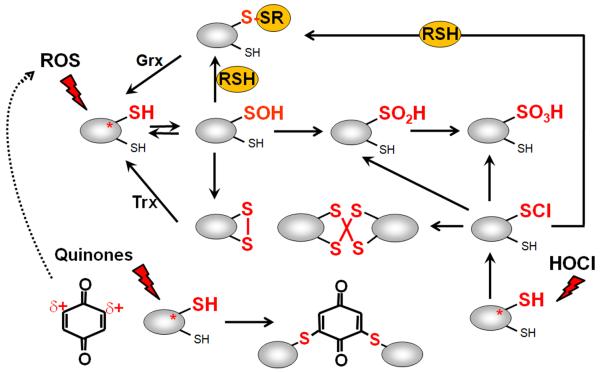
The thiol group of cysteine is the main target for ROS, RES and HOCl and subject to reversible and irreversible post-translational thiol-modifications. The thiol group can be reversibly oxidized to protein disulfides or irreversibly overoxidized to sulfinic or sulfonic acids by ROS or S-alkylated by RES (Antelmann & Helmann, 2011). ROS lead first to oxidation of protein thiols to Cys sulfenic acids (R-SOH) that rapidly react further to form intramolecular, intermolecular disulfides or mixed disulfides with LMW thiols (termed as S-thiolations) (Figure 1). Hypochloric acid (HOCl) is a strong two-electron oxidant and chlorinating agent which targets the sulfur-containing amino acids cysteine and methionine with the second-order rate constants of k=3×10⁷ M−1s−1 (Hawkins et al, 2003). HOCl first chlorinates the thiol group to form the unstable sulfenylchloride intermediate that reacts further with another thiol group to form disulfides. In the absence of another Cys residue, the chlorinated thiol group is overoxidized very rapidly to sulfinic or sulfonic acids (Gray et al, 2013a; Hawkins et al, 2003) (Figure 1). RES like quinones can react with Cys thiols via thiol-disulfide switches or thiol-(S)-alkylation. During the incomplete one-electron reduction of quinones the highly reactive semiquinone radical is produced that leads to subsequent reduction of O2 and the production of O2•−. The electrophilic reaction of quinones involves the 1,4-reductive Michael-type addition of thiols to quinones (Marnett et al, 2003). Toxic quinones lead to irreversible thiol-S-alkylation and protein aggregation to deplete protein thiols in the proteome in vivo (Liebeke et al, 2008). However, non-toxic quinones cause disulfide formation in RES-sensing redox regulators, such as YodB or NemR to up-regulate quinone detoxification pathways (Chi et al, 2010a; Gray et al, 2013b; Lee et al, 2013).
Figure 1. Thiol-chemistry of ROS, RES and HOCl with redox-sensing regulators.
Reversible thiol-oxidation by ROS leads first to a Cys sulfenic acid intermediate (R-SOH) that is unstable and reacts further to form intramolecular and intermolecular disulfides or mixed disulfides with LMW thiols, such as glutathione, bacillithiol, cysteine or CoASH, termed as S-thiolations. The Cys sulfenic acid can be also overoxidized to Cys sulfinic and sulfonic acids. Reactive electrophiles (RES) such as quinones have been shown to act via the S-alkylation and oxidation mode with quinone-sensing redox regulators. HOCl causes first chlorination of Cys thiol goups to the unstable sulfenylchloride which react further to form protein disulfides and S-thiolations in the presence of proximal thiols. In the absence of another thiol the sulfenylchloride rapidly forms irreversible Cys sulfinic or sulfonic acids (Hawkins et al, 2003).
3. Thiol-based redox sensors for ROS, RES and HOCl in bacteria
3.1. OxyR as thiol-based redox sensor for peroxides and NO in E. coli and Actinomycetes
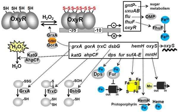
OxyR is a redox-sensor for peroxides and NO in Salmonella typhimurium and Escherichia coli and was the first discovered redox-sensitive regulator that is activated by a thiol-disulfide switch model (Storz et al, 1990b; Zheng et al, 1998). OxyR belongs to the LysR family of transcription factors that acts both as transcriptional activator of peroxide detoxification pathways and repressor of its own transcription and binds as tetramer to operator sequences (Figure 2, Table 1). Gisela Storz has shown that OxyR oxidation occurs by H2O2 at the conserved Cys199 that is oxidized to a sulfenic acid and subsequently forms an intramolecular disulfide with Cys208 in each of the four subunits of the OxyR tetramer (Storz et al, 1990b; Zheng et al, 1998). The OxyR tetramer binds to the operator sequences in the reduced and oxidized forms, but the interaction of reduced and oxidized OxyR with the DNA is different (Toledano et al, 1994). Oxidized OxyR recognizes a motif comprised of four ATAGnt elements spaced at 10 bp intervals and binds to this motif in four adjacent major grooves on one face of the DNA. Reduced OxyR binds to the DNA at two pairs of adjacent major grooves separated by on helical turn. The two modes of binding are essential for OxyR to function as both an activator and a repressor in vivo (Toledano et al, 1994). Oxidized OxyR induces the cooperative binding of the RNAP to activate transcription (Kullik et al, 1995a; Kullik et al, 1995b).
Figure 2. The thiol-disulfide-switch model of E. coli OxyR and functions of the OxyR regulon.
OxyR responds to hydrogen peroxide (H2O2) in E. coli and other bacteria. The conserved C199 and C208 residues of OxyR are essential for redox-sensing of OxyR. C199 is initially oxidized to the sulfenic acid intermediate that rapidly reacts further to form an intramolecular disulfide with C208. Oxidized OxyR binds as a tetramer to promoter regions of target genes and activates transcription of peroxide detoxification genes by contact with αCTD of RNA polymerase. OxyR positively controls genes for peroxide detoxification, such as catalase and peroxiredoxin (katG, ahpCF), Fe-storage miniferritin (dps), glutaredoxin, thioredoxin and glutathione reductase (grxA, trxC, gor), sulfenic acid oxidoreductase (dsbG), ferric uptake regulator (fur), Fe-S-cluster assembly machinery (sufABCDE), ferrochelatase (hemH), manganese import (mntH) and the small RNA (oxyS). OxyR negatively regulates its own expression and that of the genes for the ferric ion reductase (fhuF), the outer membrane protein (flu), the mannonate hydrolase (uxuAB) and gluconate permease (gntP). OxyR is regenerated by the glutaredoxin/GSH/Gor system upon return to non-stress conditions. Examples for OxyR regulon genes and their functions are also listed in Table 1.
Table 1. The LysR-family OxyR-type peroxide-sensing transcriptional regulators.
| Redox sensor | Organism | Signal | Redox-sensing mechanism | Regulon genes | Regulon functions | References |
|---|---|---|---|---|---|---|
| OxyR | Escherichia coli | H2O2 H2O2 NO GSSG |
C199*-C208 Intramolecular disulfide Cys199-SOH Cys199-SNO Cys199-SSG |
ahpCF
katG fur dps mntH sufABCDE trxC grxA gor dsbG hemH oxyR fhuF flu uxuAB gntP |
peroxiredoxin catalase Fe-uptake repressor miniferritin Mn-uptake Fe-S cluster assembly thioredoxin glutaredoxin GSH reductase Cys-SOH reduction ferrochelatase LysR-type regulator Fe 3+ reductase antigen 43 outer membrane protein mannonate hydrolase gluconate permease |
Storz et al, 1990b
Zheng et al, 1998 Choi et al, 2001 Kim et al, 2002 |
| OxyS | Mycobacterium tuberculosis | H2O2 | C25* peroxide sensing | katG | catalase |
Domenech et al, 2001
Li & He, 2012 |
| OxyR | Deinococcus radiodurans | H2O2 | C210*-SOH |
katE
drb0125 feoB dps mntH |
catalase Iron(III) dicitrate- uptake Fe2+ uptake miniferritin Mn uptake |
Chen et al, 2008 |
| OxyR | Corynebacterium glutamicum | H2O2 | C206* and C215 conserved |
katA
dps, ftn cydABCD hemH cg1292 sufABCDE proP oxyR sufR ripA |
catalase miniferritins cytochrome bd oxidase ferrochelatase flavin- monooxygenase Fe-S-cluster assembly proline-ectoine transporter LysR-type regulator FeS-cluster regulator regulator of iron- proteins |
Teramoto et al, 2013
Milse et al, 2014 |
The crystal structures of reduced and oxidized OxyR and thiol-trapping assays confirmed the Cys199-Cys208 disulfide-switch model both in vitro and in vivo. The redox-sensing Cys199 is located in a loop between the α-helix and the β8 strand and is 17Å apart from C208. OxyR oxidation to the Cys199-Cys208 intramolecular disulfide results in unwinding of the α-helix and movement of the α-helix/β8 loop causing large structural changes in the oligomeric interfaces and relative rotation among the OxyR subunits (Barford, 2004; Choi et al, 2001; Lee et al, 2004). Disulfide formation leads to the rearrangement of the N-terminal DNA-binding domains relative to the DNA to facilitate proper DNA binding of oxidized OxyR to the four adjacent major grooves to induce the cooperative interactions with the RNAP required for transcriptional activation of peroxide detoxification genes.
However, this thiol-disulfide switch model was questioned by the group of Jonathan Stamler, since mutational analyses suggested that only Cys199 is required for redox-sensing and transcriptional activation of OxyR. Different post-translational thiol-modifications were introduced at Cys199 of OxyR, including sulfenic acid formation, S-nitrosylation or S-glutathionylation that were sufficient for OxyR activation in vitro (Kim et al, 2002). These different OxyR modifications resulted in different OxyR activation states. The S-glutathionylated OxyR conferred non-cooperative DNA binding while the sulfenic acid and S-nitrosylated forms of OxyR provoke cooperative binding to the DNA.
Interestingly, S-nitrosylation of OxyR occurred specifically under conditions of anaerobic nitrate respiration. Moreover, OxyR controls widespread endogenous protein S-nitrosylation and the expression of a different anaerobic OxyR regulon during nitrate respiration (Seth et al, 2012). The anaerobically OxyR-controlled hcp gene, encoding a hybrid cluster protein, was shown to be specifically activated by S-nitrosylated OxyR. The oxyR mutant showed a growth defect with nitrate and Hcp was required for protection against endogenous nitrosative stress under anaerobic nitrate respiration. This indicates that OxyR can be activated by different post-translational thiol-modifications, including the thiol-disulfide switch under H2O2 stress and S-nitrosylation under anaerobic nitrate respiration to activate distinct regulons for protection against peroxide and nitrosative stress (Seth et al, 2012).
OxyR is conserved in Gram-negative and Gram-positive bacteria and has been studied in Proteobacteria, Bacteroidetes and Actinomycetes (Chiang & Schellhorn, 2012). In many bacteria, OxyR controls catalases and peroxiredoxins while the size of the OxyR regulon varies. In E. coli, OxyR positively regulates genes for the peroxide scavenging peroxiredoxin (ahpCF) and catalase (katG), the iron-uptake regulator (fur), the miniferritin (dps), the Mn-importer (mntH), the FeS cluster assembly machinery (sufABCDE), the ferrochelatase for ferrous ion incorporation into heme (hemH), thioredoxins (trxC), glutaredoxins (grxA), glutathione reductase (gor) and the periplasmic sulfenic acid oxidoreductase (dsbG)(Storz et al, 1990a) (Figure 2, Table 1). OxyR is both an activator and repressor and controls negatively its own transcription and that of the genes for the ferric ion reductase (fhuF), the antigen 43 outer membrane protein (flu), the mannonate hydrolase and oxidoreductase (uxuAB), the gluconate permease (gntP) and some unknown function proteins (Zheng et al, 2001). Oxidized OxyR is reduced by the glutaredoxin (GrxA)/GSH/Gor reducing system upon return to non-stress conditions. The OxyR regulon genes confer peroxide resistance in E. coli, but protect cells also against heat, UV, singlet oxygen, lipid peroxides and neutrophil killing (Chiang & Schellhorn, 2012).
OxyR homologs have been studied in Gram-positive Actinomycetes, such as Mycobacteria and Corynebacteria where they control catalases and peroxiredoxins. Interestingly, in Mycobacterium tuberculosis the catalase KatG activates the anti-tuberculosis pro-drug isoniazid (INH) upon treatment of M. tuberculosis infections (Zhang et al, 1992). However, katG expression is not regulated by OxyR and the oxyR gene has acquired several non-sense mutations and is non-functional in M. tuberculosis (Deretic et al, 1997). These nonsense oxyR mutations are conserved among most Mycobacteria, except for Mycobacterium leprae and Mycobacterium avium which encode functional oxyR genes (Sherman et al, 1995). Expression of katG is regulated by the ferric uptake regulator FurA in M. tuberculosis and M. smegmatis (Milano et al, 2001; Pym et al, 2001; Zahrt et al, 2001). The furA gene is located upstream of katG and the furA-katG operon was induced by oxidative stress in a FurA-dependent manner (Milano et al, 2001). However, in a fast-growing Mycobacterium sp. strain JC1 DSM 3803, katG was induced FurA-independently under oxidative stress (Lee et al, 2010). This peroxide-inducible expression of katG was shown to be controlled by the OxyR-homolog OxyS in M. tuberculosis (Domenech et al, 2001; Li & He, 2012). The oxyR homologous oxyS gene is located in the M. tuberculosis cosmid T919 and is highly conserved among Mycobacteria. OxyS is a repressor of katG transcription and overexpression of OxyS resulted in stronger repression of katG transcription and increased susceptibility to H2O2 stress in M. smegmatis (Domenech et al, 2001; Li & He, 2012). The operator sequence for OxyS binding was identified as GC-rich T-N(11)-A motif within the katG promoter region. OxyS has 4 Cys residues: Cys113, Cys124 and Cys293 are in the LysR-substrate-binding domain and Cys25 is located in the N-terminal DNA-binding domain. Cys25 of mycobacterial OxyS is required for peroxide-sensing which is conserved also among enteric OxyR proteins but absent from OxyR of M. leprae and M. avium (Domenech et al, 2001). The DNA-binding activity of OxyS was inhibited by H2O2 in vitro in gel-shift assays while the OxySC25A mutant did not respond to H2O2 in vitro and in vivo. These results suggest that OxyS regulation involves oxidation of the single Cys25 in the DNA-binding domain under oxidative stress, but the thiol-modification that inactivates OxyS is unknown (Li & He, 2012).
Similar to mycobacterial OxyS, a 1-Cys-type OxyR redox-sensor was characterized in Deinococcus radiodurans that is oxidized at the conserved single Cys210 to a sulfenic acid under peroxide stress. OxyR of Deinococcus radiodurans activates transcription of genes for the catalase (katE), the ferrous iron transporter (feoB) and the iron(III)dicitrate transporter (drb0125), but also operates as repressor of dps and mntH transcription to control antioxidant functions and Mn/Fe ion homeostasis (Chen et al, 2008). This indicates, that also 1-Cys-type OxyR homologs are present in other bacteria, including OxyS of Mycobacteria and OxyR of D. radiodurans that might sense peroxide stress by alternative thiol-modifications similar as has been described for OxyR of E. coli (Kim et al, 2002).
In Corynebacterium glutamicum and Corynebacterium diphtheriae, OxyR is functional as transcriptional repressor of the catalase-encoding gene. In both species, disruption of oxyR led to derepression of the catalase gene that conferred a H2O2 resistance phenotype (Kim & Holmes, 2012; Teramoto et al, 2013). DNaseI-footprinting analyses revealed the OxyR binding region that is ~ 50 bp long with multiple T-N11-A motifs but no sequence similarities, characteristic for operators recognized by LysR-type regulators. Reduced OxyR binds specifically to this operator sequence in different OxyR target gene promoters (Teramoto et al, 2013). However, DNA-binding activity of OxyR was not inhibited after oxidation and even non-specific binding of oxidized OxyR was observed. This suggests that alleviation of OxyR repression by peroxides might be due to decreased strength of its interaction with the DNA. In genome-wide transcriptome analyses, the OxyR regulon of C. glutamicum was characterized after peroxide stress (Milse et al, 2014). OxyR acts as transcriptional repressor and negatively regulates expression of 23 genes that belong to 12 transcriptional units. DNA-binding assays confirmed specific binding of OxyR to the 12 target promoters. In total, the OxyR regulon consists of genes encoding the catalase (katA), two miniferritins that function in iron homeostasis (dps and ftn), cytochrome bd oxidases (cydABCD), the heme biosynthesis enzyme ferrochelatase (hemH), a flavin-monooxygenase (cg1292), the FeS-cluster biosynthesis machinery (suf operon), the proline-ectoine transporter (proP) and several unknown function genes (Table 1) (Milse et al, 2014). In addition, transcriptional regulators were regulated by OxyR, such as oxyR, sufR and ripA. OxyR of C. glutamicum shares with E. coli OxyR the conserved redox-sensing Cys199 and Cys206 residues indicating a similar thiol-disulfide switch model for OxyR of C. glutamicum.
3.2. PerR as Fur-family metal-based peroxide sensor in Firmicutes bacteria
In B. subtilis, the Fur-family protein PerR functions as the main peroxide sensor. PerR is a dimeric repressor that binds to the PerR box (AAGTATTATTTATTATTATTA) as heptameric 7-1-7 inverted repeat in the promoter region of its target genes (Fuangthong & Helmann, 2003). PerR is inactivated by H2O2 stress leading to derepression of the PerR regulon genes. The PerR regulon includes the genes for the peroxiredoxin (ahpCF), the catalase (katA), the miniferritin (mrgA), the heme biosynthesis operon (hemAXCDBL), the iron-uptake repressor (fur), and the Zn uptake system (zosA) (Fuangthong et al, 2002; Helmann et al, 2003) (Figure 3, Table 2). Two overlapping PerR boxes are present in the perR upstream region indicating that PerR is autoregulated (Fuangthong et al, 2002). The derepression of the PerR regulon genes under peroxide stress conditions leads to peroxide resistance as adaptive response in B. subtilis (Faulkner & Helmann, 2011). In S. aureus, the PerR regulon is also peroxide-inducible and includes genes for catalase, peroxiredoxins and bacterioferritin comigratory protein (katA, ahpCF, bcp), two miniferritins (mrgA, ftn) and thioredoxin reductase (trxB) that are required for virulence (Horsburgh et al, 2001). In addition, PerR negatively regulates its own transcription and that of the gene for the ferric uptake regulator (fur). PerR is required for full virulence in a murine skin abscess model of infection. It was further shown that both katA and ahpC have compensatory roles in peroxide resistance and mediate environmental persistence and nasal colonization in S. aureus (Cosgrove et al, 2007).
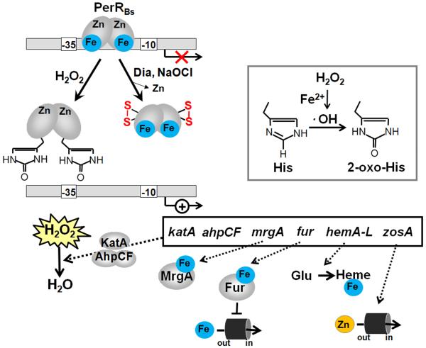
Figure 3. Redox-regulation of B. subtilis PerRBs by peroxides (metal-catalyzed histidine oxidation) and by diamide stress (intramolecular disulfides) and functions of the PerR regulon members.
PerR has a regulatory Fe2+or Mn2+-binding site with Asp and His residues as ligands and a structural Zn2+-binding site coordinated by four cysteine residues. Reaction of PerR-Fe with H2O2 leads to a Fenton reaction generating HO with subsequent oxidation of His37 and His91 to the 2-oxo-His derivatives that inactivate the PerR repressor under H2O2 stress leading to up-regulation of the PerR regulon genes. Under disulfide stress conditions provoked by diamide and NaOCl, PerR is inactivated by intramolecular disulfide formation in the Zn-binding site that also lead to derepression of the PerR regulon genes. The PerR regulon includes genes with antioxidant functions, such as the catalase and peroxiredoxin (katA, ahpCF), Fe-storage miniferritin (mrgA), ferric uptake regulator (fur), heme biosynthesis enzymes (hemAXCDBL) and zinc uptake systems (zosA). Examples for PerR regulon genes of B. subtilis and their functions are also listed in Table 2.
Table 2. The Fur/PerR-family of peroxide-specific redox regulators.
| Redox sensor | Organism | Signal | Redox-sensing mechanism | Regulon genes | Regulon functions | References |
|---|---|---|---|---|---|---|
| PerR | Bacillus subtilis | H2O2 Diamide NaOCl |
2-oxo-His37 2-oxo-His91 His93 conserved Fe-site C96, C99 C136-C139 intramolecular disulfide conserved Zn-site |
ahpCF
katA mrgA hemAXCDBL fur zosA |
peroxiredoxin catalase miniferritin heme biosynthesis Fe-uptake repressor Zn-uptake |
Fuangthong et al, 2002
Lee & Helmann, 2006 Chi et al, 2011 |
| PerR | Staphylococcus aureus | H2O2 | His43, His97, His99 conserved Fe-site C102,C105 C142,C145 conserved Zn-site |
ahpCF
katA mrgA ftn bcp trxA fur perR |
peroxiredoxin catalase miniferritin ferritin bacterioferritin comigrating protein thioredoxin Fe-uptake repressor Peroxide repressor |
Horsburgh et al, 2001 |
| PerR | Clostridium acetobutylicum | O2 H2O2 |
His32, His86, His88 conserved Fe-site C91,C94, C131,C134 conserved Zn-site |
rbr3A, rbr3B
bcp tpx bsa2 grx CAC2452 dfx fprA2 rb nror ofrAB gapN |
reverse ruberythrins bacterioferritin comigrating protein thiol peroxidase glutathione peroxidase glutaredoxin flavodoxin desulfoferredoxin flavodiiron protein rubredoxin NADH-rubredoxin oxidoreductase 2-oxoglutarate ferredoxin oxidoreductase NADP-dep. GapDH |
Hillmann et al, 2009a
Hillmann et al, 2009b Riebe et al, 2009 |
The regulatory mechanism for peroxide-sensing of PerR has been shown by the Helmann group in B. subtilis (Lee & Helmann, 2006). PerR contains two metal binding sites, a structural Zn2+ binding site coordinated by four cysteine residues (Cys96, Cys 99, Cys 136, Cys139) in the C-terminal domain and a regulatory Fe2+ or Mn2+ binding site with three histidine and two aspartic acid residues as ligands (Lee & Helmann, 2006). Both Mn2+ and Fe2+ bind competitively to the PerR regulatory site, but only iron-bound PerR is sensitive to metal-catalyzed oxidation (Mongkolsuk & Helmann, 2002). Exposure to H2O2 leads to oxidation of Fe2+ in the regulatory site by a Fenton reaction generating HO• which causes oxidation of His37 and His91 to 2-oxo-histidine and inactivation of PerR (Figure 3)(Duarte & Latour, 2010; Lee & Helmann, 2006; Traore et al, 2009). Although the 2-oxo-His37 still had affinity for the regulatory metal, no metal binding with 2-oxo-His91 was possible and PerR fails to retain the close conformation for DNA binding (Duarte & Latour, 2010; Traore et al, 2009). Thus, in contrast to OxyR which is activated by a thiol-disulfide redox-switch, the PerR transcription factor senses peroxide stress by metal-catalyzed histidine oxidation. However, the PerR regulon genes are also induced under disulfide conditions, such as diamide and hypochlorite in B. subtilis (Antelmann et al, 2008; Chi et al, 2011). Thus, the response of PerR to disulfide stress could involve thiol-redox switches in the structural Zn site of PerR leading to inactivation of its repressor function (Figure 3). In support of this thiol-based mechanism, an intramolecular C136-C139 disulfide in the Zn binding site of PerR was identified by mass spectrometry in hypochlorite-stressed B. subtilis cells in vivo (Chi et al, 2011). In addition, a thiol-redox switch was identified as peroxide-sensing mechanism of the Fur-family PerR homolog of Streptomyces coelicolor CatR that controls expression of the catalase gene catA (Hahn et al, 2000a; Hahn et al, 2000b). In CatR two CXXC motifs are present that coordinate Zn in the reduced state. In response to peroxide stress, intramolecular disulfides are formed in the Zn site of CatR that inactivate CatR’s repressor function resulting in derepression of catA. Thus, it is likely that PerR homologs respond also via thiol-based mechanisms under certain disulfide stress conditions that are different from peroxide stress.
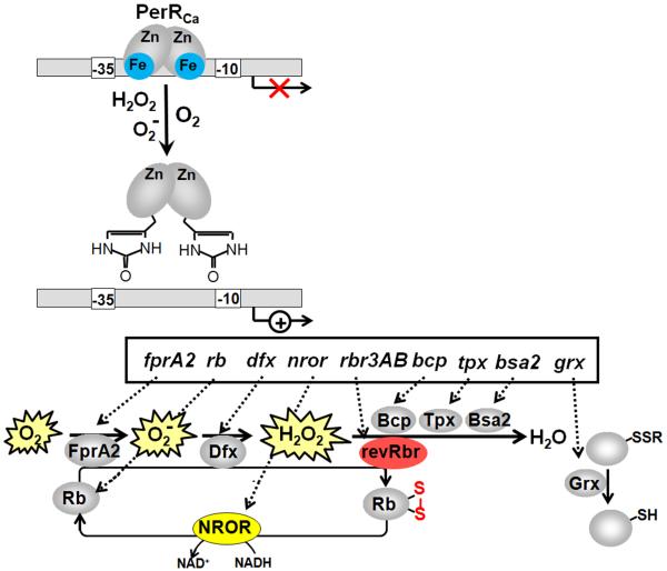
The PerR-homolog that senses peroxides and O2 has been identified in the strict anaerobic Gram-positive bacterium Clostridium acetobutylicum as defense mechanism against O2 toxicity (Hillmann et al, 2008). PerR inactivation conferred aerotolerance to C. acetobutylicum, enabled aerobic growth and O2 consumption and conferred resistance to H2O2. The PerR regulon includes genes for the reverse ruberythrins as major peroxidases for H2O2 reduction (rbr3A, rbr3B), the peroxiredoxin (bcp), the thiol peroxidase (tpx), the glutathione peroxidase (bsa2), the glutaredoxin (grx), the flavodoxin (CAC2452), the superoxide-reducing desulfoferrodoxin (dfx), the oxygen-reducing flavodiiron proteins (fprA2), rubredoxins (rd) as intermediates to regenerate the reductases FprA2, Dfx and revRbr, the NADH-dependent rubredoxin oxidoreductase (nror) to provide electrons for rubredoxin reduction, the 2-oxoglutarate ferredoxin oxidoreductase (ofrAB) and the NADPH-dependent non-phosphorylating GapDH (gapN) (Hillmann et al, 2009a; Hillmann et al, 2009b; Riebe et al, 2009)(Figure 4, Table 2). These PerR-regulon genes are all induced under O2 and peroxide stress and function collectively as anaerobic O2 and ROS detoxification pathways to promote the survival of the strict anaerobe C. acetobutylicum under short-time microaerophilic conditions.
Figure 4. Proposed redox-regulation of PerRCa in the strict anaerobe Clostridium acetobutylicum by oxygen, peroxides, superoxide and functions of the PerR regulon members.
The PerR repressor is proposed to sense O2, O2− and H2O2 by oxidation of two His residues in the conserved regulatory Fe-binding site leading to 2-oxo-His generation. This causes PerR inactivation and derepression of the PerR regulon genes. The PerR regulon controls genes for an anaerobic oxygen and ROS detoxification pathway, including the oxygen-reducing flavodiiron proteins (fprA2), reverse rubrerythrins and peroxidases (rbr3A, rbr3B), peroxiredoxin (bcp), thiol peroxidase (tpx), glutathione peroxidase (bsa2), glutaredoxin (grx), superoxide-reducing desulfoferrodoxin (dfx), rubredoxin (rd) and the NADH-dependent rubredoxin oxidoreductase (nror). Examples for PerR regulon genes of C. acetobutylicum and their functions are also listed in Table 2.
3.3. The MarR/OhrR-family regulators as sensors of organic hydroperoxides
3.3.1. The MarR/OhrR-family regulators in B. subtilis and X. campestris
MarR or Multiple antibiotics resistance-type regulators are characterized by winged helix-turn-helix (HTH) DNA binding motifs and control genes that confer resistance to antibiotics, organic solvents, detergents, ROS and RES. Several MarR-family members are important for the regulation of virulence (Ellison & Miller, 2006). Among the MarR-family regulators, the MarR/OhrR subfamily responds to organic hydroperoxides (OHP). OHP can be derived from peroxidation of unsaturated fatty acids of eukaryotic membrane lipids. Ohr-like peroxiredoxins catalyze the reduction of OHPs to their corresponding alcohols (Atichartpongkul et al, 2001; Fuangthong et al, 2001). B. subtilis has two ohr paralogs: ohrA and ohrB (Fuangthong et al, 2001). The ohrA gene is regulated by the redox-sensing OhrR repressor and ohrB is controlled by the σB alternative sigma factor in B. subtilis (Volker et al, 1998).
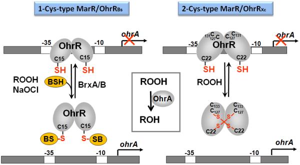
The OhrR repressor acts as a dimeric repressor that binds to inverted repeat sequences in the ohrA promoter, thereby inhibiting transcription (Fuangthong et al, 2001). OhrR harbors a conserved redox-sensing Cys residue in its N-terminal region that senses OHPs via different redox-switch mechanisms. Thiol-oxidation of OhrR results in dissociation of the protein from the operator and derepression of ohrA transcription. Based on the number of Cys residues, the OhrR family can be divided into two subfamilies: the one-Cys-type with the prototype of B. subtilis OhrRBs (Lee et al, 2007) and the two-Cys-type with the example of X. campestris OhrRXc (Figure 5, Table 3) (Antelmann & Helmann, 2011; Panmanee et al, 2006). In the two-Cys type OhrRxc, Cys22 is oxidized by OHPs to a sulfenic acid intermediate, which reacts further with Cys127 in the opposing subunit forming an intersubunit disulfide (Panmanee et al, 2006). Oxidation inactivates OhrRxc and releases the protein from the promoter DNA. X-ray crystallography reveals that disulfide formation causes a large rotation of the DNA binding domain that is not compatible with DNA-binding (Antelmann & Helmann, 2011; Newberry et al, 2007). In contrast, the one-Cys type OhrRBs of B. subtilis is oxidized at Cys15 to the sulfenic acid that reacts further to a mixed disulfide with BSH (S-bacillithiolated OhrR) in response to OHPs (Lee et al, 2007) (Figure 5). B. subtilis OhrRBs can be also converted from a one-Cys-type to a two-Cys regulator by introduction of a C-terminal Cys at a position equivalent to Cys127 of OhrRxc(Soonsanga et al, 2008).
Figure 5. Thiol-based redox sensing of organic hydroperoxides by 1-Cys and 2-Cys-type MarR/OhrR regulators.
OhrR controls the OhrA thiol-dependent peroxiredoxin and contains one conserved Cys15 residue in B. subtilis and three Cys residues (C22, C127 and C131) in X. campestris. The 1-Cys OhrR protein of B. subtilis is initially oxidized by CHP and NaOCl to the Cys sulfenic acid, which reacts further with bacillithiol (BSH) to form S-bacillithiolated OhrR. The 2-Cys OhrR protein of X. campestris is regulated by intersubunit disulfide formation between the redox-sensing C22 and C127′ of opposing subunits. These different thiol-disulfide switches inactivate OhrR proteins leading to derepression of the peroxidase OhrA that functions in detoxification of organic hydroperoxides. Regeneration of S-bacillithiolated OhrR involves the bacilliredoxins BrxA and BrxB in B. subtilis (Gaballa et al, 2014).
Table 3. The MarR-type redox-regulators of the OhrR/SarA/DUF24-subfamilies.
| Redox sensor | Organism | Signal | Redox-sensing mechanism | Regulon genes | Regulon functions | References |
|---|---|---|---|---|---|---|
| OhrR | Bacillus subtilis | ROOH NaOCl |
C15*-SSB | ohrA | 2-Cys-peroxiredoxin |
Fuangthong et al, 2001
Lee et al, 2007 Chi et al, 2011 |
| OhrR | Xanthomonas campestris | ROOH | C22*-C127 Intermolecular disulfide |
ohrA | 2-Cys-peroxiredoxin |
Panmanee et al, 2006
Newberry et al, 2007 |
| MgrA (OhrR) | Staphylococcus aureus | H2O2 ROOH | C12*-SOH C12*-Phosphate |
cap5(8) hlgABC, pvl lukED, lukMF-PV hla coa spa splABCDEF nuc lytM, lytN norA, norB tetAB agr, lytRS, arlRS, sarS and sarV |
capsule polysaccharides leukotoxins α-hemolysin coagulase, protein A serine proteases nuclease autolysis factors multidrug efflux pumps virulence regulators |
Luong et al, 2006
Chen et al, 2006 Chen et al, 2009 Sun et al, 2012 |
| SarZ (OhrR) | Staphylococcus aureus | H2O2 ROOH |
C13*-SOH C13*-SSR C13*-Phosphate |
ohr
acs, pflAB pckA argGH, ilvD, lysC, hisC fabG gntRK, lacD, malA, treC, isdC, epiEF, lrgB, efb, fib, tcaA norB, tet38 nuc |
2-Cys peroxiredoxin pyruvate metabolism amino acid metabolism fatty acid synthesis sugar metabolism cell surface proteins drug efflux pumps exonuclease |
Ballal et al, 2009
Chen et al, 2009 Poor et al, 2009 Sun et al, 2012 |
| SarA (MarR) | Staphylococcus aureus | H2O2 diamide |
C9* redox- sensitive C9*-Phosphate |
sodA trxB hla spa fnb cna sec icaRA, bap ssp, aur rot, agr, sarS, sarV, sarT |
superoxide dismutase thioredoxin reductase α-hemolysin protein A fibronectin-binding collagen-binding enterotoxin C biofilm formation proteases virulence regulators |
Ballal & Manna, 2009
Ballal & Manna, 2010 Sun et al, 2012 |
| MosR (OhrR) | Mycobacterium tuberculosis | H2O2 ROOH |
C10-C12 intramolecular disulfide |
rv1050
mosR |
exported oxidoreductase MarR/OhrR-like repressor |
Brugarolas et al, 2012 |
| RosR (MarR) | Corynebacterium glutamicum | H2O2 | C92* redox sensitive |
rosR narKGHJI cg2329 cg3085 cg1848 cg3084 cg1150 cg1850 cg1426 cg1322 |
MarR-type repressor nitrate/nitrite transporter luciferase-like monooxygenases flavin-containing monooxygenases FMN reductases glutathione-S- transferases polyisoprenoid-binding protein |
Bussmann et al, 2010 |
| HypR (MarR/DUF24) | Bacillus subtilis | Quinone Diamide NaOCl |
C14*-C49 intermolecular disulfide |
hypR
hypO |
MarR/DUF24-activator FMN-nitroreductase |
Palm et al, 2012 |
| YodB (MarR/DUF24) | Bacillus subtilis | Quinone Diamide NaOCl |
C6*-C101 intermolecular disulfide |
yodB
yodC azoR1 catDE |
MarR/DUF24-repressor nitroreductase azoreductase dioxygenase |
Leelakriangsak et al, 2008
Chi et al, 2010a Chi et al, 2010b |
| CatR (MarR/DUF24) | Bacillus subtilis | Quinone Diamide NaOCl |
C7*redox- sensitive |
catDE | dioxygenase | Chi et al, 2010b |
| QsrR (MarR/DUF24) | Staphylococcus aureus | Quinone | C5*-S-alkylation |
SAV0340
(azoR1) SAV2033 (yodC) SAV0338 SAV2522 (catE) |
FMN-dependent quinone reductase nitroreductase glyoxalase dioxygenase |
Ji et al, 2013 |
| QorR (MarR/DUF24) | Corynebacterium glutamicum | Quinone Diamide |
C17*-C17 Intermolecular disulfide |
qorA | quinone reductase | Ehira et al, 2009a |
We could show that OhrR responds also to NaOCl stress and thus, OhrR is a redox sensor for OHPs and NaOCl (Chi et al, 2011). Transcriptome analysis revealed that the ohrA gene was the most strongly up-regulated gene (220-fold) under NaOCl stress in B. subtilis. Mass spectrometry identified the S-bacillithiolation of OhrR as thiol-redox switch mechanism for OhrR inactivation. Phenotype analyses showed that the OhrA peroxiredoxin and BSH protect cells against hypochlorite stress since the growth of ohrA and bshA mutants was strongly impaired by NaOCl stress (Chi et al, 2011). Thus, we hypothesize that OhrA could be involved in NaOCl detoxification. Since hypochlorite is produced by activated neutrophils, it could be also a more physiologically oxidant for the OhrR-homologs SarZ and MgrA in the human pathogen S. aureus.
The B. subtilis OhrR repressor is redox-controlled by S-bacillithiolation which was recently shown to be reversed by bacilliredoxins (BrxA/B) which function as glutaredoxin-like enzymes in the reduction of BSH mixed protein disulfides to regenerate and re-activate the OhrR repressor in B. subtilis in vitro (Gaballa et al, 2014).
3.3.2. The MgrA/SarZ/SarA-family of virulence and antibiotic regulators
In S. aureus, two homologs of the MarR/OhrR 1-Cys-type repressor are present, including the MgrA and SarZ global regulators for antibiotic resistance and virulence (Figure 6, Table 3) (Ballal et al, 2009; Chen et al, 2009; Kaito et al, 2006; Poor et al, 2009; Truong-Bolduc et al, 2008; Truong-Bolduc et al, 2005). The Multiple gene regulator MgrA of S. aureus regulates more than 300 genes that are involved in virulence, autolysis, antibiotic resistance, biofilm formation and cell wall biosynthesis (Luong et al, 2006). MgrA controls genes for virulence factors that include enzymes for capsule polysaccharide biosynthesis (cap5(8)-locus), α-toxin (hla), coagulase (coa), protein A (spa), extracellular serine proteases (splABCDEF) and nuclease (nuc). In addition, genes encoding autolysis factors (lytM and lytN), multidrug efflux pumps (norA, norB and tetAB) and regulatory genes (agr, lytRS, arlRS, sarS and sarV) are members of the MgrA regulon (Ingavale et al, 2005; Kaatz et al, 2005; Luong et al, 2006; Truong-Bolduc et al, 2008; Truong-Bolduc et al, 2005) (Table 3). Hence, MgrA confers resistance to the antibiotics fluoroquinone, tetracycline, vancomycin and penicillin. MgrA is also required for virulence in murine abscess, septic arthritis and sepsis models. MgrA shares with OhrRBs the single conserved Cys12 and uses a thiol-based oxidation sensing mechanism to control virulence and antibiotic resistance (Chen et al, 2006). Cys12 can be oxidized by CHP, H2O2 and superoxide anion to Cys-SOH that leads to dissociation of MgrA from the operator DNA in vitro and induction of antibiotic resistance in S. aureus in vivo (Chen et al, 2006; Chen et al, 2009). However, it was shown recently that the DNA-binding activity of MgrA can be also reversibly regulated by cysteine phosphorylation via the eukaryotic-like serine/threonine kinase (Stk1) and phosphatase (Stp1)(Sun et al, 2012). Cys phosphorylation was detected as post-translational modification also in other regulatory proteins, including the MarR-family proteins SarZ and SarA and the cysteine biosynthesis regulator CymR. Moreover, Stk1 was required for full virulence and resistance to the antibiotic vancomycin by controlling Cys-phosporylation of MgrA, SarZ and SarA. Increased Cys phosphorylation of the virulence regulators in an stp1 mutant led to decreased virulence in a mouse abscess model. Interestingly, like the previously shown thiol-oxidation mechanism (Chen et al, 2006; Chen et al, 2009) also Cys phosphorylation was DTT-reversible, but the mechanism is still unknown (Sun et al, 2012).
Figure 6. Redox-sensing by the MarR/OhrR-family regulator SarZ of S. aureus.
In S. aureus, the MarR/OhrR-family regulator SarZ functions as global regulator for ROS detoxification, antibiotic resistance and virulence functions and contains a single Cys13 required for redox-sensing. The DNA-binding activity of SarZ was shown to be reversibly redox-regulated by S-thiolation with a synthetic benzene thiol (Poor et al, 2009) and by cysteine phosphorylation via the eukaryotic-like serine/threonine kinase (Stk1) and phosphatase (Stp1)(Sun et al, 2012). SarZ controls genes for the ohr peroxiredoxin, cell surface proteins, antibiotic resistance efflux pumps (norB, tet38), amino acid, sugar, fatty acid and anaerobic metabolism (pflAB). Examples for SarZ regulon genes are listed in Table 3.
The second MarR/OhrR-type regulator of S. aureus is SarZ which is also a pleiotropic virulence regulator (Kaito et al, 2006). SarZ controls the ohr peroxiredoxin, genes involved in virulence, autolysis, cell wall metabolism, antibiotic resistance, intermediary, amino acid, sugar, fatty acid and anaerobic metabolism, such as the pyruvate-formate lyase genes pflA and pflB (Figure 6, Table 3). SarZ is also transcriptionally activated by MgrA (Ballal et al, 2009; Chen et al, 2009). SarZ was shown to use a thiol-based oxidation sensing mechanism via the conserved lone Cys13 residue (Chen et al, 2009). The crystal structure of SarZ was resolved in the reduced, sulfenic acid and mixed disulfide form (Poor et al, 2009). SarZ is oxidized at Cys13 to sulfenic acid that still retains DNA binding activity. Further oxidation of SarZ with an external synthetic thiol (benzene thiol) leads to S-thiolated SarZ. These mixed SarZ disulfides cause steric clashes that contribute to an allosteric conformational change of the DNA-binding domains and release of SarZ from the operator DNA (Poor et al, 2009).
SarZ is also controlled by Cys-phosphorylation via the Stk1/Stp1 kinase/phosphatase pair (Sun et al, 2012). However, it remains yet to be shown if S-bacillithiolation can control DNA-binding activity of SarZ or MgrA in vivo also in S. aureus. Future studies should elucidate if there is a possible cross-talk between Cys phosphorylation and thiol-oxidation in these MarR-family virulence regulators of S. aureus.
The Staphylococcal accessory regulator (SarA) is another global redox-sensing regulator of the MarR-family that contains a single Cys9 residue in the dimer interface. SarA positively regulates many virulence factors including fibronectin and fibrinogen binding proteins (fnb), hemolysins (hla), enterotoxins (sec), toxic shock syndrome toxin 1, and genes involved in biofilm formation (icaRA, bap)(Cheung et al, 2008; Tamber & Cheung, 2009). SarA negatively regulates the transcription of proteases (ssp, aur), protein A (spa), and collagen-binding proteins (cna). Many virulence regulators are members of the SarA-regulon of S. aureus, such as rot, agr, sarS, sarV, sarT resulting in pleiotropic phenotypes of the sarA mutant. Furthermore, SarA was shown to control oxidative stress-related genes, such as superoxide dismutase (sodA) and thioredoxin reductase (trxB) (Ballal & Manna, 2009; Ballal & Manna, 2010) (Table 3). The redox-sensitivity of Cys9 in SarA has been analyzed in the wild type and a SarAC9G mutant in vivo and in vitro which revealed that oxidation of Cys9 by H2O2 and diamide reduced the DNA-binding activity of SarA to the trxB promoter (Ballal & Manna, 2010). SarA has been also shown to sense oxidative stress by Cys phosphorylation in vitro (Sun et al, 2012). However, the detailed thiol-switch mechanism for SarA redox regulation in vivo has yet to be elucidated.
3.3.3. The MarR/OhrR-family regulators MosR and RosR in Actinomycetes
The MarR/OhrR-family of redox regulators is also conserved among Actinomycetes. In M. tuberculosis the OhrR-family regulator MosR has been characterized as transcriptional repressor and sensor for peroxides that shares 28% sequence identity with B. subtilis OhrR and S. aureus MgrA (Brugarolas et al, 2012). Reduced MosR binds to a specific operator sequence (GTGTAnnTACAC) in its target promoters and represses its own transcription and that of the adjacent rv1050 gene, encoding an exported oxidoreductase of unknown function (Brugarolas et al, 2012). The rv1050 gene was most strongly induced by H2O2 and 352-fold derepressed in the mosR mutant. Rv1050 gene was also up-regulated during infection in INF-γ-activated macrophages suggesting a role in the host-immune defense (Schnappinger et al, 2003). In addition, arachidonic acid and linoleic acid were found to induce rv1050 indicating that Rv1050 could also function in fatty acid metabolism in macrophages.
MosR contains four Cys residues (Cys10, 12, 96, 147), but only Cys12 is conserved. The Cys10 and Cys12 residues are oxidized to intramolecular disulfides by peroxides and both Cys residues are essential for redox-sensing since C10S and C12S mutant proteins were non-responsive to H2O2 in gel-shift assays (Brugarolas et al, 2012). The structures of reduced and oxidized MosR proteins were resolved to reveal the structural mechanism for the inactivation of MosR’s repressor function upon oxidation. Disulfide formation between Cys10 and Cys12 breaks the hydrogen bond of Cys12 to Asn37′ and causes new hydrogen bonds of Arg-16 and Ser-41′. This rearrangement of hydrogen bonds results in a movement of α2 which pushes α3 ~4.5 Å toward α4. This causes rotation of α4 and α4′ that prevent them to fit into consecutive major grooves resulting in the release from the operator DNA. Consequently, MosR oxidation leads to rearrangements of hydrogen bonds resulting in large conformational changes and MosR dissociation from the DNA (Brugarolas et al, 2012).
Corynebacterium glutamicum encodes the redox-sensitive MarR-type repressor RosR that responds to peroxide stress (Bussmann et al, 2010). RosR controls positively expression of the narKGHJI operon encoding a nitrate/nitrite transporter and the dissimilatory nitrate reductase complex. RosR acts as repressor of its own transcription and represses several genes that encode luciferase-like monooxygenases (cg1848, cg2329, cg3085), flavin-containing monooxygenases (cg3084), FMN reductases (cg1150, cg1850), glutathione-S-transferases (cg1426) and a polyisoprenoid-binding protein (cg1322). The polyisoprenoid-binding protein was important under peroxide stress since the mutant showed increased H2O2 sensitivity (Bussmann et al, 2010). Reduced RosR binds to an 18-bp inverted repeat with the consensus sequence TTGTTGAYRYRTCAACWA in its target promoters. The DNA binding activity of RosR was inhibited by H2O2 and restored by DTT in gel-shift assays in vitro. RosR contains three Cys residues (Cys64, 92, 151), but only Cys92 is conserved among RosR homologs of other Corynebacteria. Cys92 was most important for redox-sensing since the DNA binding activity of the C92S mutant was not inhibited by H2O2 in vitro. In contrast, other single Cys mutants behaved like the wild type RosR, although in double and triple Cys mutants the DNA binding activity was also affected in response to H2O2 in vitro (Bussmann et al, 2010). This suggests that the RosR might be inactivated by formation of inter- or intramolecular disulfides by peroxide stress which remains to be demonstrated.
3.4. The MarR/DUF24-family regulators as sensors for RES (quinone, diamide)
3.4.1. The MarR/DUF24-family regulators YodB, CatR, HypR and HxlR of B.subtilis
The MarR/DUF24 family of transcription factors is conserved among Gram-positive bacteria (Antelmann & Helmann, 2011). In C. glutamicum, the MarR/DUF24-type QorR was first characterized as a transcriptional repressor that senses diamide and H2O2 and controls the quinone oxidoreductase QorA (Ehira et al, 2009a). Inactivation of QorR involves intersubunit disulfide formation between the conserved single Cys17 residues of both subunits (Ehira et al, 2009a). Bacillus subtilis encodes eight MarR/DUF24-family proteins: HxlR, HypR, YodB, CatR, YdeP, YdzF, YkvN, and YtcD. HxlR was identified as activator of the formaldehyde-inducible hxlAB operon that encodes enzymes of the ribulose monophosphate pathway (Yurimoto et al, 2005). HypR was characterized as positive regulator of the nitroreductase HypO that is induced by NaOCl, diamide and quinones and confers NaOCl-resistance (Palm et al, 2012) (Table 3). HypR is a two-Cys MarR/DUF24-type regulator with a redox-sensing Cys14 and a second Cys49 that are 8Å apart in the reduced HypR structure. Both Cys14 and Cys49 are essential for activation of hypO transcription by disulfide stress. HypR is activated by Cys14-Cys49′ intersubunit disulfide disulfide formation under diamide and NaOCl stress. Disulfide bond formation breaks the H-bonds of Cys14 and moves the α4 and α4′ helices of HypR ~4 Å towards each other (Palm et al, 2012).
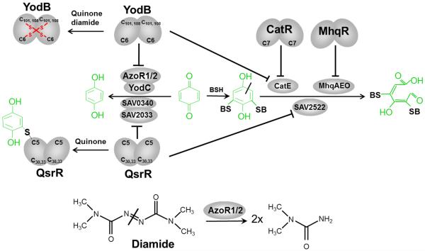
The MarR-type regulators YodB, CatR and MhqR control specific detoxification pathways that confer resistance to quinones and diamide, such as azoreductases (AzoR1 and AzoR2), nitroreductases (YodC and MhqN), and thiol-dependent dioxygenases (CatE, MhqA, MhqE, MhqO)(Antelmann et al, 2008; Chi et al, 2010b; Leelakriangsak et al, 2008; Towe et al, 2007). Azoreductases and nitroreductases reduce quinones and diamide to hydroquinones and dimethylurea, respectively (Figure 7, Table 3). Dioxygenases catalyse the ring-cleavage reaction of quinone-S-adducts. The azoreductase AzoR1 is controlled by YodB and expression of the catechol-2,3-dioxygenase CatE and oxidoreductase CatD are regulated by both YodB and CatR (Chi et al, 2010b; Leelakriangsak et al, 2008). The promoter region of the catDE operon contains two inverted repeat sequences overlapping the −35 promoter region (BS1) and the transcription start point (BS2) that are the operator sites for CatR and YodB. Both YodB and CatR are inactivated in response to quinone and diamide. YodB is inactivated by a two-Cys-type redox-switch mechanism and oxidized to intersubunit disulfides between Cys6 of one subunit and Cys101 or Cys108 of the other subunit by diamide and quinones in vivo and in vitro (Chi et al, 2010a). The conserved Cys7 is essential for redox-sensing of quinones and diamide in CatR, but its redox-sensing mechanism has yet to be explored.
Figure 7. Redox-sensing of RES by the MarR/DUF24-regulators YodB in B. subtilis and QsrR in S. aureus.
Exposure of B. subtilis to quinones induces quinone detoxification regulons controlled by the MarR-type repressors MhqR, YodB and CatR. In S. aureus, the homologous quinone-sensing YodB (QsrR) repressor responds to quinones and controls paralogous quinone reductases, dioxygenases and nitroreductases. The redox-sensing YodB and CatR repressors are inactivated by the oxidative mode of quinone leading to disulfide formation that involves the conserved Cys6 or Cys7 residues (Antelmann et al, 2008; Antelmann & Helmann, 2011; Chi et al, 2010a; Chi et al, 2010b; Towe et al, 2007). In S. aureus, YodB (QsrR) with mutated C-terminal Cys residues senses quinones by thiol-S-alkylation of Cys5 leading to up-regulation of the dioxygenase SAV2522, the quinone reductase SAV0340 and the nitroreductase SAV2033 (Ji et al, 2013). The thiol-dependent dioxygenases MhqA, MhqE, MhqO, CatE of B. subtilis and SAV2522 of S. aureus are involved in specific ring-cleavage of quinones-S-adducts. The quinone reductases AzoR1, AzoR2 of B. subtilis and SAV0340 of S. aureus and the nitroreductases YodC, MhqN of B. subtilis and SAV2033 of S. aureus catalyze the reduction of quinones to redox stable hydroquinones.
3.4.2. The MarR/DUF24-family regulator QsrR of S. aureus
The redox-sensing mechanism of the quinone-sensing MarR/DUF24-family regulator YodB (QsrR) has been characterized also in S. aureus which shares 38% sequence identity with the YodB repressor of B. subtilis (Ji et al, 2013). QsrR contains the conserved N-terminal Cys5 and two further Cys30 and Cys33 residues. QsrR and YodB control both homologous genes involved in quinone reduction and ring-cleavage that confer resistance to benzoquinone. The QsrR regulon includes genes for the FMN-dependent quinone reductase (SAV0340 or azoR1), the nitroreductase (SAV2033 or yodC), the glyoxalase (SAV0338) and the thiol-dependent dioxygenase (SAV2522 or catE). Hence, QsrR controls homologous quinone reductases and dioxygenases in S. aureus that are controlled by YodB and CatR in B. subtilis (Figure 7, Table 3). The quinone resistance QsrR regulon conferred resistance to killing by macrophages in a phagocytosis assay indicating its crucial role for virulence regulation in S. aureus. The conserved Cys5 of QsrR was shown to sense quinones by a thiol-S-alkylation mechanism. The QsrR structure shares strong similarities with the HypR structure of B. subtilis (Ji et al, 2013; Palm et al, 2012). HypR and QsrR are both dimers that have in common the wHTH motif composed of α3, α4, β1 and β2. The wHTH motifs bind to the major and minor grooves of the DNA double helix and the wing is much larger compared to OhrR-like regulators. To elucidate the structural changes upon quinone binding at Cys5, the menadione-bound QsrR structure was resolved for the QsrRC30,33S mutant (Ji et al, 2013). Menadione-binding at Cys5 causes a shift in the distance and rotation between the α4 and α4′ helices from 29.9 Å distance with 106° rotation in reduced QsrR to 39.1 Å distance and 117° rotation in the menadione-bound form. These structural changes lead to dissociation of QsrR from the operator DNA. In contrast to QsrR, YodB and HypR sense diamide and quinones by intersubunit disulfide bond formation (Palm et al, 2012). However, the in vivo mechanism of quinone sensing by wild type QsrR has yet to be explored.
3.5. Emerging thiol-based redox sensors for RES (quinones, aldehydes) and HOCl
3.5.1 The TetR-family regulator NemR as redox-sensor for RES (N-ethylmaleimide, quinones, aldehydes) and HOCl
The thiol-based TetR-family NemR regulator is conserved across Gram-negative and Gram-positive bacteria and has been characterized in E. coli as redox sensor for RES and HOCl (Gray et al, 2013b; Lee et al, 2013; Ozyamak et al, 2013) (Table 4). NemR negatively controls transcription of the nemRA operon and the gloA gene which function in detoxification of electrophiles. The nemA gene was previously shown to be strongly induced by thiol-alkylating compounds, such as N-ethylmaleimide (NEM) and iodoacetamide and shown to function as NEM reductase (Umezawa et al, 2008). The FMN-dependent reductase NemA belongs to the old-yellow-enzyme family and has a broad substrate spectrum to reduce several quinones (ubiquinone, menaquinone) and aldehydes, (glyoxal, methylglyoxal and glycolaldehyde) in vitro (Lee et al, 2013). GloA is the glyoxalase-I enzyme involved in methylglyoxal detoxification and was revealed as main methylglyoxal protection mechanism (MacLean et al, 1998). Hence, the NemR repressor responds to quinones and aldehydes, like methylglyoxal and is inactivated via thiol-based redox switches which lead to upregulation of the nemRA operon and gloA that both confer resistance to methylglyoxal and quinones in E. coli (Lee et al, 2013; Ozyamak et al, 2013). Moreover, the nemRA operon and gloA were most strongly induced by methylglyoxal in a transcriptome analyses supporting the major role of GloA and NemA as protection mechanism (Ozyamak et al, 2013). However, NemR was also shown to sense reactive chlorine species, such as HOCl and N-chlorotaurine which leads to derepression of nemRA and gloA that both confer HOCl resistance since the nemA and gloA mutants displayed HOCl sensitive phenotypes (Gray et al, 2013b). Exposure of cells to HOCl stress caused increased methylglyoxal production suggesting that detoxification of methylglyoxal is an important bacterial HOCl defense mechanism. It remains to be shown if NemA could also confer HOCl resistance by reduction of reactive chlorines as direct substrates.
Table 4. The NemR, RclR and HypT redox sensors for RES and HOCl in E. coli.
| Redox sensor | Organism | Signal | Redox-sensing mechanism | Regulon genes | Regulon functions | References |
|---|---|---|---|---|---|---|
| NemR (TetR-type) | Escherichia coli | Quinones, Glyoxal, Methylglyoxal N-ethylmaleimide Iodoacetamide HOCl |
C106* conserved C21-C116 intersubunit disulfide |
nemR
nemA gloA |
TetR-type repressor FMN-dep. reductase for aldehydes, quinones and NEM glyoxalase-I |
Umezawa et al, 2008 Gray et al, 2013 Lee et al, 2013 Ozyamak et al, 2013 |
| RcIR (AraC-ytpe) | Escherichia coli | HOCl N-chlorotaurine |
C21-C89 Intramolecular disulfide |
rclA
rclB rclC |
flavoprotein disulfide reductase, periplasmic protein quinone-binding membrane protein |
Parker et al, 2013 |
| HypT (LysR-type) | Escherichia coli | HOCl | Met123-SO Met206-SO Met230-SO C4-C4 intersubunit disulfide (in vitro) C4: HypT dodecamer formation C150: HypT stability |
metB/K/N cysH/K/N, cysPUW, sbp, sufA entC, entH, fecABCDE, fecR, fepCD, ryhB, tonB, yncE |
sulfur, Cys and Met biosynthesis and metabolism Fur-regulon genes involved in iron homeostasis |
Gebendorfer et al, 2012
Drazic et al, 2013a Drazic et al, 2013b |
NemR was shown to sense reactive electrophiles and HOCl via thiol-based oxidation mechanisms that involve intersubunit disulfides which lead to inactivation of its repressor function. NemR possesses 6 Cys residues, but only Cys106 is conserved among bacteria. However, the C106S mutant still responds to HOCl and forms intersubunit disulfides both in vitro and in vivo suggesting that other Cys residues are involved in redox-regulation of NemR which can substitute for the absence of the conserved Cys106 (Gray et al, 2013b). In another study, the glyoxal sensitivity and effect on nemRA expression was analyzed of various NemR Cys double mutants revealing significant lower responsiveness to glyoxal in the C21S, C116S double mutant both in vivo and in vitro, but no difference to the wild type in any other Cys single and double mutant (Ozyamak et al, 2013). Cys21 is located in the DNA-binding domain and Cys116 is in the dimer interface. Both Cys21 and Cys116 were involved in intersubunit disulfide formation and oligomerization of NemR. This indicates that E. coli NemR is inactivated by intersubunit disulfide formation in response to RES and HOCl to upregulate quinone and glyoxal detoxification enzymes. This thiol-disulfide switch model of NemR redox regulation resembles that of other quinone-sensing redox regulators, such as QorR, YodB and HypR suggesting that the alternative thiol-S-alkylation as shown for QsrR in vitro might be rather the exception for thiol-based quinone-sensing regulators.
3.5.2. RclR as AraC-family HOCl-specific thiol-based redox-sensor in E. coli
Recently, novel HOCl-specific redox regulators have been discovered in E. coli that are specific for chlorine species, such as HOCl but do not respond to ROS, electrophiles or other thiol-reactive compounds. RclR (formerly YkgD) is widely conserved among Gram-negative bacteria and Actinobacteria and was characterized as redox-sensing transcriptional activator of the AraC family, which uses a thiol-based oxidation mechanism for redox-sensing of HOCl (Parker et al, 2013). The redox-sensing mechanism of RclR involves both conserved Cys residues, Cys21 and Cys89 which likely form an intramolecular disulfide, which stabilizes the active RclR protein in vivo. Both Cys-21 and Cys-89 residues are required for redox-sensing of the HOCl-response in vivo, while only Cys21 is essential for redox-sensing in vitro. Oxidation of RclR by HOCl leads to specific activation of transcription of the rclABC operon which is important for survival of HOCl and N-chlorotaurine. Mutants in each single gene of the rclABC operon are sensitive to HOCl suggesting that this operon is an important HOCl protection determinant (Parker et al, 2013). However, the functions of the RclABC proteins for HOCl protection are still unknown which resemble a flavoprotein disulfide reductase, periplasmic protein and possible quinone-binding membrane protein (Table 4).
3.5.3. HypT as LysR-family HOCl-specific Met-oxidation switch in E. coli
The LysR-type regulator HypT has been discovered as another HOCl-specific redox sensor and transcriptional activator of E. coli (Gebendorfer et al, 2012). HypT belongs like OxyR to the LysR-family of transcriptional regulators which often form dimers or tetramers (OxyR) (Maddocks & Oyston, 2008), but HypT was shown to form unusual large dodecameric ring-like structures in vitro that serve as a storage form of HypT (Drazic et al, 2014). These HypT dodecamers dissociate into smaller oligomers (dimers, tetramers) in the presence of DNA in vitro and tetramers are also found in vivo in HOCl-exposed E. coli cells. Specifically, the HypT tetramers were revealed as activation-competent DNA-binding species of HypT (Drazic et al, 2014; Gebendorfer et al, 2012).
The DNA-binding activity of HypT was activated specifically by HOCl and HypT was required for the survival under HOCl stress conditions in vivo (Gebendorfer et al, 2012). HypT was shown to control positively genes that function in sulfur, Cys and Met biosynthesis (metB, metK, metN, cysH, cysK, cysN, cysPUW, sbp, sufA) while genes of the Fur-regulon are negatively regulated by HypT that function in iron homeostasis (entC, entH, fecABCDE, fecR, fepCD, ryhB, tonB, yncE) (Table 4). Since Met is rapidly oxidized by HOCl to methionine sulfoxide (Met-SO), it is suggested that HypT activates Met biosynthesis to restore the pool of reduced Met (Gebendorfer et al, 2012). Interestingly, HypT uses a reversible methionine-oxidation switch model for transcriptional activation (Drazic et al, 2013a) while the Cys residues are important for stability and oligomerization of HypT (Drazic et al, 2013b). HypT activation involves oxidation of three Met residues (Met123, 206, 230) to their Met-SO forms which were identified in HOCl-activated HypT in vitro. Mutations of the three Met to glutamines mimics the oxidized MetSO form of HypT and resulted in constitutively active HypT in vivo, while the Met-to-Ile mutation resulted in inactive HypT as revealed by the transcriptional studies of the target genes and HOCl survival assays (Drazic et al, 2013a). Furthermore, inactivation of oxidized HypT required the MetSO reductases MsrA and MsrB both in vivo and in vitro, revealing the reversibility of this Met oxidation switch model for HypT.
Surprisingly, HypT was only activated in vivo in E. coli cells by HOCl stress, but in vitro HypT rapidly lost its DNA-binding activity when treated with HOCl (Drazic et al, 2013b). HypT possesses five non-conserved Cys residues and all Cys residues are required for HypT activity in vivo and HypT stability in vitro. HypT oxidization by HOCl in vitro leads to intermolecular Cys4-Cys4 disulfides resulting in HypT inactivation. Furthermore, Cys150 was required for HypT stability and Cys4 involved in oligomerization of HypT to dodecamers (Drazic et al, 2013b). The thiol-switch model of HypT was suggested as check point in the activation of HypT preventing unwanted HypT interaction with its target promoters under oxidative stress conditions that are not sufficient to activate HypT. In conclusion, the HOCl-specific regulator HypT represents an important HOCl-protection mechanism and is activated by a reversible Met-oxidation switch to up-regulate Met biosynthesis in E. coli. It will be interesting to explore if other bacteria also use Met-oxidation switches as defense mechanisms against HOCl or ROS.
3.6. The Spx disulfide stress redox-sensors in Gram-positive Firmicutes bacteria
3.6.1. SpxA and MgsR as paralogous thiol-redox sensors for disulfide and general stress conditions in B. subtilis
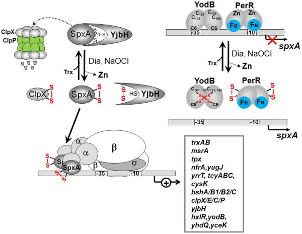
The thiol-based redox sensor SpxA is an unusual transcription factor without typical DNA-binding domains that responds to different redox stress conditions in Gram-positive bacteria (Antelmann & Helmann, 2011; Nakano et al, 2005; Zuber, 2004; Zuber, 2009). SpxA is an arsenate reductase (ArsC) family protein with a CXXC redox switch motif in its N-terminus that is essential for redox sensing and transcriptional activation. ROS, RES and HOCl lead to oxidation of the CXXC motif to an intramolecular disulfide to activate SpxA (Figure 8). Oxidized SpxA interacts with the C-terminal domain (CTD) of the α subunit of the RNA polymerase (RNAP) to recognize promoter regions of the SpxA regulon genes and thereby activates transcription (Nakano et al, 2005; Nakano et al, 2003; Zuber, 2004; Zuber, 2009). SpxA positively regulates the expression of genes that maintain the thiol redox balance in B. subtilis, including the genes for thioredoxin/thioredoxin reductases (trxAB), thiol peroxidase (tpx), FMN-dependent oxidoreductases (nfrA, yugJ), methionine sulfoxide reductase (msrA), cysteine biosynthesis and cystine transporters (yrrT operon, cysK, tcyABC) and bacillithiol biosynthesis (bshA, bshB1, bshB2 and bshC) (Antelmann & Helmann, 2011; Gaballa et al, 2013; Zuber, 2009) (Table 5). Genome-wide chromatin immunoprecipitation (ChIP-chip) analysis of RNAP-SpxA complexes revealed 275 genes that are directly controlled by SpxA. An extended −35 box was identified within SpxA-controlled promoters in which the −43/−44 positions correlated with the activation by SpxA (Rochat et al, 2012). Additional targets for SpxA control were identified among the Clp machinery as ATPase subunits (clpX, clpE and clpC) and the proteolytic subunit (clpP) and the adpaptor for ClpXP proteolysis (yjbH). Furthermore, SpxA controls thiol-based redox regulators (hxlR, yodB, yhdQ, yceK) and many other transcription factors that respond to diamide and SpxA was required for the basal level of 32 genes under non-stress conditions (Rochat et al, 2012) (Table 5).
Figure 8. Post-translational and transcriptional control of SpxA by disulfide stress in B. subtilis.
Under non-stressed conditions, SpxA is unstable and targeted by the YjbH adaptor protein to the ClpXP machinery for proteolytic degradation. The stability of SpxA is increased by diamide due to oxidation of SpxA, YjbH and ClpX that prevents SpxA degradation. SpxA is oxidized in its redox-active CXXC motif and binds to the αCTD of RNAP resulting in transcriptional activation of the SpxA disulfide stress regulon. Transcription of spxA is regulated by the repressors YodB and PerR that are oxidized and inactivated under diamide stress leading to increased spxA transcription. SpxA controls a large regulon of genes encoding thioredoxin/thioredoxin reductase (trxAB), thiol peroxidase (tpx), FMN-dependent oxidoreductases (nfrA, yugJ), methionine sulfoxide reductase (msrA), cysteine biosynthesis enzymes (yrrT-operon, cysK, tcyABC), bacillithiol biosynthesis enzymes (bshA, bshB1, bshB2 and bshC), the protein quality control Clp machinery (clpX, clpE, clpC, clpP, yjbH) and several thiol-based redox regulators (hxlR, yodB, yhdQ, yceK). Examples for SpxA regulon genes and their functions are listed in Table 5.
Table 5. The Spx disulfide stress-sensing redox-regulators of Firmicutes bacteria.
| Redox sensor | Organism | Signal | Redox-sensing mechanism | Regulon genes | Regulon functions | References |
|---|---|---|---|---|---|---|
| SpxA | Bacillus subtilis | Quinone Diamide NaOCl H2O2 |
C10*-C13 Intramolecular disulfide |
trxAB
tpx nfrA, yugJ msrA yrrT, cysK tcyABC bshA,bshB1, bshB2, bshC clpX, clp, clpC clpP yjbH hxlR,yodB yhdQ, yceK |
thioredoxin/thioredoxin reductases thiol peroxidase FMN-dependent oxidoreductases methionine sulfoxide reductase cysteine biosynthesis cystine transporter bacillithiol biosynthesis ATPase subunit of Clp protease subunit of Clp adpaptor for ClpXP thiol-redox regulators |
Nakano et al, 2003; Nakano et al, 2005; Zuber, 2004 Zuber, 2009 Rochat et al, 2012 |
| MgsR | Bacillus subtilis | Heat NaCl Ethanol |
C10*-C13 Intramolecular disulfide |
ydbD
ykuV ydaD, yhdF, yhxC,yhxD |
Mn-catalase thiol-disulfide oxidoreductase short-chain oxidoreductases |
Reder et al, 2008
Reder et al, 2012b |
| Spx | Staphylococcus aureus | Diamide H2O2 |
C10*-C13 conserved |
trxAB icaABCD trfA |
thioredoxin/thioredoxin reductases biofilm formation β-lactam resistance |
Pamp et al, 2006
Wang et al, 2010 Jousselin et al, 2013 |
| SpxAl/SpxA2 | Bacillus anthracis | Diamide H2O2 |
C10*-C13 conserved |
trxAB, ytpP
BA1263 BA0774 bshA,bshB1 bshB2, bshC ypdA yphP, ytxJ uvrC, uvrD exoA BA1951 BA0838, BA2647, BA3438 yjbH |
thioredoxin/thioredoxin reductase CoASH disulfide reductases (CoADR) bacillithiol biosynthesis NADPH-disulfide oxidoreductase bacilliredoxins DNA damage repair spore outgrowth and germination putative oxidoreductase alcohol, aldehyde, quinone dehydrogenases adpaptor for ClpXP |
Barendt et al, 2013 |
| SpxA/SpxB SpxAl/SpxA2 |
Streptococcus mutants Streptococcus suis |
Diamide H2O2 |
C10*-C13 conserved |
trxB
dpr sod ahpCF gor nox tpx |
thioredoxin reductase peroxide resistance superoxide dismutase peroxiredoxin glutathione reductase NADH oxidase thiol peroxidase |
Kajfasz et al, 2010
Zheng et al, 2014 |
Expression of SpxA is controlled at transcriptional and post-translational levels. Transcription of spxA is initiated from at least four different promoters that are recognized by three forms of RNAP containing σA, σB and σM (Leelakriangsak & Zuber, 2007). SpxA is transcriptionally regulated in response to disulfide stress provoked by diamide and NaOCl by the PerR and YodB repressors. Both PerR and YodB repressors are oxidized under disulfide stress conditions which inactivates their repressor function leading to derepression of spxA transcription (Zuber, 2009). The YodB repressor is oxidized to intermolecular disulfides between C6 and one of the C-terminal Cys residues leading the spxA, azoR1 and yodC derepression. PerR is oxidized to form intramolecular disulfides in the Zn-binding structural site that possibly lead to PerR inactivation and spxA transcription by diamide and NaOCl. The second level is the post-translational control of SpxA protein stability by proteolysis via the ClpXP proteases. The spxA gene was discovered as Suppressor of clpP and clpX mutations and it was shown that the absence of ClpXP leads to stabilization of SpxA responsible for the pleiotropic phenotypes of clpXP mutants (Nakano et al, 2001). Under non-stress condition, SpxA is targeted to the ClpXP system with the help of the adaptor YjbH for rapid degradation of SpxA (Larsson et al, 2007; Nakano et al, 2002). The YjbH adaptor protein contains a His-Cys rich N-terminal region that is oxidized by diamide resulting in loss of YjbH adaptor activity and SpxA stabilization. However, mutational analyses showed that the Cys and His residues of YjbH are not required for SpxA stability in vivo (Chan et al, 2012). Studies with the YjbHGt ortholog of Geobacillus thermodenitrificans without the Cys-His redox motif revealed that the C-terminus of YjbHGt is required for stabilization of SpxA (Chan et al, 2012). The ATPase ClpX contains a Cys-rich Zn finger motif that also functions as redox switch and is oxidized under disulfide stress conditions (Zhang & Zuber, 2007). Therefore, SpxA of B. subtilis is controlled at multiple levels, at the transcriptional level by PerR and YodB and at post-translational levels via three redox switches in SpxA itself, YjbH and ClpX that together lead to up-regulation, stabilization and activation of SpxA (Antelmann & Helmann, 2011; Zuber, 2009) (Figure 8). The mechanism how the SpxA-RNAP complex activates transcription is unknown, but a cis-acting element in the −10 promoter sequences of trxB and trxA was identified required for activation of transcription (Nakano et al, 2010; Reyes & Zuber, 2008).
While SpxA interacts with RNAP containing σA, a SpxA paralog MgsR was identified as member of the σB general stress regulon (Reder et al, 2008). Thus, B. subtilis contains Spx paralogs that interact with RNAP holoenzymes containing different sigma factors. MgsR is a Modulator of the σB general stress response and controls a sub-regulon within the σB regulon that functions in protection against secondary oxidative stress caused by ethanol, heat, salt stress (Reder et al, 2008). MgsR controls positively about 50 genes including 18 σB-dependent genes that were up-regulated in the mgsR mutant. Antioxidant and protective functions can be attributed to the genes encoding the manganese-containing catalase (ydbD), the thiol-disulfide oxidoreductase (ykuV) and paralogous short-chain oxidoreductases (ydaD, yhdF, yhxC and yhxD) that are postulated to function in NADPH production as electron source for cellular Trx/TrxR reducing systems (Reder et al, 2008) (Table 5). The mechanism of MgsR control resembles in part that of SpxA and involves a positive autoregulatory loop to increase mgsR transcription, and post-translational control of MgsR stability via ClpXP and ClpCP proteolysis (Reder et al, 2012b). In addition, ethanol stress leads to a redox switch and causes intramolecular disulfides in the CXXC motif of MgsR as activation mechanism. However, the detailed mechanism of MgsR interaction with the RNAP containing σB and activation of transcription remains to be elucidated.
The σB general stress regulon is induced after exposure to heat, salt and ethanol stress which causes non-specific resistance to secondary oxidative stress in B. subtilis (Mols & Abee, 2011; Reder et al, 2012b). The involvement of σB in the protection against secondary oxidative stress has been demonstrated by the role of the σB-dependent miniferritin Dps in stationary-phase-induced peroxide resistance (Antelmann et al, 1997). Moreover, several genes of the primary PerR-, OhrR- and SpxA-controlled oxidative stress response (katA, mrgA, ohrA, spxA) have paralogs within the σB regulon (katE, katX, ydbD, dps, ohrB, mgsR) conferring non-specific secondary oxidative stress resistance (Zuber, 2009). Other redox-stress related genes like trxA are controlled by both, SpxA and σB or by specific regulators, such as clpC that is controlled by σB and CtsR (Zuber, 2009). Moreover, phenotype screening of 94 mutants in σB-controlled genes identified 62 mutants with increased sensitivity towards paraquat or peroxides (Reder et al, 2012a). Thus, B. subtilis employs PerR, OhrR and SpxA as specific antioxidant control mechanisms to cope with ROS and MgsR and σB for protection against primary and secondary generated oxidative stress (Mols & Abee, 2011; Reder et al, 2012b).
3.6.2. Spx as thiol-redox sensor for oxidative stress in the pathogens Bacillus anthracis, S. aureus and Streptococcus
Spx homologs are highly conserved in low GC Gram-positive bacteria, such as Bacillus, Staphylococcus, Streptococcus, Lactobacillus and Listeria species where they play important functions in the oxidative stress resistance and virulence (Kajfasz et al, 2010; Pamp et al, 2006; Wang et al, 2010; Zuber, 2004). In S. aureus, Spx functions as global regulator of genes that maintain the thiol-redox balance, such as the Trx/TrxR system. The spx mutant displays growth defects and is hypersensitive to peroxide and disulfide stress, heat shock and osmotic stress (Pamp et al, 2006; Wang et al, 2010). Spx controls biofilm development in S. aureus and S. epidermidis through control of the icaABCD operon. The post-translational proteolytic control of Spx by the ClpXP system is similar like in B. subtilis, shown by the increased stability of Spx in S. aureus clpP and clpX mutants (Pamp et al, 2006). In S. aureus, YjbH functions as adaptor for ClpXP-mediated proteolysis of Spx although there is only 30% sequence identity between YjbH homologs of S. aureus and B. subtilis (Engman et al, 2012). The Cys-His rich N-terminal domain is not conserved in S. aureus YjbH and instead a CxC motif is present at another location. The B. subtilis yjbH mutant could be complemented with S. aureus yjbH to restore the Spx level and diamide susceptability to that of wild type cells (Engman et al, 2012; Gohring et al, 2011). Hence YjbH controls Spx proteolysis like in B. subtilis. The yjbH mutant further shows growth-impaired phenotypes and increased pigmentation (Engman et al, 2012), resistance to oxacillin and other β-lactam antibiotics, glycopeptides and sensitivity to desiccation stress (Chaibenjawong & Foster, 2011; Charbonnier et al, 2005; Gohring et al, 2011). This higher resistance to β-lactam antibiotics might be caused by the higher PBP4 level and increased peptidoglycan cross-linking in yjbH mutants, but the Cys residues were not required for the antibiotic resistant phenotype (Gohring et al, 2011). This indicates that YjbH regulates Spx proteolysis and antibiotic resistance mechanisms S. aureus. Spx also controls trfA, the B. subtilis mecA homolog and trfA mutants are sensitive to oxacillin and glycopeptide antibiotics (Jousselin et al, 2013). Expression of trfA was constitutively up-regulated in glycopeptide-intermediate S. aureus (GISA) derivatives of methicillin-susceptible or methicillin-resistant S. aureus (MRSA) clinical or laboratory isolates (Jousselin et al, 2013). This indicates that the up-regulation of trfA in the yjbH mutant is responsible for the β-lactam antibiotic resistance. In summary, S. aureus YjbH regulates Spx proteolysis and the Spx-dependent trfA confers antibiotic resistance S. aureus. The role of YjbH for virulence was shown in Listeria monocytogenes where yjbH and clpX mutants had hypohemolytic phenotypes indicating that YjbH and ClpX are required for the function of the pore-forming toxin listeriolysin as virulence factor (Zemansky et al, 2009).
Bacillus anthracis encodes two spx paralogs, spxA1 and spxA2. Mutants lacking spxA1 displayed increased peroxide sensitivity but only the spxA1spxA2 double mutant was hypersensitive to diamide stress suggesting overlapping roles of SpxA1 and SpxA2 in disulfide stress resistance (Barendt et al, 2013). Microarray analyses identified many genes involved in the thiol-redox homeostasis that were up-regulated when stabilized protease-resistant forms of SpxA1DD or SpxA2DD were produced. These SpxA1 and SpxA2-controlled disulfide-sress related genes encode for thioredoxins and thioredoxin reductases (trxA, trxB, ytpP), two CoASH disulfide reductases (BA1263, BA0774) to keep CoASH in its reduced state and genes for bacillithiol biosynthesis (bshA, bshB1, bshB2, bshC), bacilliredoxins (ytxJ, yphP) and the putative BSH reductase (ypdA) (Table 5). In addition, the Spx paralogs control genes involved in DNA damage repair (uvrC, uvrD), spore outgrowth and germination (exoA), detoxification of alcohols, aldehydes, and quinones (BA0838, BA2647, BA3438), unknown oxidoreductase functions (BA1951) and adapter for ClpXP proteolysis (yjbH) (Barendt et al, 2013). Furthermore, both Spx paralogs control also their own subregulon. Interestingly, spxA2 was shown to be upregulated in phagocytosis assays with infected macrophages (Bergman et al, 2007). The expression of spxA2 is negatively controlled by the Rrf2 family regulator SaiR that is conserved among the Bacillus cereus group. The Rrf2-family regulators include also IscR and NsrR with 3 conserved Cys residues that coordinate an FeS-cluster. SaiR shares C89 and C96 with IscR and NsrR and C96 was required for SaiR repressor activity and redox-regulation of spxA2. Repression of spxA2 is alleviated under NaOCl stress and in infected macrophages probably by thiol-oxidation of SaiR leading to its dissociation from the spxA2 promoter (Nakano et al, 2014). However, the role of the Cys residues and the possible involvements as ligands are still unknown in the regulation of SaiR.
Streptococci also encode two spx paralogs: spxA and spxB in Streptococcus mutans (Kajfasz et al, 2010); spxA1 and spxA2 in Streptococcus pneumoniae (Turlan et al, 2009), Streptococcus suis (Zheng et al, 2014) and Streptococcus sanguinis (Chen et al, 2012). These Spx paralogs share the conserved redox-sensing CXXC motif and also the Gly residue required for Spx-RNAP interaction. However, only SpxA and SpxA1 contain the RPI motif involved in modulation of the reactivity of the CXXC motif. In SpxB and SpxA2, the arginine is replaced by a serine residue and this may affect the sensory function (Chen et al, 2012; Kajfasz et al, 2010; Newberry et al, 2005; Turlan et al, 2009; Zheng et al, 2014). These Spx proteins are involved in the oxidative stress response and in the virulence of streptococci as demonstrated by the use of murine, rabbit or insect infection models (Chen et al, 2012; Kajfasz et al, 2010; Zheng et al, 2014). Both Spx paralogs control the expression of genes for the thioredoxin reductase (trxB), the peroxide resistance protein (dpr), the superoxide dismutase (sod), the peroxiredoxin (ahpCF), the glutathione reductase (gor), the NADH oxidase (nox) and thiol peroxidase (tpx) in S. mutans and S. suis (Kajfasz et al, 2010; Zheng et al, 2014) (Table 5). In S. sanguinis, SpxA1 regulates the genes for the pyruvate oxidase (spxB) and the NADH oxidase (nox) that are both responsible for the high production of H2O2 (Chen et al, 2012). Thus, the spxA1 mutant is more sensitive to H2O2 but produces also lower amounts of cytoplasmic H2O2 as defense mechanism against bacterial competitors.
3.7. ECF sigma factors and their cognate redox-sensitive zinc-associated anti-sigma factors (RsrA/SigR in Streptomyces; RshA/SigH in C. glutamicum and M. tuberculosis)
In Actinomycetes, the disulfide stress response is controlled by ECF sigma factors and their redox-sensitive cognate zinc-containing anti sigma factors (ZAS) that share a conserved HX(3)CX(2)C (HCC) motif (Jung et al, 2011). In addition, the region K(33)FEHH(37)FEEC(41)SPC(44)LEK(47) that includes the conserved HCC motif was identified as redox-sensitive determinant in ZAS factors. In another model, alternative Zn binding sites were identified in redox-sensitive ZAS factors which might increase the susceptibility of zinc-coordinating cysteine residues to oxidation (Heo et al, 2013). Redox-sensitive ZAS factors that are involved in the disulfide stress response include RsrA of S. coelicolor and its homologs RshA of M. tuberculosis and C. glutamicum. RsrA of S. coelicolor is the best studied ZAS factor that coordinates zinc and sequesters its cognate sigma factor SigR under reducing conditions forming a RsrA/SigR complex (Paget et al, 2001). Three of the seven cysteines in RsrA (C11, C41 and C44) are essential for anti-sigma factor activity and redox sensing (Kang et al, 1999) (Cherney et al, 2003; Zdanowski et al, 2006). Upon disulfide stress, the redox-sensitive Cys11 and Cys44 form an intramolecular disulfide resulting in zinc release and conformational changes in RsrA that lead to free SigR (Bae et al, 2004; Kang et al, 1999) (Figure 9). Free SigR interacts with the RNAP to direct transcription of the SigR disulfide stress regulon.
Figure 9. Redox regulation of the ZAS factor RsrA and its cognate sigma factor SigR in S. coelicolor and role of the SigR regulon.
RsrA is a redox-sensitive zinc-binding anti sigma (ZAS) factor in S. coelicolor that sequesters its cognate sigma factor SigR under reducing conditions. Diamide stress leads to intramolecular disulfide formation in RsrA, resulting in Zn release and relief of SigR. Free SigR activates transcription of the SigR regulon that functions to restore the thiol-redox balance. The SigR regulon includes genes for thioredoxins and thioredoxin reductase (trxAB, trxC), enzymes for mycothiol biosynthesis and recycling (mshA, mca, mtr), mycoredoxins (mrxA, mrxB), methionine sulfoxide reductase (msrA, msrB), protein quality control machinery (pepN, ssrA, clpP1P2, clpX, clpC, lon), ubiquitin-like protein-conjugation pathway and proteasomal components (pup, mpa, pafD, prcAB), Fe-S assembly components (sufA, sufU) and Fe-S containing enzymes (nadA, lipA), biosynthesis enzymes for the cofactors Fe-S, folate, CoASH and lipoic acid (moeB, coaE, folE, lipA). Examples for SigR regulon genes and their functions are also listed in Table 6.
The SigR regulon consists of more than 100 genes as revealed by DNA microarrays and genome-wide ChIP-chip analyses (Kim et al, 2012). The majority of SigR-controlled genes function in the thiol-redox homeostasis and encode for thioredoxins and thioredoxin reductase (trxAB, trxC), enzymes for mycothiol biosynthesis and recycling (mshA, mca, mtr), putative glutaredoxin-like mycoredoxins (mrxA, mrxB) and methionine sulfoxide reductase (msrA, msrB) (Newton & Fahey, 2008; Paget et al, 1998; Paget et al, 2001; Park & Roe, 2008). Targets for SigR control are also involved in protein quality control (pepN, ssrA, clpP1P2, clpX, clpC, lon), in the prokaryotic ubiquitin-like protein-conjugation and proteasomal degradation pathway (pup, pafD, mpa, prcAB), guanine biosynthesis (guaB, guaB1) and ribosome-associated functions (rpmE, rpmI, rplT, relA, engA, obgE, era, truB, rbfA, infA, infB) (Kallifidas et al, 2010; Kim et al, 2012). The SigR-regulon includes further genes that encode Fe-S assembly components (sufA, sufU), Fe-S containing enzymes (nadA, lipA), biosynthesis enzymes for redox-sensitive sulphur-containing cofactors, such as Fe-S, folate, CoASH and lipoic acid (moeB, coaE, folE, lipA), UV damage repair enzymes (uvrA) and the major housekeeping sigma factor (hrdB) (Figure 9, Table 6). This core SigR regulon and the SigR consensus sequence are conserved across 42 actinomycetes revealing an robust adapatation strategy to oxidative and disulfide stress conditions in different natural environments (Kim et al, 2012).
Table 6. The RsrA/RshA zinc-associated anti sigma factors (ZAS) for disulfide stress.
| Redox sensor | Organism | Signal | Redox-sensing mechanism | Regulon genes | Regulon functions | References |
|---|---|---|---|---|---|---|
| RsrA (ZAS of SigR) | Streptomyces coelicolor | Diamide RES | C11, C41,C44 redox sensitive, Zn-site of ZAS |
sigR-rsrA trxAB, trxC mshA, mca mtr mrxA, mrxB msrA,msrB pepN, ssrA, clpP1P2, clpX, clpC, lon pup, mpa, pafD, prcAB guaB, guaB1 rpmE, rpmI, rplT, relA, engA, obgE, era, truB, rbfA, infA, infB sufA, sufU nadA, lipA moeB, coaE, folE, lipA hrdB uvrA, uvrD |
RsrA/SigR thioredoxin/thioredoxin reductase mycothiol biosynthesis and recycling MSH reductase mycoredoxins methionine sulfoxide reductase protein quality control ubiquitin-like protein- conjugation GMP biosynthesis ribosome-associated functions Fe-S assembly machinery Fe-S enzymes cofactor synthesis (Fe-S, folate, CoASH, lipoic acid) housekeeping sigma factor UV damage repair |
Paget et al, 1998
Paget et al, 2001 Park & Roe, 2008 Kim et al, 2012 |
| RshA (ZAS of SigH) | Corynebacterium glutamicum | Diamide NaOCl | C23, C53,C56 redox sensitive, Zn-site of ZAS |
sigH-rshA
trxB, trxB1, trxC mshC,mca mtr cg2838, cg2661 msrA, msrB pup cg0378 cg1397 sigM, hspR, cglR, sufR, whcA, whcE uvrA, uvrD3 |
RshA/SigH thioredoxin/thioredoxin reductase mycothiol biosynthesis and recycling MSH reductase dsbA-like thiol-disulfide oxidoreductases methionine sulfoxide reductases ubiquitin-like protein- conjugation phage-associated tRNA-(5- methylaminomethyl-2- thiouridylate)- methyltransferase stress-responsive regulators UV damage repair |
Ehira et al, 2009b
Busche et al, 2012 Toyoda et al, 2014 |
MSH and TrxA were shown to reduce oxidized RsrA, allowing to switch-off the SigR-dependent stress response upon return to non-stress conditions (Kang et al, 1999) (Park & Roe, 2008). SigR and an unstable SigR′′ protein with an N-terminal extension are positively autoregulated under diamide stress (Kim et al, 2009). This unstable SigR′ protein is rapidly degraded by the induced ClpP1/P2 proteases which represents another negative feedback loop.
The ECF-sigma factor SigH of M. tuberculosis, an ortholog of SigR, plays an important role in the survival against oxidative stress. SigH is activated upon entry of M. tuberculosis into macrophages and has a key role in the response to oxidative stress since sigH mutants of M. tuberculosis and M. smegmatis are highly sensitive to peroxide stress (Fernandes et al, 1999; Manganelli et al, 2002). SigH is sequestered by its cognate redox-sensitive ZAS factor RshA under reduced conditions. Oxidative stress leads to RshA oxidation and release of SigH to activate transcription of its regulon (Song et al, 2003). SigH controls transcription of approximately 30 genes that include the Trx system, but also other ECF sigma factors (SigE and SigB) (Manganelli et al, 2002). Despite the presence of a ZAS motif, RshA has weak affinity for zinc and the cysteines were shown to coordinate an Fe-S cluster. However, neither the cysteines nor the Fe-S cluster in RshA were essential for RshA/SigH interaction, which is mediated by salt bridge suggesting another regulatory mechanism (Kumar et al, 2012). The serine-threonine kinase PknB that is essential for survival within the host, was shown to phosphorylate both SigH and RshA. Phosphorylation of RshA results in disruption of its interaction with SigH leading to increased activity of SigH (Park et al, 2008). These observations suggest an important role of PknB in mycobacterial adaptation to oxidative stress by activating SigH via RshA phosporylation.
SigE is another important ECF sigma factor required for M. tuberculosis survival under oxidative stress and within infected macrophages (Manganelli et al, 2004a; Manganelli et al, 2004b; Manganelli et al, 2001). Its expression is regulated by SigH and the two-component system MprAB which responds to polyphosphate stress and is implicated in the persistence of M. tuberculosis in vivo (He et al, 2006). SigE activity depends on interaction with its anti-sigma factor RseA, a ZAS protein which senses ROS and disulfide stress. This interaction requires Cys70 and Cys73 of the ZAS motif which is disrupted under oxidative stress conditions. The release of SigE leads to transcriptional activation of sigB and clgR, that controls the ClpC1/P2 system. SigE regulates also itself in a positive feed-back loop (Barik et al, 2010).
In C. glutamicum, the ECF sigma factor SigH is involved in the thiol-specific oxidative stress and heat shock response (Ehira et al, 2009b; Kim et al, 2005). SigH is sequestered by its redox-sensitive ZAS factor RshA which responds to diamide and NaOCl stress (Busche et al, 2012; Chi et al, 2014). The SigH regulon includes 83 genes that are up-regulated in the rshA mutant most of which are involved in the thiol-redox balance (Busche et al, 2012). SigH transcribes genes for the Trx/TrxR system (trxB, trxB1, trxC), for MSH biosynthesis and recycling (mshC, mca, mtr), dsbA-like thiol-disulfide oxidoreductases (cg2838, cg2661), methionine sulfoxide reductases (msrA, msrB), phage-associated functions (cg0378), the tRNA-(5-methylaminomethyl-2-thiouridylate)-methyltransferase (cg1397), the ubiquitin-like proteasomal conjugation pathway (pup) and DNA damage repair enzymes (uvrA, uvrD3) (Busche et al, 2012; Ehira et al, 2009b) (Table 6). SigH controls expression of its own sigH-rshA operon and that of genes for other stress-responsive regulators (sigM, hspR, cglR, sufR, whcA and whcE) (Ehira et al, 2009b). ChIP-chip analysis of the rshA mutant identified 75 SigH-dependent promoters that included 39 novel promoters not identified in previous transcriptome analyses (Toyoda et al, 2014). In addition, internal σH-dependent promoters within operons were identified that are involved in the pentose phosphate pathway, riboflavin biosynthesis, and Zn uptake. This resulted in increased riboflavin production and Zn overload phenotypes in the rshA mutant expanding the roles of the SigH-Regulon in C. glutamicum (Toyoda et al, 2014). In addition to SigH, also SigM is involved in the oxidative stress response. It’s transcription is increased after disulfide and peroxide stresses and the sigM mutant displayed a high sensitivity to oxidative stress. SigM controls expression of genes encoding the Trx/TrxR systems, the Fe-S-cluster assembly machinery and the disulfide stress response (Nakunst et al, 2007). These genome-wide studies have revealed an integrated network of disulfide-stress specific ECF sigma factors that can partially replace each other in functions and maintain together the thiol-redox balance among Actinomycetes. In conclusion, the functions of the SigR/SigH regulons in Actinomycetes are very similar to that of the disulfide stress responsive Spx regulons among Firmicutes bacteria.
Concluding remarks and future challenges
We have provided an overview about thiol-based redox regulators in Gram-positive bacteria and emerging redox sensors in Gram-negative bacteria that belong to different transcription factor families, including LysR(OxyR,HypT), Fur(PerR), MarR(OhrR/SarA/DUF24), TetR(NemR), AraC(RclR), Spx and zinc-associated anti sigma factors (RsrA/RshA). Many of these redox sensors employ conserved cysteine residues for redox sensing of ROS, HOCl or RES that are characterized by their lower Cys pKa value and hence are in the thiolate anion state and susceptable for versatile post-translational thiol-modifications. The redox-sensing mechanisms of many ROS, HOCl- and RES sensing redox regulators are often variations of this classical disulfide-switch model of E. coli OxyR. The 1-Cys and 2-Cys-type models of the OhrR-family regulators have been widely confirmed also for the widespread MarR/DUF24-family regulators of Firmicutes bacteria and for NemR and RclR as main sensors for RES and HOCl. However, different in vitro models have shown that thiol-based MarR-type regulators can also be inactivated by Cys-phosphorylation and Cys-alkylation as new themes of thiol-based redox regulation, yet the reversibility of these modifications still needs to be demonstrated. Another question is to what extend thiol-disulfide switches or alternative Cys phosphorylations contribute to the in vivo model of redox regulation. The burgeoning field of redox proteomics coupled with mass spectrometry and new chemical probes to analyze different types of redox-modifications, such as sulfenic acids, S-glutathionylations, S-bacillithiolations, protein disulfides, S-alkylations, S-sulfhydrations, etc., allows a perspective to analyze global thiol-oxidations in the proteome as well as for specific redox regulators under both normal and stress conditions in vivo (Leonard & Carroll, 2011; Paulsen & Carroll, 2013; Thamsen & Jakob, 2011; Zhang et al, 2014). While significant progress has been made to characterize protection mechanisms and redox sensors for reactive electrophiles (quinones and aldehydes) and reactive chlorines (HOCl), the mechanisms for the specificity of the emerging HOCl and RES-sensing redox regulators still remains a future challenge and requires more structural information.
Thiol-oxidations play also an important role in pathogens since they have to cope with ROS in the defense against the host immune system. The correlation between thiol-switches and regulation of virulence and antibiotic resistance is well established for many redox sensors of important human pathogens, including PerR, SarZ, MgrA, SarA, QsrR and Spx of S. aureus and OxyS, MosR and RshA of Mycobacterium tuberculosis. Particularly, several Spx paralogs have been characterized among pathogenic Firmicutes bacteria that contribute to virulence and to the ROS defense. While the knowledge about virulence regulation by thiol-based redox switches is emerging, the importance of S-thiolations in redox regulation of virulence functions is still unknown and represents an important challenge for future research in pathogenic bacteria.
ACKNOWLEDGEMENTS
This work was supported by grants from the Deutsche Forschungsgemeinschaft (DFG) AN746/3-1 and AN746/4-1 within the DFG priority program SPP1710, by the DFG Research Training Group GRK1947, project [C1] and by an ERC Consolidator grant (GA 615585) MYCOTHIOLOME to H.A.
References
- Antelmann H, Engelmann S, Schmid R, Sorokin A, Lapidus A, Hecker M. Expression of a stress- and starvation-induced dps/pexB-homologous gene is controlled by the alternative sigma factor sigmaB in Bacillus subtilis. J Bacteriol. 1997;179:7251–7256. doi: 10.1128/jb.179.23.7251-7256.1997. [DOI] [PMC free article] [PubMed] [Google Scholar]
- Antelmann H, Hecker M, Zuber P. Proteomic signatures uncover thiol-specific electrophile resistance mechanisms in Bacillus subtilis. Expert Rev Proteomics. 2008;5:77–90. doi: 10.1586/14789450.5.1.77. [DOI] [PubMed] [Google Scholar]
- Antelmann H, Helmann JD. Thiol-based redox switches and gene regulation. Antioxid Redox Signal. 2011;14:1049–1063. doi: 10.1089/ars.2010.3400. [DOI] [PMC free article] [PubMed] [Google Scholar]
- Atichartpongkul S, Loprasert S, Vattanaviboon P, Whangsuk W, Helmann JD, Mongkolsuk S. Bacterial Ohr and OsmC paralogues define two protein families with distinct functions and patterns of expression. Microbiology. 2001;147:1775–1782. doi: 10.1099/00221287-147-7-1775. [DOI] [PubMed] [Google Scholar]
- Bae JB, Park JH, Hahn MY, Kim MS, Roe JH. Redox-dependent changes in RsrA, an anti-sigma factor in Streptomyces coelicolor: zinc release and disulfide bond formation. J Mol Biol. 2004;335:425–435. doi: 10.1016/j.jmb.2003.10.065. [DOI] [PubMed] [Google Scholar]
- Ballal A, Manna AC. Regulation of superoxide dismutase (sod) genes by SarA in Staphylococcus aureus. J Bacteriol. 2009;191:3301–3310. doi: 10.1128/JB.01496-08. [DOI] [PMC free article] [PubMed] [Google Scholar]
- Ballal A, Manna AC. Control of thioredoxin reductase gene (trxB) transcription by SarA in Staphylococcus aureus. J Bacteriol. 2010;192:336–345. doi: 10.1128/JB.01202-09. [DOI] [PMC free article] [PubMed] [Google Scholar]
- Ballal A, Ray B, Manna AC. sarZ, a sarA family gene, is transcriptionally activated by MgrA and is involved in the regulation of genes encoding exoproteins in Staphylococcus aureus. J Bacteriol. 2009;191:1656–1665. doi: 10.1128/JB.01555-08. [DOI] [PMC free article] [PubMed] [Google Scholar]
- Barendt S, Lee H, Birch C, Nakano MM, Jones M, Zuber P. Transcriptomic and phenotypic analysis of paralogous spx gene function in Bacillus anthracis Sterne. MicrobiologyOpen. 2013;2:695–714. doi: 10.1002/mbo3.109. [DOI] [PMC free article] [PubMed] [Google Scholar]
- Barford D. The role of cysteine residues as redox-sensitive regulatory switches. Curr Opin Struct Biol. 2004;14:679–686. doi: 10.1016/j.sbi.2004.09.012. [DOI] [PubMed] [Google Scholar]
- Barik S, Sureka K, Mukherjee P, Basu J, Kundu M. RseA, the SigE specific anti-sigma factor of Mycobacterium tuberculosis, is inactivated by phosphorylation-dependent ClpC1P2 proteolysis. Mol Microbiol. 2010;75:592–606. doi: 10.1111/j.1365-2958.2009.07008.x. [DOI] [PubMed] [Google Scholar]
- Bergman NH, Anderson EC, Swenson EE, Janes BK, Fisher N, Niemeyer MM, Miyoshi AD, Hanna PC. Transcriptional profiling of Bacillus anthracis during infection of host macrophages. Infect Immun. 2007;75:3434–3444. doi: 10.1128/IAI.01345-06. [DOI] [PMC free article] [PubMed] [Google Scholar]
- Booth IR, Ferguson GP, Miller S, Li C, Gunasekera B, Kinghorn S. Bacterial production of methylglyoxal: a survival strategy or death by misadventure? Biochem Soc Trans. 2003;31:1406–1408. doi: 10.1042/bst0311406. [DOI] [PubMed] [Google Scholar]
- Brugarolas P, Movahedzadeh F, Wang Y, Zhang N, Bartek IL, Gao YN, Voskuil MI, Franzblau SG, He C. The oxidation-sensing regulator (MosR) is a new redox-dependent transcription factor in Mycobacterium tuberculosis. J Biol Chem. 2012;287:37703–37712. doi: 10.1074/jbc.M112.388611. [DOI] [PMC free article] [PubMed] [Google Scholar]
- Busche T, Silar R, Picmanova M, Patek M, Kalinowski J. Transcriptional regulation of the operon encoding stress-responsive ECF sigma factor SigH and its anti-sigma factor RshA, and control of its regulatory network in Corynebacterium glutamicum. BMC Genomics. 2012;13:445. doi: 10.1186/1471-2164-13-445. [DOI] [PMC free article] [PubMed] [Google Scholar]
- Bussmann M, Baumgart M, Bott M. RosR (Cg1324), a hydrogen peroxide-sensitive MarR-type transcriptional regulator of Corynebacterium glutamicum. J Biol Chem. 2010;285:29305–29318. doi: 10.1074/jbc.M110.156372. [DOI] [PMC free article] [PubMed] [Google Scholar]
- Chaibenjawong P, Foster SJ. Desiccation tolerance in Staphylococcus aureus. Arch Microbiol. 2011;193:125–135. doi: 10.1007/s00203-010-0653-x. [DOI] [PubMed] [Google Scholar]
- Chan CM, Garg S, Lin AA, Zuber P. Geobacillus thermodenitrificans YjbH recognizes the C-terminal end of Bacillus subtilis Spx to accelerate Spx proteolysis by ClpXP. Microbiology. 2012;158:1268–1278. doi: 10.1099/mic.0.057661-0. [DOI] [PMC free article] [PubMed] [Google Scholar]
- Charbonnier Y, Gettler B, Francois P, Bento M, Renzoni A, Vaudaux P, Schlegel W, Schrenzel J. A generic approach for the design of whole-genome oligoarrays, validated for genomotyping, deletion mapping and gene expression analysis on Staphylococcus aureus. BMC Genomics. 2005;6:95. doi: 10.1186/1471-2164-6-95. [DOI] [PMC free article] [PubMed] [Google Scholar]
- Chen H, Xu G, Zhao Y, Tian B, Lu H, Yu X, Xu Z, Ying N, Hu S, Hua Y. A novel OxyR sensor and regulator of hydrogen peroxide stress with one cysteine residue in Deinococcus radiodurans. PLoS One. 2008;3:e1602. doi: 10.1371/journal.pone.0001602. [DOI] [PMC free article] [PubMed] [Google Scholar]
- Chen L, Ge X, Wang X, Patel JR, Xu P. SpxA1 Involved in Hydrogen Peroxide Production, Stress Tolerance and Endocarditis Virulence in Streptococcus sanguinis. PLoS One. 2012;7:e40034. doi: 10.1371/journal.pone.0040034. [DOI] [PMC free article] [PubMed] [Google Scholar]
- Chen PR, Bae T, Williams WA, Duguid EM, Rice PA, Schneewind O, He C. An oxidation-sensing mechanism is used by the global regulator MgrA in Staphylococcus aureus. Nat Chem Biol. 2006;2:591–595. doi: 10.1038/nchembio820. [DOI] [PubMed] [Google Scholar]
- Chen PR, Nishida S, Poor CB, Cheng A, Bae T, Kuechenmeister L, Dunman PM, Missiakas D, He C. A new oxidative sensing and regulation pathway mediated by the MgrA homologue SarZ in Staphylococcus aureus. Mol Microbiol. 2009;71:198–211. doi: 10.1111/j.1365-2958.2008.06518.x. [DOI] [PMC free article] [PubMed] [Google Scholar]
- Cherney RJ, Mo R, Meyer DT, Wang L, Yao W, Wasserman ZR, Liu RQ, Covington MB, Tortorella MD, Arner EC, Qian M, Christ DD, Trzaskos JM, Newton RC, Magolda RL, Decicco CP. Potent and selective aggrecanase inhibitors containing cyclic P1 substituents. Bioorg Med Chem Lett. 2003;13:1297–1300. doi: 10.1016/s0960-894x(03)00124-0. [DOI] [PubMed] [Google Scholar]
- Cheung AL, Nishina KA, Trotonda MP, Tamber S. The SarA protein family of Staphylococcus aureus. Int J Biochem Cell Biol. 2008;40:355–361. doi: 10.1016/j.biocel.2007.10.032. [DOI] [PMC free article] [PubMed] [Google Scholar]
- Chi BK, Albrecht D, Gronau K, Becher D, Hecker M, Antelmann H. The redox-sensing regulator YodB senses quinones and diamide via a thiol-disulfide switch in Bacillus subtilis. Proteomics. 2010a;10:3155–3164. doi: 10.1002/pmic.201000230. [DOI] [PubMed] [Google Scholar]
- Chi BK, Busche T, Van Laer K, Basell K, Becher D, Clermont L, Seibold GM, Persicke M, Kalinowski J, Messens J, Antelmann H. Protein S-mycothiolation functions as redox-switch and thiol protection mechanism in Corynebacterium glutamicum under hypochlorite stress. Antioxid Redox Signal. 2014;20:589–605. doi: 10.1089/ars.2013.5423. [DOI] [PMC free article] [PubMed] [Google Scholar]
- Chi BK, Gronau K, Mader U, Hessling B, Becher D, Antelmann H. S-bacillithiolation protects against hypochlorite stress in Bacillus subtilis as revealed by transcriptomics and redox proteomics. Mol Cell Proteomics. 2011;10 doi: 10.1074/mcp.M111.009506. M111 009506. [DOI] [PMC free article] [PubMed] [Google Scholar]
- Chi BK, Kobayashi K, Albrecht D, Hecker M, Antelmann H. The paralogous MarR/DUF24-family repressors YodB and CatR control expression of the catechol dioxygenase CatE in Bacillus subtilis. J Bacteriol. 2010b;192:4571–4581. doi: 10.1128/JB.00409-10. [DOI] [PMC free article] [PubMed] [Google Scholar]
- Chiang SM, Schellhorn HE. Regulators of oxidative stress response genes in Escherichia coli and their functional conservation in bacteria. Arch Biochem Biophys. 2012;525:161–169. doi: 10.1016/j.abb.2012.02.007. [DOI] [PubMed] [Google Scholar]
- Choi H, Kim S, Mukhopadhyay P, Cho S, Woo J, Storz G, Ryu SE. Structural basis of the redox switch in the OxyR transcription factor. Cell. 2001;105:103–113. doi: 10.1016/s0092-8674(01)00300-2. [DOI] [PubMed] [Google Scholar]
- Cosgrove K, Coutts G, Jonsson IM, Tarkowski A, Kokai-Kun JF, Mond JJ, Foster SJ. Catalase (KatA) and alkyl hydroperoxide reductase (AhpC) have compensatory roles in peroxide stress resistance and are required for survival, persistence, and nasal colonization in Staphylococcus aureus. J Bacteriol. 2007;189:1025–1035. doi: 10.1128/JB.01524-06. [DOI] [PMC free article] [PubMed] [Google Scholar]
- Deretic V, Song J, Pagan-Ramos E. Loss of oxyR in Mycobacterium tuberculosis. Trends in Microbiology. 1997;5 doi: 10.1016/S0966-842X(97)01112-8. [DOI] [PubMed] [Google Scholar]
- Domenech P, Honore N, Heym B, Cole ST. Role of OxyS of Mycobacterium tuberculosis in oxidative stress: overexpression confers increased sensitivity to organic hydroperoxides. Microbes and Infection. 2001;3:713–721. doi: 10.1016/s1286-4579(01)01422-8. [DOI] [PubMed] [Google Scholar]
- Drazic A, Gebendorfer KM, Mak S, Steiner A, Krause M, Bepperling A, Winter J. Tetramers are the activation-competent species of the HOCl-specific transcription factor HypT. J Biol Chem. 2014;289:977–986. doi: 10.1074/jbc.M113.521401. [DOI] [PMC free article] [PubMed] [Google Scholar]
- Drazic A, Miura H, Peschek J, Le Y, Bach NC, Kriehuber T, Winter J. Methionine oxidation activates a transcription factor in response to oxidative stress. Proc Natl Acad Sci U S A. 2013a;110:9493–9498. doi: 10.1073/pnas.1300578110. [DOI] [PMC free article] [PubMed] [Google Scholar]
- Drazic A, Tsoutsoulopoulos A, Peschek J, Gundlach J, Krause M, Bach NC, Gebendorfer KM, Winter J. Role of cysteines in the stability and DNA-binding activity of the hypochlorite-specific transcription factor HypT. PLoS One. 2013b;8:e75683. doi: 10.1371/journal.pone.0075683. [DOI] [PMC free article] [PubMed] [Google Scholar]
- Duarte V, Latour JM. PerR vs OhrR: selective peroxide sensing in Bacillus subtilis. Mol Biosyst. 2010;6:316–323. doi: 10.1039/b915042k. [DOI] [PubMed] [Google Scholar]
- Ehira S, Ogino H, Teramoto H, Inui M, Yukawa H. Regulation of quinone oxidoreductase by the redox-sensing transcriptional regulator QorR in Corynebacterium glutamicum. J Biol Chem. 2009a;284:16736–16742. doi: 10.1074/jbc.M109.009027. [DOI] [PMC free article] [PubMed] [Google Scholar]
- Ehira S, Teramoto H, Inui M, Yukawa H. Regulation of Corynebacterium glutamicum heat shock response by the extracytoplasmic-function sigma factor SigH and transcriptional regulators HspR and HrcA. J Bacteriol. 2009b;191:2964–2972. doi: 10.1128/JB.00112-09. [DOI] [PMC free article] [PubMed] [Google Scholar]
- Ellison DW, Miller VL. Regulation of virulence by members of the MarR/SlyA family. Curr Opin Microbiol. 2006;9:153–159. doi: 10.1016/j.mib.2006.02.003. [DOI] [PubMed] [Google Scholar]
- Engman J, Rogstam A, Frees D, Ingmer H, von Wachenfeldt C. The YjbH adaptor protein enhances proteolysis of the transcriptional regulator Spx in Staphylococcus aureus. J Bacteriol. 2012;194:1186–1194. doi: 10.1128/JB.06414-11. [DOI] [PMC free article] [PubMed] [Google Scholar]
- Faulkner MJ, Helmann JD. Peroxide stress elicits adaptive changes in bacterial metal ion homeostasis. Antioxid Redox Signal. 2011;15:175–189. doi: 10.1089/ars.2010.3682. [DOI] [PMC free article] [PubMed] [Google Scholar]
- Ferguson GP, Totemeyer S, MacLean MJ, Booth IR. Methylglyoxal production in bacteria: suicide or survival? Arch Microbiol. 1998;170:209–218. doi: 10.1007/s002030050635. [DOI] [PubMed] [Google Scholar]
- Fernandes ND, Wu QL, Kong D, Puyang X, Garg S, Husson RN. A Mycobacterial Extracytoplasmic Sigma Factor Involved in Survival following Heat Shock and Oxidative Stress. J Bacteriol. 1999;181:4266–4274. doi: 10.1128/jb.181.14.4266-4274.1999. [DOI] [PMC free article] [PubMed] [Google Scholar]
- Forman HJ, Torres M. Redox signaling in macrophages. Mol Aspects Med. 2001;22:189–216. doi: 10.1016/s0098-2997(01)00010-3. [DOI] [PubMed] [Google Scholar]
- Fuangthong M, Atichartpongkul S, Mongkolsuk S, Helmann JD. OhrR is a repressor of ohrA, a key organic hydroperoxide resistance determinant in Bacillus subtilis. J Bacteriol. 2001;183:4134–4141. doi: 10.1128/JB.183.14.4134-4141.2001. [DOI] [PMC free article] [PubMed] [Google Scholar]
- Fuangthong M, Helmann JD. Recognition of DNA by three ferric uptake regulator (Fur) homologs in Bacillus subtilis. J Bacteriol. 2003;185:6348–6357. doi: 10.1128/JB.185.21.6348-6357.2003. [DOI] [PMC free article] [PubMed] [Google Scholar]
- Fuangthong M, Herbig AF, Bsat N, Helmann JD. Regulation of the Bacillus subtilis fur and perR genes by PerR: not all members of the PerR regulon are peroxide inducible. J Bacteriol. 2002;184:3276–3286. doi: 10.1128/JB.184.12.3276-3286.2002. [DOI] [PMC free article] [PubMed] [Google Scholar]
- Gaballa A, Antelmann H, Hamilton CJ, Helmann JD. Regulation of Bacillus subtilis bacillithiol biosynthesis operons by Spx. Microbiology. 2013;159:2025–2035. doi: 10.1099/mic.0.070482-0. [DOI] [PubMed] [Google Scholar]
- Gaballa A, Chi BK, Roberts AA, Becher D, Hamilton CJ, Antelmann H, Helmann JD. Redox regulation in Bacillus subtilis: The bacilliredoxins BrxA(YphP) and BrxB(YqiW) function in de-bacillithiolation of S-bacillithiolated OhrR and MetE. Antioxid Redox Signal. 2014;21:357–367. doi: 10.1089/ars.2013.5327. [DOI] [PMC free article] [PubMed] [Google Scholar]
- Gebendorfer KM, Drazic A, Le Y, Gundlach J, Bepperling A, Kastenmuller A, Ganzinger KA, Braun N, Franzmann TM, Winter J. Identification of a hypochlorite-specific transcription factor from Escherichia coli. J Biol Chem. 2012;287:6892–6903. doi: 10.1074/jbc.M111.287219. [DOI] [PMC free article] [PubMed] [Google Scholar]
- Gohring N, Fedtke I, Xia G, Jorge AM, Pinho MG, Bertsche U, Peschel A. New role of the disulfide stress effector YjbH in beta-lactam susceptibility of Staphylococcus aureus. Antimicrob Agents Chemother. 2011;55:5452–5458. doi: 10.1128/AAC.00286-11. [DOI] [PMC free article] [PubMed] [Google Scholar]
- Gray MJ, Wholey WY, Jakob U. Bacterial Responses to Reactive Chlorine Species. Annu Rev Microbiol. 2013a doi: 10.1146/annurev-micro-102912-142520. [DOI] [PMC free article] [PubMed] [Google Scholar]
- Gray MJ, Wholey WY, Parker BW, Kim M, Jakob U. NemR is a bleach-sensing transcription factor. J Biol Chem. 2013b;288:13789–13798. doi: 10.1074/jbc.M113.454421. [DOI] [PMC free article] [PubMed] [Google Scholar]
- Hahn JS, Oh SY, Chater KF, Cho YH, Roe JH. H2O2-sensitive fur-like repressor CatR regulating the major catalase gene in Streptomyces coelicolor. J Biol Chem. 2000a;275:38254–38260. doi: 10.1074/jbc.M006079200. [DOI] [PubMed] [Google Scholar]
- Hahn JS, Oh SY, Roe JH. Regulation of the furA and catC operon, encoding a ferric uptake regulator homologue and catalase-peroxidase, respectively, in Streptomyces coelicolor A3(2) J Bacteriol. 2000b;182:3767–3774. doi: 10.1128/jb.182.13.3767-3774.2000. [DOI] [PMC free article] [PubMed] [Google Scholar]
- Hawkins CL, Pattison DI, Davies MJ. Hypochlorite-induced oxidation of amino acids, peptides and proteins. Amino Acids. 2003;25:259–274. doi: 10.1007/s00726-003-0016-x. [DOI] [PubMed] [Google Scholar]
- He H, Hovey R, Kane J, Singh V, Zahrt TC. MprAB is a stress-responsive two-component system that directly regulates expression of sigma factors SigB and SigE in Mycobacterium tuberculosis. J Bacteriol. 2006;188:2134–2143. doi: 10.1128/JB.188.6.2134-2143.2006. [DOI] [PMC free article] [PubMed] [Google Scholar]
- Helmann JD, Wu MF, Gaballa A, Kobel PA, Morshedi MM, Fawcett P, Paddon C. The global transcriptional response of Bacillus subtilis to peroxide stress is coordinated by three transcription factors. J Bacteriol. 2003;185:243–253. doi: 10.1128/JB.185.1.243-253.2003. [DOI] [PMC free article] [PubMed] [Google Scholar]
- Heo L, Cho YB, Lee MS, Roe JH, Seok C. Alternative zinc-binding sites explain the redox sensitivity of zinc-containing anti-sigma factors. Proteins. 2013;81:1644–1652. doi: 10.1002/prot.24323. [DOI] [PubMed] [Google Scholar]
- Hillmann F, Doring C, Riebe O, Ehrenreich A, Fischer RJ, Bahl H. The role of PerR in O2-affected gene expression of Clostridium acetobutylicum. J Bacteriol. 2009a;191:6082–6093. doi: 10.1128/JB.00351-09. [DOI] [PMC free article] [PubMed] [Google Scholar]
- Hillmann F, Fischer RJ, Saint-Prix F, Girbal L, Bahl H. PerR acts as a switch for oxygen tolerance in the strict anaerobe Clostridium acetobutylicum. Mol Microbiol. 2008;68:848–860. doi: 10.1111/j.1365-2958.2008.06192.x. [DOI] [PubMed] [Google Scholar]
- Hillmann F, Riebe O, Fischer RJ, Mot A, Caranto JD, Kurtz DM, Jr., Bahl H. Reductive dioxygen scavenging by flavo-diiron proteins of Clostridium acetobutylicum. FEBS Lett. 2009b;583:241–245. doi: 10.1016/j.febslet.2008.12.004. [DOI] [PMC free article] [PubMed] [Google Scholar]
- Horsburgh MJ, Clements MO, Crossley H, Ingham E, Foster SJ. PerR controls oxidative stress resistance and iron storage proteins and is required for virulence in Staphylococcus aureus. Infect Immun. 2001;69:3744–3754. doi: 10.1128/IAI.69.6.3744-3754.2001. [DOI] [PMC free article] [PubMed] [Google Scholar]
- Imlay JA. Pathways of oxidative damage. Annu Rev Microbiol. 2003;57:395–418. doi: 10.1146/annurev.micro.57.030502.090938. [DOI] [PubMed] [Google Scholar]
- Imlay JA. Cellular defenses against superoxide and hydrogen peroxide. Annu Rev Biochem. 2008;77:755–776. doi: 10.1146/annurev.biochem.77.061606.161055. [DOI] [PMC free article] [PubMed] [Google Scholar]
- Imlay JA. The molecular mechanisms and physiological consequences of oxidative stress: lessons from a model bacterium. Nat Rev Microbiol. 2013;11:443–454. doi: 10.1038/nrmicro3032. [DOI] [PMC free article] [PubMed] [Google Scholar]
- Ingavale S, van Wamel W, Luong TT, Lee CY, Cheung AL. Rat/MgrA, a regulator of autolysis, is a regulator of virulence genes in Staphylococcus aureus. Infect Immun. 2005;73:1423–1431. doi: 10.1128/IAI.73.3.1423-1431.2005. [DOI] [PMC free article] [PubMed] [Google Scholar]
- Jacobs AT, Marnett LJ. Systems analysis of protein modification and cellular responses induced by electrophile stress. Acc Chem Res. 2010;43:673–683. doi: 10.1021/ar900286y. [DOI] [PMC free article] [PubMed] [Google Scholar]
- Ji Q, Zhang L, Jones MB, Sun F, Deng X, Liang H, Cho H, Brugarolas P, Gao YN, Peterson SN, Lan L, Bae T, He C. Molecular mechanism of quinone signaling mediated through S-quinonization of a YodB family repressor QsrR. Proc Natl Acad Sci U S A. 2013;110:5010–5015. doi: 10.1073/pnas.1219446110. [DOI] [PMC free article] [PubMed] [Google Scholar]
- Jousselin A, Kelley WL, Barras C, Lew DP, Renzoni A. The Staphylococcus aureus Thiol/Oxidative Stress Global Regulator Spx Controls trfA, a Gene Implicated in Cell Wall Antibiotic Resistance. Antimicrob Agents Chemother. 2013;57:3283–3292. doi: 10.1128/AAC.00220-13. [DOI] [PMC free article] [PubMed] [Google Scholar]
- Jung YG, Cho YB, Kim MS, Yoo JS, Hong SH, Roe JH. Determinants of redox sensitivity in RsrA, a zinc-containing anti-sigma factor for regulating thiol oxidative stress response. Nucleic Acids Res. 2011;39:7586–7597. doi: 10.1093/nar/gkr477. [DOI] [PMC free article] [PubMed] [Google Scholar]
- Kaatz GW, Thyagarajan RV, Seo SM. Effect of promoter region mutations and mgrA overexpression on transcription of norA, which encodes a Staphylococcus aureus multidrug efflux transporter. Antimicrob Agents Chemother. 2005;49:161–169. doi: 10.1128/AAC.49.1.161-169.2005. [DOI] [PMC free article] [PubMed] [Google Scholar]
- Kaito C, Morishita D, Matsumoto Y, Kurokawa K, Sekimizu K. Novel DNA binding protein SarZ contributes to virulence in Staphylococcus aureus. Mol Microbiol. 2006;62:1601–1617. doi: 10.1111/j.1365-2958.2006.05480.x. [DOI] [PubMed] [Google Scholar]
- Kajfasz JK, Rivera-Ramos I, Abranches J, Martinez AR, Rosalen PL, Derr AM, Quivey RG, Lemos JA. Two Spx proteins modulate stress tolerance, survival, and virulence in Streptococcus mutans. J Bacteriol. 2010;192:2546–2556. doi: 10.1128/JB.00028-10. [DOI] [PMC free article] [PubMed] [Google Scholar]
- Kalapos MP. The tandem of free radicals and methylglyoxal. Chem Biol Interact. 2008;171:251–271. doi: 10.1016/j.cbi.2007.11.009. [DOI] [PubMed] [Google Scholar]
- Kallifidas D, Thomas D, Doughty P, Paget MS. The sigmaR regulon of Streptomyces coelicolor A32 reveals a key role in protein quality control during disulphide stress. Microbiology. 2010;156:1661–1672. doi: 10.1099/mic.0.037804-0. [DOI] [PubMed] [Google Scholar]
- Kang JG, Paget MS, Seok YJ, Hahn MY, Bae JB, Hahn JS, Kleanthous C, Buttner MJ, Roe JH. RsrA, an anti-sigma factor regulated by redox change. EMBO J. 1999;18:4292–4298. doi: 10.1093/emboj/18.15.4292. [DOI] [PMC free article] [PubMed] [Google Scholar]
- Kim JS, Holmes RK. Characterization of OxyR as a negative transcriptional regulator that represses catalase production in Corynebacterium diphtheriae. PLoS One. 2012;7:e31709. doi: 10.1371/journal.pone.0031709. [DOI] [PMC free article] [PubMed] [Google Scholar]
- Kim MS, Dufour YS, Yoo JS, Cho YB, Park JH, Nam GB, Kim HM, Lee KL, Donohue TJ, Roe JH. Conservation of thiol-oxidative stress responses regulated by SigR orthologues in actinomycetes. Mol Microbiol. 2012;85:326–344. doi: 10.1111/j.1365-2958.2012.08115.x. [DOI] [PMC free article] [PubMed] [Google Scholar]
- Kim MS, Hahn MY, Cho Y, Cho SN, Roe JH. Positive and negative feedback regulatory loops of thiol-oxidative stress response mediated by an unstable isoform of sigmaR in actinomycetes. Mol Microbiol. 2009;73:815–825. doi: 10.1111/j.1365-2958.2009.06824.x. [DOI] [PubMed] [Google Scholar]
- Kim SO, Merchant K, Nudelman R, Beyer WF, Jr., Keng T, DeAngelo J, Hausladen A, Stamler JS. OxyR: a molecular code for redox-related signaling. Cell. 2002;109:383–396. doi: 10.1016/s0092-8674(02)00723-7. [DOI] [PubMed] [Google Scholar]
- Kim TH, Kim HJ, Park JS, Kim Y, Kim P, Lee HS. Functional analysis of sigH expression in Corynebacterium glutamicum. Biochem Biophys Res Commun. 2005;331:1542–1547. doi: 10.1016/j.bbrc.2005.04.073. [DOI] [PubMed] [Google Scholar]
- Kullik I, Stevens J, Toledano MB, Storz G. Mutational analysis of the redox-sensitive transcriptional regulator OxyR: regions important for DNA binding and multimerization. J Bacteriol. 1995a;177:1285–1291. doi: 10.1128/jb.177.5.1285-1291.1995. [DOI] [PMC free article] [PubMed] [Google Scholar]
- Kullik I, Toledano MB, Tartaglia LA, Storz G. Mutational analysis of the redox-sensitive transcriptional regulator OxyR: regions important for oxidation and transcriptional activation. J Bacteriol. 1995b;177:1275–1284. doi: 10.1128/jb.177.5.1275-1284.1995. [DOI] [PMC free article] [PubMed] [Google Scholar]
- Kumar S, Badireddy S, Pal K, Sharma S, Arora C, Garg SK, Alam MS, Agrawal P, Anand GS, Swaminathan K. Interaction of Mycobacterium tuberculosis RshA and SigH is mediated by salt bridges. PLoS One. 2012;7:e43676. doi: 10.1371/journal.pone.0043676. [DOI] [PMC free article] [PubMed] [Google Scholar]
- Larsson JT, Rogstam A, von Wachenfeldt C. YjbH is a novel negative effector of the disulphide stress regulator, Spx, in Bacillus subtilis. Mol Microbiol. 2007;66:669–684. doi: 10.1111/j.1365-2958.2007.05949.x. [DOI] [PubMed] [Google Scholar]
- Lee C, Lee SM, Mukhopadhyay P, Kim SJ, Lee SC, Ahn WS, Yu MH, Storz G, Ryu SE. Redox regulation of OxyR requires specific disulfide bond formation involving a rapid kinetic reaction path. Nat Struct Mol Biol. 2004;11:1179–1185. doi: 10.1038/nsmb856. [DOI] [PubMed] [Google Scholar]
- Lee C, Shin J, Park C. Novel regulatory system nemRA-gloA for electrophile reduction in Escherichia coli K-12. Mol Microbiol. 2013;88:395–412. doi: 10.1111/mmi.12192. [DOI] [PubMed] [Google Scholar]
- Lee HI, Yoon JH, Nam JS, Kim YM, Ro YT. Cloning, expression and characterization of the catalase-peroxidase (KatG) gene from a fast-growing Mycobacterium sp. strain JC1 DSM 3803. J Biochem. 2010;147:511–522. doi: 10.1093/jb/mvp197. [DOI] [PubMed] [Google Scholar]
- Lee JW, Helmann JD. The PerR transcription factor senses H2O2 by metal-catalysed histidine oxidation. Nature. 2006;440:363–367. doi: 10.1038/nature04537. [DOI] [PubMed] [Google Scholar]
- Lee JW, Soonsanga S, Helmann JD. A complex thiolate switch regulates the Bacillus subtilis organic peroxide sensor OhrR. Proc Natl Acad Sci U S A. 2007;104:8743–8748. doi: 10.1073/pnas.0702081104. [DOI] [PMC free article] [PubMed] [Google Scholar]
- Leelakriangsak M, Huyen NT, Towe S, van Duy N, Becher D, Hecker M, Antelmann H, Zuber P. Regulation of quinone detoxification by the thiol stress sensing DUF24/MarR-like repressor, YodB in Bacillus subtilis. Mol Microbiol. 2008;67:1108–1124. doi: 10.1111/j.1365-2958.2008.06110.x. [DOI] [PubMed] [Google Scholar]
- Leelakriangsak M, Zuber P. Transcription from the P3 promoter of the Bacillus subtilis spx gene is induced in response to disulfide stress. J Bacteriol. 2007;189:1727–1735. doi: 10.1128/JB.01519-06. [DOI] [PMC free article] [PubMed] [Google Scholar]
- Leonard SE, Carroll KS. Chemical ‘omics’ approaches for understanding protein cysteine oxidation in biology. Curr Opin Chem Biol. 2011;15:88–102. doi: 10.1016/j.cbpa.2010.11.012. [DOI] [PubMed] [Google Scholar]
- Li Y, He ZG. The Mycobacterial LysR-Type Regulator OxyS Responds to Oxidative Stress and Negatively Regulates Expression of the Catalase-Peroxidase Gene. PlosOne. 2012;7:e30186. doi: 10.1371/journal.pone.0030186. [DOI] [PMC free article] [PubMed] [Google Scholar]
- Liebeke M, Pother DC, van Duy N, Albrecht D, Becher D, Hochgrafe F, Lalk M, Hecker M, Antelmann H. Depletion of thiol-containing proteins in response to quinones in Bacillus subtilis. Mol Microbiol. 2008;69:1513–1529. doi: 10.1111/j.1365-2958.2008.06382.x. [DOI] [PubMed] [Google Scholar]
- Luong TT, Dunman PM, Murphy E, Projan SJ, Lee CY. Transcription Profiling of the mgrA Regulon in Staphylococcus aureus. J Bacteriol. 2006;188:1899–1910. doi: 10.1128/JB.188.5.1899-1910.2006. [DOI] [PMC free article] [PubMed] [Google Scholar]
- MacLean MJ, Ness LS, Ferguson GP, Booth IR. The role of glyoxalase I in the detoxification of methylglyoxal and in the activation of the KefB K+ efflux system in Escherichia coli. Mol Microbiol. 1998;27:563–571. doi: 10.1046/j.1365-2958.1998.00701.x. [DOI] [PubMed] [Google Scholar]
- Maddocks SE, Oyston PC. Structure and function of the LysR-type transcriptional regulator (LTTR) family proteins. Microbiology. 2008;154:3609–3623. doi: 10.1099/mic.0.2008/022772-0. [DOI] [PubMed] [Google Scholar]
- Manganelli R, Fattorini L, Tan D, Iona E, Orefici G, Altavilla G, Cusatelli P, Smith I. The extra cytoplasmic function sigma factor sigma(E) is essential for Mycobacterium tuberculosis virulence in mice. Infect Immun. 2004a;72:3038–3041. doi: 10.1128/IAI.72.5.3038-3041.2004. [DOI] [PMC free article] [PubMed] [Google Scholar]
- Manganelli R, Provvedi R, Rodrigue S, Beaucher J, Gaudreau L, Smith I. Sigma factors and global gene regulation in Mycobacterium tuberculosis. J Bacteriol. 2004b;186:895–902. doi: 10.1128/JB.186.4.895-902.2004. [DOI] [PMC free article] [PubMed] [Google Scholar]
- Manganelli R, Voskuil MI, Schoolnik GK, Smith I. The Mycobacterium tuberculosis ECF sigma factor sigmaE: role in global gene expression and survival in macrophages. Mol Microbiol. 2001;41:423–437. doi: 10.1046/j.1365-2958.2001.02525.x. [DOI] [PubMed] [Google Scholar]
- Manganelli R, Voskuil MI, Schoolnik GK, Smith I. Role of the extracytoplasmic-function sigma factor sigH in Mycobacterium tuberculosis global gene expression. Molecular Microbiology. 2002;45:365–374. doi: 10.1046/j.1365-2958.2002.03005.x. [DOI] [PubMed] [Google Scholar]
- Marnett LJ, Riggins JN, West JD. Endogenous generation of reactive oxidants and electrophiles and their reactions with DNA and protein. J Clin Invest. 2003;111:583–593. doi: 10.1172/JCI18022. [DOI] [PMC free article] [PubMed] [Google Scholar]
- Milano A, Forti F, Sala C, Riccardi G, Ghisotti D. Transcriptional regulation of furA and katG upon oxidative stress in Mycobacterium smegmatis. J Bacteriol. 2001;183:6801–6806. doi: 10.1128/JB.183.23.6801-6806.2001. [DOI] [PMC free article] [PubMed] [Google Scholar]
- Milse J, Petri K, Ruckert C, Kalinowski J. Transcriptional response of Corynebacterium glutamicum ATCC 13032 to hydrogen peroxide stress and characterization of the OxyR regulon. J Biotechnol. 2014 doi: 10.1016/j.jbiotec.2014.07.452. [DOI] [PubMed] [Google Scholar]
- Mols M, Abee T. Primary and secondary oxidative stress in Bacillus. Environ Microbiol. 2011;13:1387–1394. doi: 10.1111/j.1462-2920.2011.02433.x. [DOI] [PubMed] [Google Scholar]
- Mongkolsuk S, Helmann JD. Regulation of inducible peroxide stress responses. Mol Microbiol. 2002;45:9–15. doi: 10.1046/j.1365-2958.2002.03015.x. [DOI] [PubMed] [Google Scholar]
- Nakano MM, Hajarizadeh F, Zhu Y, Zuber P. Loss-of-function mutations in yjbD result in ClpX- and ClpP-independent competence development of Bacillus subtilis. Mol Microbiol. 2001;42:383–394. doi: 10.1046/j.1365-2958.2001.02639.x. [DOI] [PubMed] [Google Scholar]
- Nakano MM, Kominos-Marvell W, Sane B, Nader YM, Barendt SM, Jones MB, Zuber P. spxA2, encoding a regulator of stress resistance in Bacillus anthracis, is controlled by SaiR, a new member of the Rrf2 protein family. Mol Microbiol. 2014;94:815–827. doi: 10.1111/mmi.12798. [DOI] [PMC free article] [PubMed] [Google Scholar]
- Nakano MM, Lin A, Zuber CS, Newberry KJ, Brennan RG, Zuber P. Promoter recognition by a complex of Spx and the C-terminal domain of the RNA polymerase alpha subunit. PLoS One. 2010;5:e8664. doi: 10.1371/journal.pone.0008664. [DOI] [PMC free article] [PubMed] [Google Scholar]
- Nakano S, Erwin KN, Ralle M, Zuber P. Redox-sensitive transcriptional control by a thiol/disulphide switch in the global regulator, Spx. Mol Microbiol. 2005;55:498–510. doi: 10.1111/j.1365-2958.2004.04395.x. [DOI] [PubMed] [Google Scholar]
- Nakano S, Kuster-Schock E, Grossman AD, Zuber P. Spx-dependent global transcriptional control is induced by thiol-specific oxidative stress in Bacillus subtilis. Proc Natl Acad Sci U S A. 2003;100:13603–13608. doi: 10.1073/pnas.2235180100. [DOI] [PMC free article] [PubMed] [Google Scholar]
- Nakano S, Zheng G, Nakano MM, Zuber P. Multiple pathways of Spx (YjbD) proteolysis in Bacillus subtilis. J Bacteriol. 2002;184:3664–3670. doi: 10.1128/JB.184.13.3664-3670.2002. [DOI] [PMC free article] [PubMed] [Google Scholar]
- Nakunst D, Larisch C, Huser AT, Tauch A, Puhler A, Kalinowski J. The extracytoplasmic function-type sigma factor SigM of Corynebacterium glutamicum ATCC 13032 is involved in transcription of disulfide stress-related genes. J Bacteriol. 2007;189:4696–4707. doi: 10.1128/JB.00382-07. [DOI] [PMC free article] [PubMed] [Google Scholar]
- Newberry KJ, Fuangthong M, Panmanee W, Mongkolsuk S, Brennan RG. Structural mechanism of organic hydroperoxide induction of the transcription regulator OhrR. Mol Cell. 2007;28:652–664. doi: 10.1016/j.molcel.2007.09.016. [DOI] [PubMed] [Google Scholar]
- Newberry KJ, Nakano S, Zuber P, Brennan RG. Crystal structure of the Bacillus subtilis anti-alpha, global transcriptional regulator, Spx, in complex with the alpha C-terminal domain of RNA polymerase. Proc Natl Acad Sci U S A. 2005;102:15839–15844. doi: 10.1073/pnas.0506592102. [DOI] [PMC free article] [PubMed] [Google Scholar]
- Newton GL, Fahey RC. Regulation of mycothiol metabolism by sigma(R) and the thiol redox sensor anti-sigma factor RsrA. Mol Microbiol. 2008;68:805–809. doi: 10.1111/j.1365-2958.2008.06222.x. [DOI] [PubMed] [Google Scholar]
- Ozyamak E, de Almeida C, de Moura AP, Miller S, Booth IR. Integrated stress response of Escherichia coli to methylglyoxal: transcriptional readthrough from the nemRA operon enhances protection through increased expression of glyoxalase I. Mol Microbiol. 2013;88:936–950. doi: 10.1111/mmi.12234. [DOI] [PMC free article] [PubMed] [Google Scholar]
- Paget MS, Kang JG, Roe JH, Buttner MJ. sigmaR, an RNA polymerase sigma factor that modulates expression of the thioredoxin system in response to oxidative stress in Streptomyces coelicolor A3(2) EMBO J. 1998;17:5776–5782. doi: 10.1093/emboj/17.19.5776. [DOI] [PMC free article] [PubMed] [Google Scholar]
- Paget MS, Molle V, Cohen G, Aharonowitz Y, Buttner MJ. Defining the disulphide stress response in Streptomyces coelicolor A3(2): identification of the sigmaR regulon. Mol Microbiol. 2001;42:1007–1020. doi: 10.1046/j.1365-2958.2001.02675.x. [DOI] [PubMed] [Google Scholar]
- Palm GJ, Khanh Chi B, Waack P, Gronau K, Becher D, Albrecht D, Hinrichs W, Read RJ, Antelmann H. Structural insights into the redox-switch mechanism of the MarR/DUF24-type regulator HypR. Nucleic Acids Res. 2012;40:4178–4192. doi: 10.1093/nar/gkr1316. [DOI] [PMC free article] [PubMed] [Google Scholar]
- Pamp SJ, Frees D, Engelmann S, Hecker M, Ingmer H. Spx is a global effector impacting stress tolerance and biofilm formation in Staphylococcus aureus. J Bacteriol. 2006;188:4861–4870. doi: 10.1128/JB.00194-06. [DOI] [PMC free article] [PubMed] [Google Scholar]
- Panmanee W, Vattanaviboon P, Poole LB, Mongkolsuk S. Novel organic hydroperoxide-sensing and responding mechanisms for OhrR, a major bacterial sensor and regulator of organic hydroperoxide stress. J Bacteriol. 2006;188:1389–1395. doi: 10.1128/JB.188.4.1389-1395.2006. [DOI] [PMC free article] [PubMed] [Google Scholar]
- Park JH, Roe JH. Mycothiol regulates and is regulated by a thiol-specific antisigma factor RsrA and sigma(R) in Streptomyces coelicolor. Mol Microbiol. 2008;68:861–870. doi: 10.1111/j.1365-2958.2008.06191.x. [DOI] [PubMed] [Google Scholar]
- Park ST, Kang CM, Husson RN. Regulation of the SigR stress response regulon by an essential protein kinase in Mycobacterium tuberculosis. PNAS. 2008;105:13105–13110. doi: 10.1073/pnas.0801143105. [DOI] [PMC free article] [PubMed] [Google Scholar]
- Parker BW, Schwessinger EA, Jakob U, Gray MJ. The RclR protein is a reactive chlorine-specific transcription factor in Escherichia coli. J Biol Chem. 2013;288:32574–32584. doi: 10.1074/jbc.M113.503516. [DOI] [PMC free article] [PubMed] [Google Scholar]
- Paulsen CE, Carroll KS. Cysteine-mediated redox signaling: chemistry, biology, and tools for discovery. Chem Rev. 2013;113:4633–4679. doi: 10.1021/cr300163e. [DOI] [PMC free article] [PubMed] [Google Scholar]
- Poor CB, Chen PR, Duguid E, Rice PA, He C. Crystal structures of the reduced, sulfenic acid, and mixed disulfide forms of SarZ, a redox active global regulator in Staphylococcus aureus. J Biol Chem. 2009;284:23517–23524. doi: 10.1074/jbc.M109.015826. [DOI] [PMC free article] [PubMed] [Google Scholar]
- Pym AS, Domenech P, Honore N, Song J, Deretic V, Cole ST. Regulation of catalase-peroxidase (KatG) expression, isoniazid sensitivity and virulence by furA of Mycobacterium tuberculosis. Molecular Microbiology. 2001;40:879–889. doi: 10.1046/j.1365-2958.2001.02427.x. [DOI] [PubMed] [Google Scholar]
- Reder A, Hoper D, Gerth U, Hecker M. Contributions of individual sigmaB-dependent general stress genes to oxidative stress resistance of Bacillus subtilis. J Bacteriol. 2012a;194:3601–3610. doi: 10.1128/JB.00528-12. [DOI] [PMC free article] [PubMed] [Google Scholar]
- Reder A, Hoper D, Weinberg C, Gerth U, Fraunholz M, Hecker M. The Spx paralogue MgsR (YqgZ) controls a subregulon within the general stress response of Bacillus subtilis. Mol Microbiol. 2008;69:1104–1120. doi: 10.1111/j.1365-2958.2008.06332.x. [DOI] [PubMed] [Google Scholar]
- Reder A, Pother DC, Gerth U, Hecker M. The modulator of the general stress response, MgsR, of Bacillus subtilis is subject to multiple and complex control mechanisms. Environ Microbiol. 2012b;14:2838–2850. doi: 10.1111/j.1462-2920.2012.02829.x. [DOI] [PubMed] [Google Scholar]
- Reyes DY, Zuber P. Activation of transcription initiation by Spx: formation of transcription complex and identification of a Cis-acting element required for transcriptional activation. Mol Microbiol. 2008;69:765–779. doi: 10.1111/j.1365-2958.2008.06330.x. [DOI] [PMC free article] [PubMed] [Google Scholar]
- Riebe O, Fischer RJ, Wampler DA, Kurtz DM, Jr., Bahl H. Pathway for H2O2 and O2 detoxification in Clostridium acetobutylicum. Microbiology. 2009;155:16–24. doi: 10.1099/mic.0.022756-0. [DOI] [PMC free article] [PubMed] [Google Scholar]
- Rochat T, Nicolas P, Delumeau O, Rabatinova A, Korelusova J, Leduc A, Bessieres P, Dervyn E, Krasny L, Noirot P. Genome-wide identification of genes directly regulated by the pleiotropic transcription factor Spx in Bacillus subtilis. Nucleic Acids Res. 2012;40:9571–9583. doi: 10.1093/nar/gks755. [DOI] [PMC free article] [PubMed] [Google Scholar]
- Rudolph TK, Freeman BA. Transduction of redox signaling by electrophile-protein reactions. Sci Signal. 2009;2:re7. doi: 10.1126/scisignal.290re7. [DOI] [PMC free article] [PubMed] [Google Scholar]
- Schnappinger D, Ehrt S, Voskuil MI, Liu Y, Mangan JA, Monahan IM, Dolganov G, Efron B, Butcher PD, Nathan C, Schoolnik GK. Transcriptional Adaptation of Mycobacterium tuberculosis within Macrophages: Insights into the Phagosomal Environment. J Exp Med. 2003;198:693–704. doi: 10.1084/jem.20030846. [DOI] [PMC free article] [PubMed] [Google Scholar]
- Seth D, Hausladen A, Wang YJ, Stamler JS. Endogenous protein S-Nitrosylation in E. coli: regulation by OxyR. Science. 2012;336:470–473. doi: 10.1126/science.1215643. [DOI] [PMC free article] [PubMed] [Google Scholar]
- Sherman DR, Sabo PJ, Hickey MJ, Arain TM, Mahairas GG, Yuan Y, Barry CE, 3rd, Stover CK. Disparate responses to oxidative stress in saprophytic and pathogenic mycobacteria. Proc Natl Acad Sci U S A. 1995;92:6625–6629. doi: 10.1073/pnas.92.14.6625. [DOI] [PMC free article] [PubMed] [Google Scholar]
- Song T, Dove SL, Lee KH, Husson RN. RshA, an anti-sigma factor that regulates the activity of the mycobacterial stress response sigma factor SigH. Molecular Microbiology. 2003;50:949–959. doi: 10.1046/j.1365-2958.2003.03739.x. [DOI] [PubMed] [Google Scholar]
- Soonsanga S, Lee JW, Helmann JD. Conversion of Bacillus subtilis OhrR from a 1-Cys to a 2-Cys peroxide sensor. J Bacteriol. 2008;190:5738–5745. doi: 10.1128/JB.00576-08. [DOI] [PMC free article] [PubMed] [Google Scholar]
- Storz G, Tartaglia LA, Ames BN. The OxyR regulon. Antonie Van Leeuwenhoek. 1990a;58:157–161. doi: 10.1007/BF00548927. [DOI] [PubMed] [Google Scholar]
- Storz G, Tartaglia LA, Ames BN. Transcriptional regulator of oxidative stress-inducible genes: direct activation by oxidation. Science. 1990b;248:189–194. doi: 10.1126/science.2183352. [DOI] [PubMed] [Google Scholar]
- Sun F, Ding Y, Ji Q, Liang Z, Deng X, Wong CC, Yi C, Zhang L, Xie S, Alvarez S, Hicks LM, Luo C, Jiang H, Lan L, He C. Protein cysteine phosphorylation of SarA/MgrA family transcriptional regulators mediates bacterial virulence and antibiotic resistance. Proc Natl Acad Sci U S A. 2012;109:15461–15466. doi: 10.1073/pnas.1205952109. [DOI] [PMC free article] [PubMed] [Google Scholar]
- Tamber S, Cheung AL. SarZ promotes the expression of virulence factors and represses biofilm formation by modulating SarA and agr in Staphylococcus aureus. Infect Immun. 2009;77:419–428. doi: 10.1128/IAI.00859-08. [DOI] [PMC free article] [PubMed] [Google Scholar]
- Teramoto H, Inui M, Yukawa H. OxyR acts as a transcriptional repressor of hydrogen peroxide-inducible antioxidant genes in Corynebacterium glutamicum R. FEBS J. 2013;280:3298–3312. doi: 10.1111/febs.12312. [DOI] [PubMed] [Google Scholar]
- Thamsen M, Jakob U. The redoxome: Proteomic analysis of cellular redox networks. Curr Opin Chem Biol. 2011;15:113–119. doi: 10.1016/j.cbpa.2010.11.013. [DOI] [PMC free article] [PubMed] [Google Scholar]
- Toledano MB, Kullik I, Trinh F, Baird PT, Schneider TD, Storz G. Redox-dependent shift of OxyR-DNA contacts along an extended DNA-binding site: a mechanism for differential promoter selection. Cell. 1994;78:897–909. doi: 10.1016/s0092-8674(94)90702-1. [DOI] [PubMed] [Google Scholar]
- Towe S, Leelakriangsak M, Kobayashi K, Van Duy N, Hecker M, Zuber P, Antelmann H. The MarR-type repressor MhqR (YkvE) regulates multiple dioxygenases/glyoxalases and an azoreductase which confer resistance to 2-methylhydroquinone and catechol in Bacillus subtilis. Mol Microbiol. 2007;66:40–54. doi: 10.1111/j.1365-2958.2007.05891.x. [DOI] [PubMed] [Google Scholar]
- Toyoda K, Teramoto H, Yukawa H, Inui M. Expanding the Regulatory Network Governed by the ECF-family Sigma factor σH in Corynebacterium glutamicum. J Bacteriol. 2014 doi: 10.1128/JB.02248-14. Epub ahead of print. [DOI] [PMC free article] [PubMed] [Google Scholar]
- Traore DA, El Ghazouani A, Jacquamet L, Borel F, Ferrer JL, Lascoux D, Ravanat JL, Jaquinod M, Blondin G, Caux-Thang C, Duarte V, Latour JM. Structural and functional characterization of 2-oxo-histidine in oxidized PerR protein. Nat Chem Biol. 2009;5:53–59. doi: 10.1038/nchembio.133. [DOI] [PubMed] [Google Scholar]
- Truong-Bolduc QC, Ding Y, Hooper DC. Posttranslational modification influences the effects of MgrA on norA expression in Staphylococcus aureus. J Bacteriol. 2008;190:7375–7381. doi: 10.1128/JB.01068-08. [DOI] [PMC free article] [PubMed] [Google Scholar]
- Truong-Bolduc QC, Dunman PM, Strahilevitz J, Projan SJ, Hooper DC. MgrA is a multiple regulator of two new efflux pumps in Staphylococcus aureus. J Bacteriol. 2005;187:2395–2405. doi: 10.1128/JB.187.7.2395-2405.2005. [DOI] [PMC free article] [PubMed] [Google Scholar]
- Turlan C, Prudhomme M, Fichant G, Martin B, Gutierrez C. SpxA1, a novel transcriptional regulator involved in X-state (competence) development in Streptococcus pneumoniae. Mol Microbiol. 2009;73:492–506. doi: 10.1111/j.1365-2958.2009.06789.x. [DOI] [PubMed] [Google Scholar]
- Umezawa Y, Shimada T, Kori A, Yamada K, Ishihama A. The uncharacterized transcription factor YdhM is the regulator of the nemA gene, encoding N-ethylmaleimide reductase. J Bacteriol. 2008;190:5890–5897. doi: 10.1128/JB.00459-08. [DOI] [PMC free article] [PubMed] [Google Scholar]
- Vazquez-Torres A. Redox active thiol sensors of oxidative and nitrosative stress. Antioxid Redox Signal. 2012;17:1201–1214. doi: 10.1089/ars.2012.4522. [DOI] [PMC free article] [PubMed] [Google Scholar]
- Volker U, Andersen KK, Antelmann H, Devine KM, Hecker M. One of two osmC homologs in Bacillus subtilis is part of the sigmaB-dependent general stress regulon. J Bacteriol. 1998;180:4212–4218. doi: 10.1128/jb.180.16.4212-4218.1998. [DOI] [PMC free article] [PubMed] [Google Scholar]
- Wang C, Fan J, Niu C, Wang C, Villaruz AE, Otto M, Gao Q. Role of spx in biofilm formation of Staphylococcus epidermidis. FEMS Immunol Med Microbiol. 2010;59:152–160. doi: 10.1111/j.1574-695X.2010.00673.x. [DOI] [PMC free article] [PubMed] [Google Scholar]
- Winterbourn CC, Kettle AJ. Redox Reactions and Microbial Killing in the Neutrophil Phagosome. Antioxid Redox Signal. 2012 doi: 10.1089/ars.2012.4827. [DOI] [PubMed] [Google Scholar]
- Yurimoto H, Hirai R, Matsuno N, Yasueda H, Kato N, Sakai Y. HxlR, a member of the DUF24 protein family, is a DNA-binding protein that acts as a positive regulator of the formaldehyde-inducible hxlAB operon in Bacillus subtilis. Mol Microbiol. 2005;57:511–519. doi: 10.1111/j.1365-2958.2005.04702.x. [DOI] [PubMed] [Google Scholar]
- Zahrt TC, Song J, Siple J, Deretic V. Mycobacterial FurA is a negative regulator of catalase-peroxidase gene katG. Molecular Microbiology. 2001;39:1174–1185. doi: 10.1111/j.1365-2958.2001.02321.x. [DOI] [PubMed] [Google Scholar]
- Zdanowski K, Doughty P, Jakimowicz P, O’Hara L, Buttner MJ, Paget MS, Kleanthous C. Assignment of the zinc ligands in RsrA, a redox-sensing ZAS protein from Streptomyces coelicolor. Biochemistry. 2006;45:8294–8300. doi: 10.1021/bi060711v. [DOI] [PubMed] [Google Scholar]
- Zemansky J, Kline BC, Woodward JJ, Leber JH, Marquis H, Portnoy DA. Development of a mariner-based transposon and identification of Listeria monocytogenes determinants, including the peptidyl-prolyl isomerase PrsA2, that contribute to its hemolytic phenotype. J Bacteriol. 2009;191:3950–3964. doi: 10.1128/JB.00016-09. [DOI] [PMC free article] [PubMed] [Google Scholar]
- Zhang D, Macinkovic I, Devarie-Baez NO, Pan J, Park CM, Carroll KS, Filipovic MR, Xian M. Detection of protein S-sulfhydration by a tag-switch technique. Angew Chem Int Ed Engl. 2014;53:575–581. doi: 10.1002/anie.201305876. [DOI] [PMC free article] [PubMed] [Google Scholar]
- Zhang Y, Heym B, Allen B, Young D, Cole S. The catalase-peroxidase gene and isoniazid resistance of Mycobacterium tuberculosis. Nature. 1992;358:591–593. doi: 10.1038/358591a0. [DOI] [PubMed] [Google Scholar]
- Zhang Y, Zuber P. Requirement of the zinc-binding domain of ClpX for Spx proteolysis in Bacillus subtilis and effects of disulfide stress on ClpXP activity. J Bacteriol. 2007;189:7669–7680. doi: 10.1128/JB.00745-07. [DOI] [PMC free article] [PubMed] [Google Scholar]
- Zheng C, Xu J, Li J, Hu L, Xia J, Fan J, Guo W, Chen H, Bei W. Two Spx regulators modulate stress tolerance and virulence in Streptococcus suis serotype 2. PLoS One. 2014;9:e108197. doi: 10.1371/journal.pone.0108197. [DOI] [PMC free article] [PubMed] [Google Scholar]
- Zheng M, Aslund F, Storz G. Activation of the OxyR transcription factor by reversible disulfide bond formation. Science. 1998;279:1718–1721. doi: 10.1126/science.279.5357.1718. [DOI] [PubMed] [Google Scholar]
- Zheng M, Wang X, Doan B, Lewis KA, Schneider TD, Storz G. Computation-directed identification of OxyR DNA binding sites in Escherichia coli. J Bacteriol. 2001;183:4571–4579. doi: 10.1128/JB.183.15.4571-4579.2001. [DOI] [PMC free article] [PubMed] [Google Scholar]
- Zuber P. Spx-RNA polymerase interaction and global transcriptional control during oxidative stress. J Bacteriol. 2004;186:1911–1918. doi: 10.1128/JB.186.7.1911-1918.2004. [DOI] [PMC free article] [PubMed] [Google Scholar]
- Zuber P. Management of oxidative stress in Bacillus. Annu Rev Microbiol. 2009;63:575–597. doi: 10.1146/annurev.micro.091208.073241. [DOI] [PubMed] [Google Scholar]


