



Abstract
Purpose of review
Deregulated proteolysis is increasingly being implicated in pathogenesis of lymphoma. In this review, we highlight the major cellular processes that are affected by deregulated proteolysis of critical substrates that promote lymphoproliferative disorders.
Recent findings
Emerging evidences support the role of aberrant proteolysis by the ubiquitin proteasome system (UPS) in lymphoproliferative disorders. Several UPS mediators are identified to be altered in lymphomagenesis. However, the precise role of their alteration and comprehensive knowledge of their target substrate critical for lymphomagenesis is far from complete.
Summary
Many E3 ligase and Deubiquitinases that contribute to regulated proteolysis of substrates critical for major cellular processes are altered in various lineages of lymphoma. Understanding of the proteolytic regulatory mechanisms of these major cellular pathways may offer novel biomarkers and targets for lymphoma therapy.
Keywords: deubiquitinase, E3 ligase, lymphoma, ubiquitin proteasome system
INTRODUCTION
Lymphomas are clonal proliferations that arise from B,T or natural killer lymphoid cells at various stages of maturation. Lymphomas represent about 4% of the new malignancies in the western countries, making it the fifth most common cancer and the fifth leading cause of mortality. Lymphomas are classified on the basis of the clinical, histologic, immunophenotypic, and genetic features. B-cell lymphomas make up over 85% of lymphoma in the western hemisphere. Whereas the genetic derangements underlying the pathogenesis of malignant lymphomas are relatively well studied, the role of deregulation of the ubiquitin proteasome system in the pathogenesis of these neoplasms has not been fully characterized. In this review, we discuss derangement in the proteolytic system that contributes to lymphoma pathogenesis and evolution.
UBIQUITIN–PROTEASOME SYSTEM
The ubiquitin proteasome system (UPS) is the major proteolytic machinery that controls the site and context-specific levels of cellular regulatory proteins via a stepwise cascade of action of proteolytic enzymes, leading to the ligation of multiple ubiquitin to a protein target [1,2]. Figure 1 illustrates a schematic of the series of reactions mediated by the cascade of enzymes in the ubiquitin–proteasome system for degradation of a target substrate and their protection by deubiquitinase enzymes.
FIGURE 1.
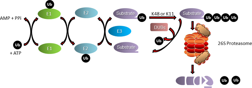
A multistep enzymatic cascade attaches ubiquitin (Ub) to its Substrate. An E1 (ubiquitin activating enzyme) activate ubiquitin using energy from ATP hydrolysis and transfers it to an E2 (ubiquitin conjugating enzyme. The E2 subsequently transfer the activated ubiquitin to a substrate that is specifically recruited by an E3 (ubiquitin ligase). Polyubiquitination comprising four or more ubiquitin protein linked either through K48 or K11 undergo degradation in 26S proteasome. The ubiquitination mark can be reversed by deubiquitinase enzymes (DUBs) to protect substrate from degradation. The dynamic equilibrium of E3 ligase and DUBs dictates the level of substrate steady state levels and turnover.
First, the reaction begins with ATP-dependent ubiquitin activation by the ubiquitin activating enzyme (E1) followed by transfer of the activated ubiquitin to a cysteine residue of ubiquitin conjugating enzyme (E2) and finally, transfer of ubiquitin to a free amine group in either the amino terminus or an internal lysine of a protein substrate that is mediated via a ubiquitin ligase (E3) enzyme. This cascade can proceed repeatedly to result in long chain of ubiquitin (polyubiquitin) on substrate. Ubiquitin has seven internal lysines (K6, K11, K27, K29, K33, K48, and K63). Each of these lysine residues can be utilized to mark substrates with monoubiquitination or polyubiquitination to feature different topology and consequently different biochemical consequences. Polyubiquitination at K48 and rarely at K11 marks substrates for degradation by the proteasome. The process of ubiquitination is counterbalanced by the process of deubiquitination mediated by deubiquitinating enzymes. The counteracting activities of E3 ligases and deubiquitinases control the cellular homeostasis. Deregulated degradation of critical cellular substrate proteins via UPS interrupts normal proteostasis of the cell, and triggers aberrant progression of cell-division cycle, transcriptional regulation, DNA-damage response, and apoptosis (Table 1 [3–44]) [11,43,45,46,47▪▪]. Deregulations of these critical cellular pathways culminate into multistep process of lymphoid transformation (Fig. 2).
Table 1.
Cellular substrates and associated E3 ligases/deubiquitinases implicated in lymphoma pathogenesis
| Substrates | Lymphoma subtype associated |
E 3 ligase and Deubiquitinases |
References |
|---|---|---|---|
| Transcription factors and epigenetic modulators | |||
| NFκB components | T-PLL, ABC-DLBCL, | SCFFbxw7α, SCFβTrCP, | [3–8] |
| Notch signaling components | T-ALL, B-ALL SMZL, | SCFFbxw7α | [9,10] |
| Myc | BL, DLBCL, FL, ALL, BCLU, MCL, CLL, PBL | SCFFbxw7α, SCFβTrCP, SCFSkp2, Truss, Huwe1, USP28 | [11–14] |
| Polycomb group proteins | GCB DLBCL, FL, | SCFβTrCP, Smurf2 | [15–18] |
| MLL | B-ALL, DLBCL, AML, MCL | SCFSkp2, APCCDC20 | [19] |
| DNMT1 | TCL, ALL | UHRF1, HAUSP | [20,21] |
| BCL6 | GCB-DLBCL, FL | SCFFbxo11 | [22] |
| Genotoxic stress and Apoptosis | |||
| p53 | TCL, ATLL, FL, DLBCL, MCL, MZL,CLL | MDM2, Pirh2, SCFβTrCP, USP10 and HAUSP | [11,23] |
| BCL2 | FL, DLBCL, CLL, MCL, ALL, TCL, MM | SCFFbxo10 | [24] |
| BIM | SCFβTrCP, APCCDC20 | [25,26] | |
| MCL1 | MCL, DLBCL, BL, CLL, AML, ALL and MM | SCFβTrCP, SCFFbxw7α, USP9X | [27] |
| Cell-cycle regulators | |||
| Cyclins | BL,MCL,DLBCL, | APC/CCdh1,SCFFbxw7α, SCFFbxo4, SCFFbxo8, SCFFbxo31, SCFSkp2 and SCFκTrCP | [28–36] |
| CKI | BL, DLBCL, FL, ALL, MCL, CLL, PBL, MZL,cHL, TCL | SCFSkp2 | [37–42] |
| CDK | TCL | SCFSkp2, SCFβTrCP | [43,44] |
ALL, Acute Lymphoblastic Leukemia; BCLU, B-cell lymphoma unclassifiable; BL, Burkitt lymphoma; CDK, cyclin-dependent kinases; CKI, cyclin-dependent kinase inhibitors; FL, follicular lymphoma; MCL, mantle-cell lymphoma; MLL, mixed-lineage leukemia; SMZL, splenic marginal zone lymphoma.
FIGURE 2.
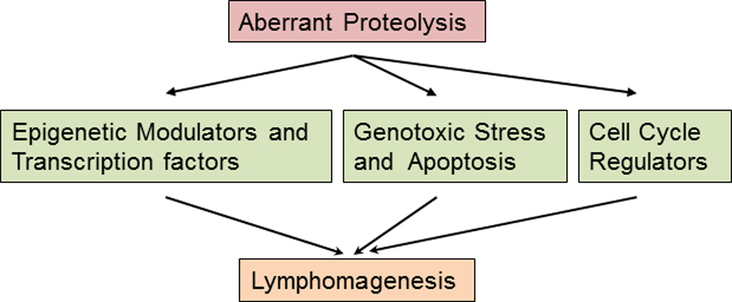
Aberrant proteolysis of critical substrates involved in cell-cycle progression, genotoxic stress, apoptosis, or cellular machinery such as epigenetic modulators and transcription factors that control gene expression contribute to lymphoma development and evolution.
REGULATION OF TRANSCRIPTION FACTORS
Hematopoiesis is a multistep process sustained by coordinated regulation of proliferation, self-renewal, and differentiation of stem and progenitor and lineage-committed cells to generate mature and functional lymphoid cells. The process of maturation of hematopoietic cells largely depends upon the establishment of correct gene expression patterns under the control of a limited set of transcription factors. These factors act as important cell-fate switches in the hematopoietic system. Hence their physiologic levels are tightly controlled by the ubiquitin proteasome system-mediated protein turnover. Deregulated proteolysis of these transcription factors leads to aberrant gene expression and contributes to lymphomagenesis. In this section, we will discuss the transcription factors and epigenetic modulators that significantly contribute to hematopoietic- cell lineage commitment, lymphoid maturation and proliferation, and how their aberrant proteolysis contribute to lymphoma pathogenesis.
Nuclear Factor κB signaling components
Nuclear factor (NF)-κB is a structurally and evolutionally conserved family of transcription factors that are critical for physiologic immune response and lineage commitment of lymphocytes into T cells or B cells [48▪▪]. The family is comprised of five structurally related activators (RelA or p65, RelB, c-Rel, p50, and p52) and five inhibitors [p100, p105 and Inhibitor of κB (IκBα, IκBβ and IκBε)] [49].
Two cullin-RING type SCF E3 ligases Fbxw1 (βTrCP) and Fbxw7α have been majorly implicated in the proteolytic regulation of the NFκB components [3–5,48▪▪]. βTrCP polyubiquitinates IκBα as well as p105 for proteasomal degradation. It also activates the proteolytic processing of p100 to generate p52, the active component of NFκB transcription factor, which directs activation of canonical as well as noncanonical NFκB signaling [6–8]. The Fbxw7α regulates the degradation of nuclear p100 and favors noncanonical pathway activation [3]. The functional dynamics of βTrCP and Fbxw7α regulate the activation of canonical or noncanonical NFκB pathway. However, the context or maturation stage of lymphoid lineage cells at which the specific E3 ligase activity prevails for canonical or noncanonical NFκB pathway activation is not yet completely understood. Aberrant activation of NFκB signaling is associated with lymphomagenesis and hence, a clear understanding of proteolytic mechanisms in this pathways is crucial to the design of specific therapies against NFκB-driven lymphomas.
Notch signaling proteins
Notch signaling plays critical roles in lineage commitment of T lymphocytes and B lymphocytes [50]. Accordingly, aberrant activation of Notch signaling has been associated with several hematologic malignancies [51–54]. Somatic mutations leading to gain-of-function of Notch signaling is associated with oncogenesis in many lymphoid neoplasms. In myeloid malignancies, however, loss-of-function mutations implicate a tumor suppressor role of Notch signaling components [55]. From this we can surmise that oncogenic or tumor suppressor activities of Notch signaling pathways are highly context dependent [55]. Notch signaling is significantly regulated by the UPS. Aberrant proteolysis of Notch1 due to activating mutations in Notch1 receptors impair its recognition and degradation by SCFFbxw7 E3 ligase and promote lymphomagenesis [9,10]. The C-terminal PEST domain of Notch is highly conserved among different Notch proteins. Independent studies have identified Notch2 mutations in splenic marginal zone lymphoma (SMZL) patients using next-generation sequencing suggesting a mechanism analogous to that observed with Notch1 mutations. Most of the identified mutations targeted the conserved C terminal PEST region and are predicted to be recognition sites for E3 ligase(s) [51,52]. Accordingly, frameshift and deletion mutations targeting the PEST domain could increase the stability of mutant Notch2 protein with attendant Notch hyperactivity contributing to SMZL pathogenesis. Although Notch1 proteolysis is well understood, the machinery and regulation of Notch2 are not completely characterized. Thus, the identification of E3 ligase(s) and molecular mechanism that regulate Notch2 proteolysis might offer novel insights for therapeutic intervention against SMZL.
Myc
The Myc family of transcription factors is one of the most frequently deregulated oncogenes in human cancer [56]. It is a leucine zipper transcription factor that modulates several critical cellular processes including growth, metabolism, cell proliferation, apoptosis, genomic instability, stem cell self-renewal, and differentiation [57]. With the cooperation of other genetic lesions, myc deregulation derives tumorigenesis in several subtypes of hematopoietic malignancy and human cancer. Overexpression and chromosomal translocation-mediated hyperactivation of c-Myc oncoprotein has been observed in large number of hematologic malignancies including Burkitt lymphoma, Diffuse Large B-Cell Lymphoma (DLBCL), follicular lymphoma, Acute Lymphoblastic Leukemia (ALL), B-cell lymphoma unclassifiable with features intermediates between DLBCL and Burkitt lymphoma (BCLU), plasma-cell myeloma, mantle-cell lymphoma (MCL), chronic lymphocytic leukemia, and plasmablastic lymphoma [58–74]. Myc levels are regulated posttranslationally by SCFFbxw7 E3 ligase. Several mutations identified in the coding region of c-Myc center around Thr 58, which undergoes phosphorylation by GSK3β and facilitates recognition and polyubiquitination of c-Myc by Fbxw7 for degradation [12]. Degradation of c-Myc is significantly impaired in Burkitt lymphoma harboring Thr58 or other mutations critical for Fbxw7-mediated recognition of the transcription factor [12,13]. The counteracting activity of c-Myc ubiquitination has been shown to be mediated by the USP28 deubiquitinase in colorectal cancer [14]. However, the deubiquitinase that regulates c-Myc abundance in hematologic malignancy has not been clearly established and offers a potential avenue for further studies. Apart from c-Myc, SCFFbxw7 also regulate several other transcription factors involved oncogenic transcription, such as c-Myb, Jun, Notch, and KLF5 and thereby control growth-regulating pathways [75–81]. Consequently, mutational inactivation of Fbxw7 has been observed and implicated in the development and evolution of many subsets of malignant lymphoma [82▪▪].
Epigenetic regulators
The polycomb group (PcG) proteins are evolutionarily conserved epigenetic regulators of development [83]. Deregulated expression of PcG proteins is associated with several forms of cancer including hematologic malignancies [84–86]. The critical catalytic subunits of PcG protein; BMI1 and EZH2 are most frequently altered in several hematologic malignancies. BMI1 is recurrently upregulated in MCL and chronic myelogenous leukemia and a novel gene translocation t(10;14)(p12;q32) resulting in the IGH-BMI1 juxtaposition has been reported in chronic lymphocytic leukemia [87–89]. EZH2, a SET domain containing histone methyltransferase is a critical enzymatic subunit of the polycomb repressive complex 2 (PRC2), which trimethylates histone H3 (H3K27) to mediate gene repression [90]. Somatic mutations, overexpression, and hyper activation of EZH2 have been reported in the pathogenesis of several types of lymphoma [84]. Genome wide sequencing studies identified heterozygous somatic and recurrent gain-of-function mutations targeting EZH2Y641 occurring most frequently in germinal center derived follicular and aggressive diffuse large B-cell lymphoma. This mutation is associated with H3K27me3 hyperactivation promoting lymphomagenesis [91–94]. These mutations have been reported to alter the enzymatic activity of EZH2 (with preference for trimethylation of H3K27) and increase its stability, resulting in a protein with prolonged half-life. SCFβTrCP E3 ubiquitin ligase controls the rapid proteolysis of BMI1 and EZH2 [15,16,82▪▪]. Although the complete molecular mechanism and context of their proteolysis is not completely understood, SCFβTrCP E3 ligase has been shown to recognize Y641 phosphorylated EZH2 for polyubiquitination and degradation [15]. The mutations at Y641 abrogate EZH2 phosphorylation by JAK2 tyrosine kinase and compromise its recognition by SCFβTrCP rendering the EZH2 protein long-lived with attendant increase in H3K27 trimethylation activity. Similarly, the polycomb group protein Yin Yang 1 is targeted for degradation by smurf2 (smad ubiquitination regulatory factor 2) E3 ligase in germinal center and smurf2 is reported to be downregulated in germinal center derived DLBCL[17]. Mice deficient in smurf2 develop B-cell lymphoma. In a different context, smurf2 is also reported to regulate EZH2 proteolysis [18]. The possibility of novel smurf2 targets in the germinal center that may contribute to lymphomagenesis remains an intriguing. Further studies on the detail molecular mechanism of proteolytic regulation of PRC components may provide potential new therapeutic opportunities for treatment of hematopoietic malignancies driven by aberrant activity of PcG proteins.
PcG group-mediated transcriptional repression is counteracted by the multisubunit complex of trithorax group (TrxG) proteins. Mutational inactivation of TrxG complex components are identified in several B-cell and T-cell malignancies. The trithorax group histone methyl transferase mixed-lineage leukemia (MLL) is posttranslationally regulated by E3 ligases SCFSkp2 and anaphase-promoting complex, APCCDC20 during cell-cycle progression [19]. However, the posttranslational regulations of other TrxG proteins are poorly understood. As several hematologic malignancies are associated with mutational inactivation of TrxG proteins [95–98], understanding their proteolytic mechanism might provide significant insight on lymphomagenesis driven by increased stability of these epigenetic modifiers.
In concert with histone modifiers, DNA methyl transferases also play significant role in epigenetic regulation of gene expression. Several DNA methyl transferases have been implicated in hematologic malignancies [99]. DNA methyl transferase DNMT1, implicated in acute lymphoblastic leukemia and malignant T-cell lymphoma is regulated at the posttranslational level by UHRF1 E3 ligase and HAUSP deubiquitinase in response to acetylation [20,21]. Given the importance of the field, the identification of posttranslational regulatory mechanisms for DNA methyl transferases and demethylases warrants detailed investigation in relation to their possible roles in the pathogenesis of hematopoietic neoplasms.
BCL6
BCL6 is a transcriptional repressor required for the establishment of the germinal center during B-cell maturation [100]. BCL6 deregulation has also been associated with B-cell lymphomagenesis [100]. BCL6 regulates the germinal center reaction by transcriptional repression of genes required for cellular proliferation and differentiation, and thus prevent premature activation and differentiation of B-cell toward memory and plasma cells [101–109]. Overexpression of BCL6 through promoter hypermutation (~15%) and chromosomal translocations involving BCL6 (~40%) have been identified in DLBCL [22,110–113]. Another regulatory mechanism involving proteasomal degradation of BCL6 has been recently identified. [22]. The SCFFbxo11 E3 ubiquitin ligase regulates BCL6 degradation, and thus controls its levels required for germinal center formation [22]. Fbxo11 silencing in a DLBCL-cell line promotes tumorigenesis in immunodeficient mice and its reconstitution suppresses tumor growth [22]. Fbxo11 deletion and mutations are found in several DLBCL-cell lines and primary DLBCL-patient samples. Loss of Fbxo11 promotes BCL6 stability. Fbxo11 mutations identified in DLBCL are unable to trigger BCL6 degradation, and thus potentially contribute to lymphomagenesis through BCL6 stabilization. The identification of regulatory mechanisms that control BCL6 abundance in GC B-cell may offer a potential avenue for targeted therapeutic intervention against BCL6-deregulated lymphoma.
GENOTOXIC STRESS AND CELLULAR APOPTOSIS
P53
The cellular response following DNA damage is largely controlled by tumor suppressor p53 by blocking cell-cycle arrest for repair or alternatively inducing apoptosis. P53 is mutated in, approximately, 50% of human malignancies; however, its mutational inactivation is relatively less frequent in hematologic malignancies [23]. Instead, p53 levels are regulated at other levels including proteasomal degradation. Whereas MDM2 is the major E3 ligase for p53, other E3 ligases are also involved in p53 stability and activity [11]. Similarly, genotoxic stress also triggers the phosphorylation and proteolysis of cyclin D1 to regulate cell-cycle progression in order for DNA damage repair machinery to detect and repair [114]. The proteolytic regulatory mechanism of cyclin D1 is discussed in later section of this review.
BCL2 family
Two major signaling pathways namely the extrinsic or death receptor and intrinsic or mitochondrial pathway trigger apoptosis [115]. The intrinsic pathways are more commonly perturbed in lymphoid malignancies. This pathway is largely regulated by BCL2 family proteins.
This family constitutes three subfamilies: prosurvival (BCL2, BCLxl, BCLw, MCL1, and BFL1/A1), the subfamily that promote cell death; the initiator BH3-only proteins (BIM, PUMA, BAD, or NOXA); and the cell death mediators (BAX and BAK) [116,117].
Overexpression of BCL2 induced by the t(14;18)(q32;21) is characteristic of follicular lymphoma [118,119]. However, it is also overexpressed in CLL [120,121], MCL [122], DLBCL [123], ALL [124], MM and other plasma-cell dyscrasias [125], and some T-cell lymphomas [126]. Proteins of this family are regulated at posttranslational level by several E3 ligases and deubiquitinases. The deregulation of corresponding degradation machinery components has been documented in several forms of lymphoma. SCFFbxo10 has been implicated in BCL2 protein degradation and reported to be compromised by mutational inactivation or deletion in germinal center type DLBCL [24]. Similarly, the BCL2 family protein BIM is targeted for degradation by two E3 ligases namely βTrCP and APCCDC20 in different cellular contexts [25,26]. Overexpression of the BCL2 family protein myeloid-cell leukemia sequence 1 (MCL1), cooperates with Myc and to promote lymphomagenesis [127]. Four E3 ligases: Mule, βTrCP, Fbxw7, and Trim17 trigger MCL1 proteolysis and its degradation is prevented by the deubiquitination activity of USP9X [27]. Targeting MCL1 through increased proteolysis might be a potential avenue of investigation for MCL1 overexpression-mediated lymphomagenesis. Thus, while several apoptosis intermediates are regulated by the UPS, the regulatory mechanisms of many are yet to be explored and may offer novel opportunities for targeted therapeutic interventions in malignant lymphomas.
TNFAIP3
The ubiquitin ligase TNFAIP3 (A20), a known effector of apoptosis, is a unique ubiquitin ligase that also possesses deubiqutinating activity and controls its own degradation in a negative feedback loop [128]. TNFAIP3 is primarily responsible for attenuation of NFκB signaling and thereby inhibits inflammation and tumorigenesis and TNF-mediated mediated apoptosis [128]. Mutational inactivation of TNFAIP3 by deletions, frameshift or nonsense mutations, and by promoter hypermethylation has been reported in several forms of B-cell and T-cell lymphoma [129–137]. TNFAIP3 inactivation by any mechanism contributes to lymphoma pathogenesis by promoting unchecked NFκB signaling and enhanced-cell survival [132]. Considering the critical role played by TNFAIP3 in hematopoiesis, the studies involving its regulatory mechanisms and other novel substrates targeted by this enzyme might offer an opportunity for therapeutic interventions in TNFAIP3 dysregulated lymphomas.
CELL-CYCLE CONTROL
Deregulation of the cell cycle fundamentally underlies the development and evolution of various forms of malignant lymphoma. Cell-cycle progression is precisely regulated by timely synthesis and degradation of cell-cycle regulatory proteins; cyclin-dependent kinases (CDK1, CDK2, CDK4, and CDK6), their essential activating coenzymes, the cyclins (cyclin A, B, D, and E), interaction of CDK-inhibitory proteins (CDKIs) and the signaling networks that control their function via posttranslational modifications. The abundance of these intermediates during the different phases of cell-division cycle is tightly controlled by the ubiquitin proteasome system [,138]. Mutational inactivation of proteolytic machinery components that control critical cell-cycle regulators contribute to the pathogenesis of malignant lymphoma. Additionally, the genetic lesions in cell-cycle regulators that enable them to bypass proteolytic turnover also promote aberrant lymphoid-cell growth and malignant transformation. Two major ubiquitin ligase complexes; the APC or cyclosome (APC/C) and the SCF complex are primarily involved in the regulated proteolysis of several cell-cycle regulatory proteins [43,139].
Cyclin and cyclin-dependent kinases
Cell-cycle progression is principally driven by cyclins and their respective CDK and regulated by cyclin-dependent kinase inhibitors (CKIs) in different phases of cell cycle. Several E3 ligases that participate in physiologic cell-cycle progression have been shown either to be deregulated in specific types of lymphoma or contribute to lymphoma pathogenesis in mouse models. APC/CCdh1 E3 ligase targets cyclin A and B for degradation along with many other factors associated with M to G1 phase transition [140]. Cdh1 heterozygous mice develop B-cell lymphoma and a myelodysplastic syndrome-like disorder [141]. Further, the reduced expression of CDH1 has been described in the malignant progression of B lymphoma cell line suggesting a plausible role APC/C complex components in lymphomagenesis [142]. Cyclins are often overexpressed in several types of cancer and promote cancer progression by accelerating the cell-division cycle [143–147]. Thus, failure to degrade cyclins is associated with tumor development and progression. For example, neoplasia of diverse lineages overexpress cyclin E and this overexpression correlates with increased tumor aggressiveness. Cyclin E is known to promote entry into S phase and the cyclin E-CDK2 complex phosphorylates retinoblastoma to promote G1 progression. The cyclin E is a prototypic substrate of SCFFbxw7 E3 ligase [28]. The regulation of cyclin E protein abundance is compromised by mutational inactivation of Fbw7 that contributes to several forms of lymphoid neoplasia [29]. Frequent inactivation of Fbw7 by deletions or point mutations occurs in diverse hematopoietic malignancies [9,10,30,148]. Specifically, the most frequent Fbw7 mutations are identified in T-ALL (~30%) and cholangiocarcinomas (~35%) [149]. Commonly, missense mutations of Fbw7 occurring at R465C, R479Q, and R505C withinWD40 repeats 3 and 4 (from Fbw7α) diminish its capacity to recruit substrates to the E3 ligase complex by loss of interaction [10]. Further, during G1 to S phase transition cyclin D1 is targeted for degradation by several SCF E3 ligases including FBXO4, FBXO8, FBXO31, SKP2, βTRCP, and APC [31–36,150]. It is anticipated that studies investigating the pathogenic deregulation of these E3 ligases will be informative on their role in the pathogenesis of lymphoid malignancies.
Cyclin-dependent kinase inhibitors
CKIs are the critical regulator of cyclin-CDK activities. CKIs function by inhibiting CDK activity and promoting cell-cycle arrest and/or delaying the response to antimitogenic stimuli. These CKIs fall into two families: the INK4 inhibitors and the CIP/KIP inhibitors. There are four known INK4 family members: p16INK4A, p15INK4B, p19INK4D, and p18INK4C. The three mammalian CIP/KIP CKIs: namely p21CIP1, p27KIP1, and p57KIP2 play different role in cell-cycle regulation [138,151]. The SCFSKP2 E3 ligase targets all three CKIs: p21CIP1, p27KIP1, and p57KIP2 (all widely accepted tumor suppressors) at different stages of the cell cycle for degradation [37–42]. However, p27 seems to be a principal target of SKP2 as the prominent phenotypes apparent in Skp2 −/− mice including nuclear enlargement, polyploidy, and an increased number of centrosomes. This phenotype is ameliorated in Skp2−/− p27−/− double mutant mice [2,153]. As SKP2 target p27 and other CKIs for degradation, it can be considered an oncogene. Apart from CKIs, SKP2 also targets p130, cyclin A, cyclin D1, free cyclin E, ORC1, CDK9, MYC, B, Myb, SMAD4, RAG2, UBP43, FOXO1, and papillomavirus E7(139). SKP2 over expression has been associated with poor prognosis in human lymphoma patients [154,155].
CONCLUSION
The controlled degradation of cellular proteins involved in cell-cycle progression, response to genotoxic stress, signaling pathways, and transcriptional control of gene expression is prerequisite for normal hematopoiesis. In this context, the UPS plays a pivotal role by degradation of regulatory proteins in a precisely timed and context-dependent manner. Deregulation of the UPS results in complex outcomes in term of tumorigenesis. Ubiquitin ligases control a remarkable diversity of substrates for the control of cellular homeostasis and hence, it is not surprising that these E3 ligases are deregulated by multiple mechanisms cumulatively resulting in lymphoproliferative disorders. Although E3 ligases have been implicated in the regulation of a diverse array of substrates, the knowledge on the subcellular location, context, and cofactors that dictate the degradation of these substrates in particular metabolic states, as well as, lineage and cellular contexts is far from complete. The main goal of studying proteolytic mechanisms in lymphopoiesis and lymphoid malignancies is to develop a more sophisticated understanding of the regulation of these processes and the phenomena that play a role in their perturbation resulting in malignancy. Such knowledge could be exploited in the development of therapeutic strategies targeting lymphomas driven by aberrant proteolysis of critical cellular substrates. Advances in this arena include the FDA approval of Velcade (Bortezomib, a general proteasome inhibitor) and the use of the neddylation inhibitor (MLN4924) in the management of cancers. Inhibition of specific components of the proteolytic machinery in cell lineage and context-dependent settings offers plausible options for novel lymphoma therapies.
KEY POINTS.
Proteolytic regulatory mechanisms that control several critical cellular processes maintain cellular homeostasis.
Deregulation of the proteolytic machinery by mutational inactivation or hyperactivation contributes to lymphoma pathogenesis.
Somatic mutations in critical cellular substrates that result in failure to degrade the substrates also contribute to lymphoma pathogenesis.
Acknowledgements
None.
Financial support and sponsorship
This work was supported in part by NIH Grants R01 DE119249, and R01 CA136905 to K.S.J. E.-J.
Footnotes
Uncited reference
[152].
Conflicts of interest
There are no conflicts of interest to declare.
REFERENCES AND RECOMMENDED READING
Papers of particular interest, published within the annual period of review, have been highlighted as:
▪ of special interest
▪▪ of outstanding interest
- 1.Hershko A. The ubiquitin system for protein degradation and some of its roles in the control of the cell-division cycle (Nobel lecture) Angew Chem Int Ed Engl. 2005;44:5932–5943. doi: 10.1002/anie.200501724. [DOI] [PubMed] [Google Scholar]
- 2.Hershko A, Ciechanover A. The ubiquitin system. Annu Rev Biochem. 1998;67:425–479. doi: 10.1146/annurev.biochem.67.1.425. [DOI] [PubMed] [Google Scholar]
- 3.Busino L, Millman SE, Scotto L, et al. Fbxw7alpha- and GSK3-mediated degradation of p100 is a pro-survival mechanism in multiple myeloma. Nat Cell Biol. 2012;14:375–385. doi: 10.1038/ncb2463. [DOI] [PMC free article] [PubMed] [Google Scholar]
- 4.Kanarek N, London N, Schueler-Furman O, Ben-Neriah Y. Ubiquitination and degradation of the inhibitors of NF-kappaB. Cold Spring HarbPerspect Biol. 2010;2:a000166. doi: 10.1101/cshperspect.a000166. [DOI] [PMC free article] [PubMed] [Google Scholar]
- 5.Busino L, Millman SE, Pagano M. SCF-mediated degradation of p100 (NF-kappaB2): mechanisms and relevance in multiple myeloma. Sci Signal. 2012;5:t14. doi: 10.1126/scisignal.2003408. [DOI] [PMC free article] [PubMed] [Google Scholar]
- 6.Amir RE, Haecker H, Karin M, Ciechanover A. Mechanism of processing of the NF-kappa B2 p100 precursor: identification of the specific polyubiquitin chain-anchoring lysine residue and analysis of the role of NEDD8-modification on the SCF(beta-TrCP) ubiquitin ligase. Oncogene. 2004;23:2540–2547. doi: 10.1038/sj.onc.1207366. [DOI] [PubMed] [Google Scholar]
- 7.Cohen S, Achbert-Weiner H, Ciechanover A. Dual effects of IkappaB kinase beta-mediated phosphorylation on p105 Fate: SCF(beta-TrCP)-dependent degradation and SCF(beta-TrCP)-independent processing. Mol Cell Biol. 2004;24:475–486. doi: 10.1128/MCB.24.1.475-486.2004. [DOI] [PMC free article] [PubMed] [Google Scholar]
- 8.Orian A, Gonen H, Bercovich B, et al. SCF(beta)(-TrCP) ubiquitin ligase-mediated processing of NF-kappaB p105 requires phosphorylation of its C-terminus by IkappaB kinase. EMBO J. 2000;19:2580–2591. doi: 10.1093/emboj/19.11.2580. [DOI] [PMC free article] [PubMed] [Google Scholar]
- 9.O’Neil J, Grim J, Strack P, et al. FBW7 mutations in leukemic cells mediate NOTCH pathway activation and resistance to gamma-secretase inhibitors. J Exp Med. 2007;204:1813–1824. doi: 10.1084/jem.20070876. [DOI] [PMC free article] [PubMed] [Google Scholar]
- 10.Thompson BJ, Buonamici S, Sulis ML, et al. The SCFFBW7 ubiquitin ligase complex as a tumor suppressor in T cell leukemia. J Exp Med. 2007;204:1825–1835. doi: 10.1084/jem.20070872. [DOI] [PMC free article] [PubMed] [Google Scholar]
- 11.Hammond-Martel I, Yu H, Affar el B. Roles of ubiquitin signaling in transcription regulation. Cell Signal. 2012;24:410–421. doi: 10.1016/j.cellsig.2011.10.009. [DOI] [PubMed] [Google Scholar]
- 12.Bahram F, von der Lehr N, Cetinkaya C, Larsson LG. c-Myc hot spot mutations in lymphomas result in inefficient ubiquitination and decreased proteasome-mediated turnover. Blood. 2000;95:2104–2110. [PubMed] [Google Scholar]
- 13.Gregory MA, Hann SR. c-Myc proteolysis by the ubiquitin-proteasome pathway: stabilization of c-Myc in Burkitt’s lymphoma cells. Mol Cell Biol. 2000;20:2423–2435. doi: 10.1128/mcb.20.7.2423-2435.2000. [DOI] [PMC free article] [PubMed] [Google Scholar]
- 14.Diefenbacher ME, Popov N, Blake SM, et al. The deubiquitinase USP28 controls intestinal homeostasis and promotes colorectal cancer. J Clin Invest. 2014 Jun 24; doi: 10.1172/JCI73733. PubMed PMID: 24960159. [DOI] [PMC free article] [PubMed] [Google Scholar]
- 15.Sahasrabuddhe AA, Chen X, Chung F, et al. Oncogenic Y641 mutations in EZH2 prevent Jak2/beta-TrCP-mediated degradation. Oncogene. 2015;34:445–454. doi: 10.1038/onc.2013.571. [DOI] [PubMed] [Google Scholar]
- 16.Sahasrabuddhe AA, Dimri M, Bommi PV, Dimri GP. betaTrCP regulates BMI1 protein turnover via ubiquitination and degradation. Cell Cycle. 2011;10:1322–1330. doi: 10.4161/cc.10.8.15372. [DOI] [PMC free article] [PubMed] [Google Scholar]
- 17.Ramkumar C, Cui H, Kong Y, et al. Smurf2 suppresses B-cell proliferation and lymphomagenesis by mediating ubiquitination and degradation of YY1. Nat Commun. 2013;4:2598. doi: 10.1038/ncomms3598. [DOI] [PMC free article] [PubMed] [Google Scholar]
- 18.Yu YL, Chou RH, Shyu WC, et al. Smurf2-mediated degradation of EZH2 enhances neuron differentiation and improves functional recovery after ischaemic stroke. EMBO Mol Med. 2013;5:531–547. doi: 10.1002/emmm.201201783. [DOI] [PMC free article] [PubMed] [Google Scholar]
- 19.Liu H, Cheng EH, Hsieh JJ. Bimodal degradation of MLL by SCFSkp2 and APCCdc20 assures cell cycle execution: a critical regulatory circuit lost in leukemogenic MLL fusions. Genes Dev. 2007;21:2385–2398. doi: 10.1101/gad.1574507. [DOI] [PMC free article] [PubMed] [Google Scholar]
- 20.Du Z, Song J, Wang Y, et al. DNMT1 stability is regulated by proteins coordinating deubiquitination and acetylation-driven ubiquitination. Sci Signal. 2010;3:ra80. doi: 10.1126/scisignal.2001462. [DOI] [PMC free article] [PubMed] [Google Scholar]
- 21.Yuan B, Zhang J, Wang H, et al. 6-Thioguanine reactivates epigenetically silenced genes in acute lymphoblastic leukemia cells by facilitating proteasome- mediated degradation of DNMT1. Cancer Res. 2011;71:1904–1911. doi: 10.1158/0008-5472.CAN-10-3430. [DOI] [PMC free article] [PubMed] [Google Scholar]
- 22.Duan S, Cermak L, Pagan JK, et al. FBXO11 targets BCL6 for degradation and is inactivated in diffuse large B-cell lymphomas. Nature. 2012;481:90–93. doi: 10.1038/nature10688. [DOI] [PMC free article] [PubMed] [Google Scholar]
- 23.Xu-Monette ZY, Medeiros LJ, Li Y, et al. Dysfunction of the TP53 tumor suppressor gene in lymphoid malignancies. Blood. 2012;119:3668–3683. doi: 10.1182/blood-2011-11-366062. [DOI] [PMC free article] [PubMed] [Google Scholar]
- 24.Chiorazzi M, Rui L, Yang Y, et al. Related F-box proteins control cell death in Caenorhabditis elegans and human lymphoma. Proc Natl Acad Sci U S A. 2013;110:3943–3948. doi: 10.1073/pnas.1217271110. [DOI] [PMC free article] [PubMed] [Google Scholar]
- 25.Moustafa-Kamal M, Gamache I, Lu Y, et al. BimEL is phosphorylated at mitosis by Aurora A and targeted for degradation by betaTrCP1. Cell Death Differ. 2013;20:1393–1403. doi: 10.1038/cdd.2013.93. [DOI] [PMC free article] [PubMed] [Google Scholar]
- 26.Wan L, Tan M, Yang J, et al. APC(Cdc20) suppresses apoptosis through targeting Bim for ubiquitination and destruction. Dev Cell. 2014;29:377–391. doi: 10.1016/j.devcel.2014.04.022. [DOI] [PMC free article] [PubMed] [Google Scholar]
- 27.Mojsa B, Lassot I, Desagher S. Mcl-1 ubiquitination: unique regulation of an essential survival protein. Cells. 2014;3:418–437. doi: 10.3390/cells3020418. [DOI] [PMC free article] [PubMed] [Google Scholar]
- 28.Koepp DM, Schaefer LK, Ye X, et al. Phosphorylation-dependent ubiquitination of cyclin E by the SCFFbw7 ubiquitin ligase. Science. 2001;294:173–177. doi: 10.1126/science.1065203. [DOI] [PubMed] [Google Scholar]
- 29.Siu KT, Xu Y, Swartz KL, et al. Chromosome instability underlies hematopoietic stem cell dysfunction and lymphoid neoplasia associated with impaired Fbw7-mediated cyclin E regulation. Mol Cell Biol. 2014;34:3244–3258. doi: 10.1128/MCB.01528-13. [DOI] [PMC free article] [PubMed] [Google Scholar]
- 30.Onoyama I, Tsunematsu R, Matsumoto A, et al. Conditional inactivation of Fbxw7 impairs cell-cycle exit during T cell differentiation and results in lymphomatogenesis. J Exp Med. 2007;204:2875–2888. doi: 10.1084/jem.20062299. [DOI] [PMC free article] [PubMed] [Google Scholar]
- 31.Barbash O, Zamfirova P, Lin DI, et al. Mutations in Fbx4 inhibit dimerization of the SCF(Fbx4) ligase and contribute to cyclin D1 overexpression in human cancer. Cancer Cell. 2008;14:68–78. doi: 10.1016/j.ccr.2008.05.017. [DOI] [PMC free article] [PubMed] [Google Scholar]
- 32.Frescas D, Pagano M. Deregulated proteolysis by the F-box proteins SKP2 and beta-TrCP: tipping the scales of cancer. Nat Rev Cancer. 2008;8:438–449. doi: 10.1038/nrc2396. [DOI] [PMC free article] [PubMed] [Google Scholar]
- 33.Lin DI, Barbash O, Kumar KG, et al. Phosphorylation-dependent ubiquitination of cyclin D1 by the SCF(FBX4-alphaB crystallin) complex. Mol Cell. 2006;24:355–366. doi: 10.1016/j.molcel.2006.09.007. [DOI] [PMC free article] [PubMed] [Google Scholar]
- 34.Okabe H, Lee SH, Phuchareon J, et al. A critical role for FBXW8 and MAPK in cyclin D1 degradation and cancer cell proliferation. PLoS One. 2006;1:e128. doi: 10.1371/journal.pone.0000128. [DOI] [PMC free article] [PubMed] [Google Scholar]
- 35.Pawar SA, Sarkar TR, Balamurugan K, et al. C/EBP{delta} targets cyclin D1 for proteasome-mediated degradation via induction of CDC27/APC3 expression. Proc Natl Acad Sci U S A. 2010;107:9210–9215. doi: 10.1073/pnas.0913813107. [DOI] [PMC free article] [PubMed] [Google Scholar]
- 36.Santra MK, Wajapeyee N, Green MR. F-box protein FBXO31mediates cyclin D1 degradation to induce G1 arrest after DNA damage. Nature. 2009;459:722–725. doi: 10.1038/nature08011. [DOI] [PMC free article] [PubMed] [Google Scholar]
- 37.Carrano AC, Eytan E, Hershko A, Pagano M. SKP2 is required for ubiquitin-mediated degradation of the CDK inhibitor p27. Nat Cell Biol. 1999;1:193–199. doi: 10.1038/12013. [DOI] [PubMed] [Google Scholar]
- 38.Sutterluty H, Chatelain E, Marti A, et al. p45SKP2 promotes p27Kip1 degradation and induces S phase in quiescent cells. Nat Cell Biol. 1999;1:207–214. doi: 10.1038/12027. [DOI] [PubMed] [Google Scholar]
- 39.Tsvetkov LM, Yeh KH, Lee SJ, Sun H, Zhang H. p27(Kip1) ubiquitination and degradation is regulated by the SCF(Skp2) complex through phosphorylated Thr187 in p27. Curr Biol. 1999;9:661–664. doi: 10.1016/s0960-9822(99)80290-5. [DOI] [PubMed] [Google Scholar]
- 40.Yu ZK, Gervais JL, Zhang H. Human CUL-1 associates with the SKP1/SKP2 complex and regulates p21(CIP1/WAF1) and cyclin D proteins. Proc Natl Acad Sci U S A. 1998;95:11324–11329. doi: 10.1073/pnas.95.19.11324. [DOI] [PMC free article] [PubMed] [Google Scholar]
- 41.Bornstein G, Bloom J, Sitry-Shevah D, et al. Role of the SCFSkp2 ubiquitin ligase in the degradation of p21Cip1 in S phase. J Biol Chem. 2003;278:25752–25757. doi: 10.1074/jbc.M301774200. [DOI] [PubMed] [Google Scholar]
- 42.Kamura T, Hara T, Kotoshiba S, et al. Degradation of p57Kip2 mediated by SCFSkp2-dependent ubiquitylation. Proc Natl Acad Sci U S A. 2003;100:10231–10236. doi: 10.1073/pnas.1831009100. [DOI] [PMC free article] [PubMed] [Google Scholar]
- 43.Nakayama KI, Nakayama K. Ubiquitin ligases: cell-cycle control and cancer. Nat Rev Cancer. 2006;6:369–381. doi: 10.1038/nrc1881. [DOI] [PubMed] [Google Scholar]
- 44.Herrero-Ruiz J, Mora-Santos M, Giraldez S, et al. betaTrCP controls the lysosome-mediated degradation of CDK1, whose accumulation correlates with tumor malignancy. Oncotarget. 2014;5:7563–7574. doi: 10.18632/oncotarget.2274. [DOI] [PMC free article] [PubMed] [Google Scholar]
- 45.Silverman JS, Skaar JR, Pagano M. SCF ubiquitin ligases in the maintenance of genome stability. Trends Biochem Sci. 2011 Nov 16; doi: 10.1016/j.tibs.2011.10.004. Epub 2011/11/22. Eng. [DOI] [PMC free article] [PubMed] [Google Scholar]
- 46.Vucic D, Dixit VM, Wertz IE. Ubiquitylation in apoptosis: a posttranslational modification at the edge of life and death. Nat Rev Cell Mol Biol. 2011;12:439–452. doi: 10.1038/nrm3143. [DOI] [PubMed] [Google Scholar]
- 47. Wang Z, Liu P, Inuzuka H, Wei W. Roles of F-box proteins in cancer. Nat Rev Cancer. 2014;14:233–247. doi: 10.1038/nrc3700. PubMed PMID: 24658274. Pubmed Central PMCID: 4306233.
- 48. Gasparini C, Celeghini C, Monasta L, Zauli G. NF-kappaB pathways in hematological malignancies. Cell Mol Life Sci. 2014;71:2083–2102. doi: 10.1007/s00018-013-1545-4. The authors show the diversity of cellular processes regulated by various E3 ligases.
- 49.Oeckinghaus A, Ghosh S. The NF-kappaB family of transcription factors and its regulation. Cold Spring Harb Perspect Biol. 2009;1:a000034. doi: 10.1101/cshperspect.a000034. [DOI] [PMC free article] [PubMed] [Google Scholar]
- 50.Lobry C, Oh P, Mansour MR, et al. Notch signaling: switching an oncogene to a tumor suppressor. Blood. 2014;123:2451–2459. doi: 10.1182/blood-2013-08-355818. [DOI] [PMC free article] [PubMed] [Google Scholar]
- 51.Kiel MJ, Velusamy T, Betz BL, et al. Whole-genome sequencing identifies recurrent somatic NOTCH2 mutations in splenic marginal zone lymphoma. J Exp Med. 2012;209:1553–1565. doi: 10.1084/jem.20120910. [DOI] [PMC free article] [PubMed] [Google Scholar]
- 52.Rossi D, Trifonov V, Fangazio M, et al. The coding genome of splenic marginal zone lymphoma: activation of NOTCH2 and other pathways regulating marginal zone development. J Exp Med. 2012;209:1537–1551. doi: 10.1084/jem.20120904. [DOI] [PMC free article] [PubMed] [Google Scholar]
- 53.Rosati E, Sabatini R, Rampino G, et al. Constitutively activated Notch signaling is involved in survival and apoptosis resistance of B-CLL cells. Blood. 2009;113:856–865. doi: 10.1182/blood-2008-02-139725. [DOI] [PubMed] [Google Scholar]
- 54.Weng AP, Ferrando AA, Lee W, et al. Activating mutations of NOTCH1 in human T cell acute lymphoblastic leukemia. Science. 2004;306:269–271. doi: 10.1126/science.1102160. [DOI] [PubMed] [Google Scholar]
- 55.Klinakis A, Lobry C, Abdel-Wahab O, et al. A novel tumour-suppressor function for the Notch pathway in myeloid leukaemia. Nature. 2011;473:230–233. doi: 10.1038/nature09999. [DOI] [PMC free article] [PubMed] [Google Scholar]
- 56.Sewastianik T, Prochorec-Sobieszek M, Chapuy B, Juszczynski P. MYC deregulation in lymphoid tumors: molecular mechanisms, clinical consequences and therapeutic implications. Biochim Biophys Acta. 2014;1846:457–467. doi: 10.1016/j.bbcan.2014.08.006. [DOI] [PubMed] [Google Scholar]
- 57.Pelengaris S, Khan M, Evan G. c-MYC: more than just a matter of life and death. Nat Rev Cancer. 2002;2:764–776. doi: 10.1038/nrc904. [DOI] [PubMed] [Google Scholar]
- 58.Avet-Loiseau H, Gerson F, Magrangeas F, et al. Rearrangements of the c-myc oncogene are present in 15% of primary human multiple myeloma tumors. Blood. 2001;98:3082–3086. doi: 10.1182/blood.v98.10.3082. [DOI] [PubMed] [Google Scholar]
- 59.Barrans S, Crouch S, Smith A, et al. Rearrangement of MYC is associated with poor prognosis in patients with diffuse large B-cell lymphoma treated in the era of rituximab. J Clin Oncol. 2010;28:3360–3365. doi: 10.1200/JCO.2009.26.3947. [DOI] [PubMed] [Google Scholar]
- 60.Bertrand P, Bastard C, Maingonnat C, et al. Mapping of MYC breakpoints in 8q24 rearrangements involving nonimmunoglobulin partners in B-cell lymphomas. Leukemia. 2007;21:515–523. doi: 10.1038/sj.leu.2404529. [DOI] [PubMed] [Google Scholar]
- 61.Boerma EG, Siebert R, Kluin PM, Baudis M. Translocations involving 8q24 in Burkitt lymphoma and other malignant lymphomas: a historical review of cytogenetics in the light of todays knowledge. Leukemia. 2009;23:225–234. doi: 10.1038/leu.2008.281. [DOI] [PubMed] [Google Scholar]
- 62.De Jong D, Voetdijk BM, Beverstock GC, et al. Activation of the c-myc oncogene in a precursor-B-cell blast crisis of follicular lymphoma, presenting as composite lymphoma. N Engl J Med. 1988;318:1373–1378. doi: 10.1056/NEJM198805263182106. [DOI] [PubMed] [Google Scholar]
- 63.Faderl S, O’Brien S, Pui CH, et al. Adult acute lymphoblastic leukemia: concepts and strategies. Cancer. 2010;116:1165–1176. doi: 10.1002/cncr.24862. [DOI] [PMC free article] [PubMed] [Google Scholar]
- 64.Haralambieva E, Boerma EJ, van Imhoff GW, et al. Clinical, immunophenotypic, and genetic analysis of adult lymphomas with morphologic features of Burkitt lymphoma. Am J Surg Pathol. 2005;29:1086–1094. [PubMed] [Google Scholar]
- 65.Hummel M, Bentink S, Berger H, et al. A biologic definition of Burkitt’s lymphoma from transcriptional and genomic profiling. N Engl J Med. 2006;354:2419–2430. doi: 10.1056/NEJMoa055351. [DOI] [PubMed] [Google Scholar]
- 66.Klapper W, Stoecklein H, Zeynalova S, et al. Structural aberrations affecting the MYC locus indicate a poor prognosis independent of clinical risk factors in diffuse large B-cell lymphomas treated within randomized trials of the German High-Grade Non-Hodgkin’s Lymphoma Study Group (DSHNHL) Leukemia. 2008;22:2226–2229. doi: 10.1038/leu.2008.230. [DOI] [PubMed] [Google Scholar]
- 67.Niitsu N, Okamoto M, Miura I, Hirano M. Clinical features and prognosis of de novo diffuse large B-cell lymphoma with t(14;18) and 8q24/c-MYC translocations. Leukemia. 2009;23:777–783. doi: 10.1038/leu.2008.344. [DOI] [PubMed] [Google Scholar]
- 68.Put N, Van Roosbroeck K, Konings P, et al. Chronic lymphocytic leukemia and prolymphocytic leukemia with MYC translocations: a subgroup with an aggressive disease course. Ann Hematol. 2012;91:863–873. doi: 10.1007/s00277-011-1393-y. [DOI] [PubMed] [Google Scholar]
- 69.Savage KJ, Johnson NA, Ben-Neriah S, et al. MYC gene rearrangements are associated with a poor prognosis in diffuse large B-cell lymphoma patients treated with R-CHOP chemotherapy. Blood. 2009;114:3533–3537. doi: 10.1182/blood-2009-05-220095. [DOI] [PubMed] [Google Scholar]
- 70.Setoodeh R, Schwartz S, Papenhausen P, et al. Double-hit mantle cell lymphoma with MYC gene rearrangement or amplification: a report of four cases and review of the literature. Int J Clin Exp Pathol. 2013;6:155–167. [PMC free article] [PubMed] [Google Scholar]
- 71.Shou Y, Martelli ML, Gabrea A, et al. Diverse karyotypic abnormalities of the c-myc locus associated with c-myc dysregulation and tumor progression in multiple myeloma. Proc Natl Acad Sci U S A. 2000;97:228–233. doi: 10.1073/pnas.97.1.228. [DOI] [PMC free article] [PubMed] [Google Scholar]
- 72.Slack GW, Gascoyne RD. MYC and aggressive B-cell lymphomas. Adv Anat Pathol. 2011;18:219–228. doi: 10.1097/PAP.0b013e3182169948. [DOI] [PubMed] [Google Scholar]
- 73.Valera A, Balague O, Colomo L, et al. IG/MYC rearrangements are the main cytogenetic alteration in plasmablastic lymphomas. Am J Surg Pathol. 2010;34:1686–1694. doi: 10.1097/PAS.0b013e3181f3e29f. [DOI] [PMC free article] [PubMed] [Google Scholar]
- 74.Young KH, Xie Q, Zhou G, et al. Transformation of follicular lymphoma to precursor B-cell lymphoblastic lymphoma with c-myc gene rearrangement as a critical event. Am J Clin Pathol. 2008;129:157–166. doi: 10.1309/NKK3FEX2BE5L7EKB. [DOI] [PubMed] [Google Scholar]
- 75.Matsumoto A, Onoyama I, Sunabori T, et al. Fbxw7-dependent degradation of Notch is required for control of “stemness” and neuronal-glial differentiation in neural stem cells. J Biol Chem. 2011;286:13754–13764. doi: 10.1074/jbc.M110.194936. [DOI] [PMC free article] [PubMed] [Google Scholar]
- 76.Sundqvist A, Bengoechea-Alonso MT, Ye X, et al. Control of lipid metabolism by phosphorylation-dependent degradation of the SREBP family of transcription factors by SCF(Fbw7) Cell Metab. 2005;1:379–391. doi: 10.1016/j.cmet.2005.04.010. [DOI] [PubMed] [Google Scholar]
- 77.Wei W, Jin J, Schlisio S, et al. The v-Jun point mutation allows c-Jun to escape GSK3-dependent recognition and destruction by the Fbw7 ubiquitin ligase. Cancer Cell. 2005;8:25–33. doi: 10.1016/j.ccr.2005.06.005. [DOI] [PubMed] [Google Scholar]
- 78.Welcker M, Orian A, Jin J, et al. The Fbw7 tumor suppressor regulates glycogen synthase kinase 3 phosphorylation-dependent c-Myc protein degradation. Proc Natl Acad Sci U S A. 2004;101:9085–9090. doi: 10.1073/pnas.0402770101. [DOI] [PMC free article] [PubMed] [Google Scholar]
- 79.Wu G, Lyapina S, Das I, et al. SEL-10 is an inhibitor of notch signaling that targets notch for ubiquitin-mediated protein degradation. Mol Cell Biol. 2001;21:7403–7415. doi: 10.1128/MCB.21.21.7403-7415.2001. [DOI] [PMC free article] [PubMed] [Google Scholar]
- 80.Yada M, Hatakeyama S, Kamura T, et al. Phosphorylation-dependent degradation of c-Myc is mediated by the F-box protein Fbw7. EMBO J. 2004;23:2116–2125. doi: 10.1038/sj.emboj.7600217. [DOI] [PMC free article] [PubMed] [Google Scholar]
- 81.Zhao D, Zheng HQ, Zhou Z, Chen C. The Fbw7 tumor suppressor targets KLF5 for ubiquitin-mediated degradation and suppresses breast cell proliferation. Cancer Res. 2010;70:4728–4738. doi: 10.1158/0008-5472.CAN-10-0040. [DOI] [PubMed] [Google Scholar]
- 82. Sahasrabuddhe AA, Elenitoba-Johnson KS. Role of the ubiquitin proteasome system in hematologic malignancies. Immunol Rev. 2015;263:224–239. doi: 10.1111/imr.12236. The authors identify mutational inactivation or hyperactivation of ubiquitin proteasome system components and their role in hematologic malignancies.
- 83.Laugesen A, Helin K. Chromatin repressive complexes in stem cells, development, and cancer. Cell Stem Cell. 2014;14:735–751. doi: 10.1016/j.stem.2014.05.006. [DOI] [PubMed] [Google Scholar]
- 84.Lund K, Adams PD, Copland M. EZH2 in normal and malignant hematopoiesis. Leukemia. 2014;28:44–49. doi: 10.1038/leu.2013.288. [DOI] [PubMed] [Google Scholar]
- 85.Martin-Perez D, Piris MA, Sanchez-Beato M. Polycomb proteins in hematologic malignancies. Blood. 2010;116:5465–5475. doi: 10.1182/blood-2010-05-267096. [DOI] [PubMed] [Google Scholar]
- 86.Radulovic V, de Haan G, Klauke K. Polycomb-group proteins in hematopoietic stem cell regulation and hematopoietic neoplasms. Leukemia. 2013;27:523–533. doi: 10.1038/leu.2012.368. [DOI] [PubMed] [Google Scholar]
- 87.Bea S, Tort F, Pinyol M, et al. BMI-1 gene amplification and overexpression in hematological malignancies occur mainly in mantle cell lymphomas. Cancer Res. 2001;61:2409–2412. [PubMed] [Google Scholar]
- 88.Bhattacharyya J, Mihara K, Yasunaga S, et al. BMI-1 expression is enhanced through transcriptional and posttranscriptional regulation during the progression of chronic myeloid leukemia. Ann Hematol. 2009;88:333–340. doi: 10.1007/s00277-008-0603-8. [DOI] [PubMed] [Google Scholar]
- 89.Rouhigharabaei L, Ferreiro JF, Put N, et al. BMI1, the polycomb-group gene, is recurrently targeted by genomic rearrangements in progressive B-cell leukemia/lymphoma. Genes Chromosomes Cancer. 2013;52:928–944. doi: 10.1002/gcc.22088. [DOI] [PubMed] [Google Scholar]
- 90.Yamaguchi H, Hung MC. Regulation and Role of EZH2 in Cancer. Cancer Res Treat. 2014;46:209–222. doi: 10.4143/crt.2014.46.3.209. [DOI] [PMC free article] [PubMed] [Google Scholar]
- 91.Caganova M, Carrisi C, Varano G, et al. Germinal center dysregulation by histone methyltransferase EZH2 promotes lymphomagenesis. J Clin Invest. 2013;123:5009–5022. doi: 10.1172/JCI70626. [DOI] [PMC free article] [PubMed] [Google Scholar]
- 92.Bodor C, Grossmann V, Popov N, et al. EZH2 mutations are frequent and represent an early event in follicular lymphoma. Blood. 2013;122:3165–3168. doi: 10.1182/blood-2013-04-496893. [DOI] [PMC free article] [PubMed] [Google Scholar]
- 93.Beguelin W, Popovic R, Teater M, et al. EZH2 is required for germinal center formation and somatic EZH2 mutations promote lymphoid transformation. Cancer Cell. 2013;23:677–692. doi: 10.1016/j.ccr.2013.04.011. [DOI] [PMC free article] [PubMed] [Google Scholar]
- 94.Morin RD, Johnson NA, Severson TM, et al. Somatic mutations altering EZH2 (Tyr641) in follicular and diffuse large B-cell lymphomas of germinal-center origin. Nat Genet. 2010;42:181–185. doi: 10.1038/ng.518. [DOI] [PMC free article] [PubMed] [Google Scholar]
- 95.Li H, Kaminski MS, Li Y, et al. Mutations in linker histone genes HIST1H1 B, C, D, and E; OCT2 (POU2F2); IRF8; and ARID1A underlying the pathogenesis of follicular lymphoma. Blood. 2014;123:1487–1498. doi: 10.1182/blood-2013-05-500264. [DOI] [PMC free article] [PubMed] [Google Scholar]
- 96.Liedtke M, Cleary ML. Therapeutic targeting of MLL. Blood. 2009;113:6061–6068. doi: 10.1182/blood-2008-12-197061. [DOI] [PMC free article] [PubMed] [Google Scholar]
- 97.Love C, Sun Z, Jima D, et al. The genetic landscape of mutations in Burkitt lymphoma. Nat Genet. 2012;44:1321–1325. doi: 10.1038/ng.2468. [DOI] [PMC free article] [PubMed] [Google Scholar]
- 98.Zhang J, Grubor V, Love CL, et al. Genetic heterogeneity of diffuse large B-cell lymphoma. Proc Natl Acad Sci U S A. 2013;110:1398–1403. doi: 10.1073/pnas.1205299110. [DOI] [PMC free article] [PubMed] [Google Scholar]
- 99.Li KK, Luo LF, Shen Y, et al. DNA methyltransferases in hematologic malignancies. Semin Hematol. 2013;50:48–60. doi: 10.1053/j.seminhematol.2013.01.005. [DOI] [PubMed] [Google Scholar]
- 100.Basso K, Dalla-Favera R. Roles of BCL6 in normal and transformed germinal center B cells. Immunol Rev. 2012;247:172–183. doi: 10.1111/j.1600-065X.2012.01112.x. [DOI] [PubMed] [Google Scholar]
- 101.Angelin-Duclos C, Cattoretti G, Lin KI, Calame K. Commitment of B lymphocytes to a plasma cell fate is associated with Blimp-1 expression in vivo. J Immunol. 2000;165:5462–5471. doi: 10.4049/jimmunol.165.10.5462. [DOI] [PubMed] [Google Scholar]
- 102.Basso K, Saito M, Sumazin P, et al. Integrated biochemical and computational approach identifies BCL6 direct target genes controlling multiple pathways in normal germinal center B cells. Blood. 2010;115:975–984. doi: 10.1182/blood-2009-06-227017. [DOI] [PMC free article] [PubMed] [Google Scholar]
- 103.Ci W, Polo JM, Cerchietti L, et al. The BCL6 transcriptional program features repression of multiple oncogenes in primary B cells and is deregulated in DLBCL. Blood. 2009;113:5536–5548. doi: 10.1182/blood-2008-12-193037. [DOI] [PMC free article] [PubMed] [Google Scholar]
- 104.De Silva NS, Simonetti G, Heise N, Klein U. The diverse roles of IRF4 in late germinal center B-cell differentiation. Immunol Rev. 2012;247:73–92. doi: 10.1111/j.1600-065X.2012.01113.x. [DOI] [PubMed] [Google Scholar]
- 105.Ding BB, Yu JJ, Yu RY, et al. Constitutively activated STAT3 promotes cell proliferation and survival in the activated B-cell subtype of diffuse large B-cell lymphomas. Blood. 2008;111:1515–1523. doi: 10.1182/blood-2007-04-087734. [DOI] [PMC free article] [PubMed] [Google Scholar]
- 106.Ranuncolo SM, Polo JM, Dierov J, et al. Bcl-6 mediates the germinal center B cell phenotype and lymphomagenesis through transcriptional repression of the DNA-damage sensor ATR. Nat Immunol. 2007;8:705–714. doi: 10.1038/ni1478. [DOI] [PubMed] [Google Scholar]
- 107.Ranuncolo SM, Polo JM, Melnick A. BCL6 represses CHEK1 and suppresses DNA damage pathways in normal and malignant B-cells. Blood Cells Mol Dis. 2008;41:95–99. doi: 10.1016/j.bcmd.2008.02.003. [DOI] [PMC free article] [PubMed] [Google Scholar]
- 108.Shaffer AL, Yu X, He Y, et al. BCL-6 represses genes that function in lymphocyte differentiation, inflammation, and cell cycle control. Immunity. 2000;13:199–212. doi: 10.1016/s1074-7613(00)00020-0. [DOI] [PubMed] [Google Scholar]
- 109.Tunyaplin C, Shaffer AL, Angelin-Duclos CD, et al. Direct repression of prdm1 by Bcl-6 inhibits plasmacytic differentiation. J Immunol. 2004;173:1158–1165. doi: 10.4049/jimmunol.173.2.1158. [DOI] [PubMed] [Google Scholar]
- 110.Pasqualucci L, Migliazza A, Fracchiolla N, et al. BCL-6 mutations in normal germinal center B cells: evidence of somatic hypermutation acting outside Ig loci. Proc Natl Acad Sci U S A. 1998;95:11816–11821. doi: 10.1073/pnas.95.20.11816. [DOI] [PMC free article] [PubMed] [Google Scholar]
- 111.Migliazza A, Martinotti S, Chen W, et al. Frequent somatic hypermutation of the 5’ noncoding region of the BCL6 gene in B-cell lymphoma. Proc Natl Acad Sci U S A. 1995;92:12520–12524. doi: 10.1073/pnas.92.26.12520. [DOI] [PMC free article] [PubMed] [Google Scholar]
- 112.Ye BH, Rao PH, Chaganti RS, Dalla-Favera R. Cloning of bcl-6, the locus involved in chromosome translocations affecting band 3q27 in B-cell lymphoma. Cancer Res. 1993;53:2732–2735. [PubMed] [Google Scholar]
- 113.Baron BW, Nucifora G, McCabe N, et al. Identification of the gene associated with the recurring chromosomal translocations t(3;14)(q27;q32) and t(3;22)(q27;q11) in B-cell lymphomas. Proc Natl Acad Sci U S A. 1993;90:5262–5266. doi: 10.1073/pnas.90.11.5262. [DOI] [PMC free article] [PubMed] [Google Scholar]
- 114.Pontano LL, Aggarwal P, Barbash O, et al. Genotoxic stress-induced cyclin D1 phosphorylation and proteolysis are required for genomic stability. Mol Cell Biol. 2008;28:7245–7258. doi: 10.1128/MCB.01085-08. [DOI] [PMC free article] [PubMed] [Google Scholar]
- 115.Hotchkiss RS, Strasser A, McDunn JE, et al. Cell death. N Engl J Med. 2009;361:1570–1583. doi: 10.1056/NEJMra0901217. [DOI] [PMC free article] [PubMed] [Google Scholar]
- 116.Anderson MA, Huang D, Roberts A. Targeting BCL2 for the treatment of lymphoid malignancies. Semin Hematol. 2014;51:219–227. doi: 10.1053/j.seminhematol.2014.05.008. [DOI] [PubMed] [Google Scholar]
- 117.Brinkmann K, Kashkar H. Targeting the mitochondrial apoptotic pathway: a preferred approach in hematologic malignancies? Cell Death Dis. 2014;5:e1098. doi: 10.1038/cddis.2014.61. [DOI] [PMC free article] [PubMed] [Google Scholar]
- 118.Tsujimoto Y, Cossman J, Jaffe E, Croce CM. Involvement of the bcl-2 gene in human follicular lymphoma. Science. 1985;228:1440–1443. doi: 10.1126/science.3874430. [DOI] [PubMed] [Google Scholar]
- 119.Graham C, LeBrun DP. Tumor suppressors in follicular lymphoma. Leuk Lymphoma. 2015:1–8. doi: 10.3109/10428194.2014.982638. [DOI] [PubMed] [Google Scholar]
- 120.Davids MS, Letai A, Brown JR. Overcoming stroma-mediated treatment resistance in chronic lymphocytic leukemia through BCL-2 inhibition. Leuk Lymphoma. 2013;54:1823–1825. doi: 10.3109/10428194.2013.796051. [DOI] [PMC free article] [PubMed] [Google Scholar]
- 121.Robertson LE, Plunkett W, McConnell K, et al. Bcl-2 expression in chronic lymphocytic leukemia and its correlation with the induction of apoptosis and clinical outcome. Leukemia. 1996;10:456–459. [PubMed] [Google Scholar]
- 122.Agarwal B, Naresh KN. Bcl-2 family of proteins in indolent B-cell non-Hodgkin’s lymphoma: study of 116 cases. Am J Hematol. 2002;70:278–282. doi: 10.1002/ajh.10139. [DOI] [PubMed] [Google Scholar]
- 123.Aisenberg AC, Wilkes BM, Jacobson JO. The bcl-2 gene is rearranged in many diffuse B-cell lymphomas. Blood. 1988;71:969–972. [PubMed] [Google Scholar]
- 124.Sanda T, Tyner JW, Gutierrez A, et al. TYK2-STAT1-BCL2 pathway dependence in T-cell acute lymphoblastic leukemia. Cancer Discov. 2013;3:564–577. doi: 10.1158/2159-8290.CD-12-0504. [DOI] [PMC free article] [PubMed] [Google Scholar]
- 125.Pettersson M, Jernberg-Wiklund H, Larsson LG, et al. Expression of the bcl-2 gene in human multiple myeloma cell lines and normal plasma cells. Blood. 1992;79:495–502. [PubMed] [Google Scholar]
- 126.Rassidakis GZ, Jones D, Lai R, et al. BCL-2 family proteins in peripheral T-cell lymphomas: correlation with tumour apoptosis and proliferation. J Pathol. 2003;200:240–248. doi: 10.1002/path.1346. [DOI] [PubMed] [Google Scholar]
- 127.Kelly GL, Grabow S, Glaser SP, et al. Targeting of MCL-1 kills MYC-driven mouse and human lymphomas even when they bear mutations in p53. Genes Dev. 2014;28:58–70. doi: 10.1101/gad.232009.113. [DOI] [PMC free article] [PubMed] [Google Scholar]
- 128.Ma A, Malynn BA. A20: linking a complex regulator of ubiquitylation to immunity and human disease. Nat Rev Immunol. 2012;12:774–785. doi: 10.1038/nri3313. [DOI] [PMC free article] [PubMed] [Google Scholar]
- 129.Chanudet E, Huang Y, Ichimura K, et al. A20 is targeted by promoter methylation, deletion and inactivating mutation in MALT lymphoma. Leukemia. 2010;24:483–487. doi: 10.1038/leu.2009.234. [DOI] [PubMed] [Google Scholar]
- 130.Compagno M, Lim WK, Grunn A, et al. Mutations of multiple genes cause deregulation of NF-kappaB in diffuse large B-cell lymphoma. Nature. 2009;459:717–721. doi: 10.1038/nature07968. [DOI] [PMC free article] [PubMed] [Google Scholar]
- 131.Honma K, Tsuzuki S, Nakagawa M, et al. TNFAIP3/A20 functions as a novel tumor suppressor gene in several subtypes of non-Hodgkin lymphomas. Blood. 2009;114:2467–2475. doi: 10.1182/blood-2008-12-194852. [DOI] [PubMed] [Google Scholar]
- 132.Kato M, Sanada M, Kato I, et al. Frequent inactivation of A20 in B-cell lymphomas. Nature. 2009;459:712–716. doi: 10.1038/nature07969. [DOI] [PubMed] [Google Scholar]
- 133.Nomoto J, Hiramoto N, Kato M, et al. Deletion of the TNFAIP3/A20 gene detected by FICTION analysis in classical Hodgkin lymphoma. BMC cancer. 2012;12:457. doi: 10.1186/1471-2407-12-457. [DOI] [PMC free article] [PubMed] [Google Scholar]
- 134.Novak U, Rinaldi A, Kwee I, et al. The NF-{kappa}B negative regulator TNFAIP3 (A20) is inactivated by somatic mutations and genomic deletions in marginal zone lymphomas. Blood. 2009;113:4918–4921. doi: 10.1182/blood-2008-08-174110. [DOI] [PMC free article] [PubMed] [Google Scholar]
- 135.Schmitz R, Hansmann ML, Bohle V, et al. TNFAIP3 (A20) is a tumor suppressor gene in Hodgkin lymphoma and primary mediastinal B cell lymphoma. J Exp Med. 2009;206:981–989. doi: 10.1084/jem.20090528. [DOI] [PMC free article] [PubMed] [Google Scholar]
- 136.Braun FC, Grabarczyk P, Mobs M, et al. Tumor suppressor TNFAIP3 (A20) is frequently deleted in Sezary syndrome. Leukemia. 2011;25:1494–1501. doi: 10.1038/leu.2011.101. [DOI] [PubMed] [Google Scholar]
- 137.Zhu L, Zhang F, Shen Q, et al. Characteristics of A20 gene polymorphisms in T-cell acute lymphocytic leukemia. Hematology. 2014 Mar 11; doi: 10.1179/1607845414Y.0000000160. PubMed PMID: 24611736. [DOI] [PubMed] [Google Scholar]
- 138.Starostina NG, Kipreos ET. Multiple degradation pathways regulate versatile CIP/KIP CDK inhibitors. Trends Cell Biol. 2011 Dec 8; doi: 10.1016/j.tcb.2011.10.004. PubMed PMID: 22154077. Epub 2011/12/14. Eng. [DOI] [PMC free article] [PubMed] [Google Scholar]
- 139.Nakayama KI, Nakayama K. Regulation of the cell cycle by SCF-type ubiquitin ligases. Semin Cell Dev Biol. 2005;16:323–333. doi: 10.1016/j.semcdb.2005.02.010. [DOI] [PubMed] [Google Scholar]
- 140.Penas C, Ramachandran V, Ayad NG. The APC/C Ubiquitin Ligase: From Cell Biology to Tumorigenesis. Front Oncol. 2011;1:60. doi: 10.3389/fonc.2011.00060. [DOI] [PMC free article] [PubMed] [Google Scholar]
- 141.Garcia-Higuera I, Manchado E, Dubus P, et al. Genomic stability and tumour suppression by the APC/C cofactor Cdh1. Nat Cell Biol. 2008;10:802–811. doi: 10.1038/ncb1742. [DOI] [PubMed] [Google Scholar]
- 142.Wang CX, Fisk BC, Wadehra M, et al. Overexpression of murine fizzy-related (fzr) increases natural killer cell-mediated cell death and suppresses tumor growth. Blood. 2000;96:259–263. [PubMed] [Google Scholar]
- 143.Callanan M, Leroux D, Magaud JP, Rimokh R. Implication of cyclin D1 in malignant lymphoma. Crit Rev Oncog. 1996;7:191–203. doi: 10.1615/critrevoncog.v7.i3-4.30. [DOI] [PubMed] [Google Scholar]
- 144.Frei E, Visco C, Xu-Monette ZY, et al. Addition of rituximab to chemotherapy overcomes the negative prognostic impact of cyclin E expression in diffuse large B-cell lymphoma. J Clin Pathol. 2013;66:956–961. doi: 10.1136/jclinpath-2013-201619. [DOI] [PubMed] [Google Scholar]
- 145.Gladkikh A, Potashnikova D, Korneva E, et al. Cyclin D1 expression in B-cell lymphomas. Exp Hematol. 2010;38:1047–1057. doi: 10.1016/j.exphem.2010.08.002. [DOI] [PubMed] [Google Scholar]
- 146.Moller MB, Nielsen O, Pedersen NT. Cyclin D3 expression in non-Hodgkin lymphoma. Correlation with other cell cycle regulators and clinical features. Am J Clin Pathol. 2001;115:404–412. doi: 10.1309/8KF0-0Y0C-2F4L-UHXL. [DOI] [PubMed] [Google Scholar]
- 147.Nagel I, Akasaka T, Klapper W, et al. Identification of the gene encoding cyclin E1 (CCNE1) as a novel IGH translocation partner in t(14;19)(q32;q12) in diffuse large B-cell lymphoma. Haematologica. 2009;94:1020–1023. doi: 10.3324/haematol.2008.000968. [DOI] [PMC free article] [PubMed] [Google Scholar]
- 148.Song JH, Schnittke N, Zaat A, et al. FBXW7 mutation in adult T-cell and B-cell acute lymphocytic leukemias. Leuk Res. 2008;32:1751–1755. doi: 10.1016/j.leukres.2008.03.040. [DOI] [PubMed] [Google Scholar]
- 149.Crusio KM, King B, Reavie LB, Aifantis I. The ubiquitous nature of cancer: the role of the SCF(Fbw7) complex in development and transformation. Oncogene. 2010;29:4865–4873. doi: 10.1038/onc.2010.222. [DOI] [PMC free article] [PubMed] [Google Scholar]
- 150.Agami R, Bernards R. Distinct initiation and maintenance mechanisms cooperate to induce G1 cell cycle arrest in response to DNA damage. Cell. 2000;102:55–66. doi: 10.1016/s0092-8674(00)00010-6. [DOI] [PubMed] [Google Scholar]
- 151.Pietenpol JA, Stewart ZA. Cell cycle checkpoint signaling: cell cycle arrest versus apoptosis. Toxicology. 2002;181–182:475–481. doi: 10.1016/s0300-483x(02)00460-2. [DOI] [PubMed] [Google Scholar]
- 152.Nakayama K, Nagahama H, Minamishima YA, et al. Skp2-mediated degradation of p27 regulates progression into mitosis. Dev Cell. 2004;6:661–672. doi: 10.1016/s1534-5807(04)00131-5. [DOI] [PubMed] [Google Scholar]
- 153.Kossatz U, Dietrich N, Zender L, et al. Skp2-dependent degradation of p27kip1 is essential for cell cycle progression. Genes Dev. 2004;18:2602–2607. doi: 10.1101/gad.321004. [DOI] [PMC free article] [PubMed] [Google Scholar]
- 154.Latres E, Chiarle R, Schulman BA, et al. Role of the F-box protein Skp2 in lymphomagenesis. Proc Natl Acad Sci U S A. 2001;98:2515–2520. doi: 10.1073/pnas.041475098. [DOI] [PMC free article] [PubMed] [Google Scholar]
- 155.Seki R,Okamura T, Koga H, et al. Prognostic significance of the F-box protein Skp2 expression in diffuse large B-cell lymphoma. Am J Hematol. 2003;73:230–235. doi: 10.1002/ajh.10379. [DOI] [PubMed] [Google Scholar]