


Abstract
Two decades after the discovery that heterozygous mutations within and around SOX9 cause campomelic dysplasia, a generalized skeleton malformation syndrome, it is well established that SOX9 is a master transcription factor in chondrocytes. In contrast, the mechanisms whereby translocations in the –350/–50-kb region 5′ of SOX9 cause severe disease and whereby SOX9 expression is specified in chondrocytes remain scarcely known. We here screen this upstream region and uncover multiple enhancers that activate Sox9-promoter transgenes in the SOX9 expression domain. Three of them are primarily active in chondrocytes. E250 (located at –250 kb) confines its activity to condensed prechondrocytes, E195 mainly targets proliferating chondrocytes, and E84 is potent in all differentiated chondrocytes. E84 and E195 synergize with E70, previously shown to be active in most Sox9-expressing somatic tissues, including cartilage. While SOX9 protein powerfully activates E70, it does not control E250. It requires its SOX5/SOX6 chondrogenic partners to robustly activate E195 and additional factors to activate E84. Altogether, these results indicate that SOX9 expression in chondrocytes relies on widely spread transcriptional modules whose synergistic and overlapping activities are driven by SOX9, SOX5/SOX6 and other factors. They help elucidate mechanisms underlying campomelic dysplasia and will likely help uncover other disease mechanisms.
INTRODUCTION
The critical importance of SOX9 in development came to light in 1994 with demonstration that heterozygous mutations within and around human SOX9 cause campomelic dysplasia (1,2). This severe malformation syndrome is characterized by defects in all skeletal structures made of and derived from cartilage (3). Besides characteristic bending (campo-) of the limbs (-melic), skeletal manifestations include, but are not limited to, a small thoracic cage, cleft secondary palate, micrognathia and underdeveloped airway cartilage structures. These defects contribute to frequent neonatal death from respiratory distress. In addition, the disease is often accompanied with XY sex reversal and occasionally with cardiac and other malformations. Subsequent genetic and molecular studies in vivo and in vitro have demonstrated that SOX9 encodes a transcription factor that exerts pivotal roles in determining the lineage fate and differentiation program of many cell types. These cell types include chondrocytes, Sertoli cells, neuronal and glial cells, pancreas endocrine cells and heart valve cells (4–9). SOX9 is expressed in the chondrocyte lineage from the multipotent mesenchymal precursor stage through most subsequent cell differentiation stages. Sox9 inactivation in chondrocyte precursors precludes cartilage formation in the mouse embryo (10,11). Its inactivation in differentiated chondrocytes blocks cartilage primordia and growth plate development in mouse fetuses and severely impairs permanent cartilage maintenance in adult mice (11–13). In line with these animal studies, translational studies have linked positive and negative changes in SOX9 expression in chondrocytes to several forms of cartilage malformation and degeneration diseases, namely achondroplasia and osteoarthritis (14–16). They have also linked changes in SOX9 expression in nonchondrocytic cells to other diseases, including various cancer types (17).
All studies concur that the temporal, spatial, and quantitative expression of SOX9 must be precisely regulated to ensure proper development and adult maintenance of cartilage. It is thus imperative to decipher the mechanisms that underlie SOX9 expression in this tissue in order to pin down the molecular basis of campomelic dysplasia and other cartilage diseases and to fill a large void currently existing in strategies available to prevent and treat these diseases. Post-transcriptional and post-translational mechanisms undoubtedly participate in modulating SOX9 protein level and activity, but above that, it is clear that transcription is a first and critical level of regulation of SOX9 (6). Various signaling pathways have been shown that induce, modulate or repress SOX9 expression, and like many other master transcription factors, SOX9 protein has been proposed to positively control its own gene (18,19). However, the precise transcriptional mechanisms that directly and effectively regulate SOX9 expression in the chondrocyte lineage remain largely undefined. It is well established that campomelic dysplasia is due to SOX9 haploinsufficiency. Not only do mice lacking one allele of Sox9 reproduce most clinical features of the human disease (20), but heterozygous mutations in the SOX9 coding region found in children with campomelic dysplasia have been shown to impair the stability or activity of the SOX9 protein (21). Hence, the heterozygous mutations found around SOX9 in campomelic dysplasia patients likely cause the disease by reducing the level of SOX9 transcription. These mutations present as chromosomal translocations, inversions or deletions (22,23). Most of those occurring between 50 and 350 kb upstream of SOX9 have been associated with severe campomelic dysplasia, whereas most of those occurring 375 to 932 kb upstream of SOX9 have been linked to mild campomelic or acampomelic (no limb bending) dysplasia (1,2,23,24). Aberrations occurring ∼1.1 Mb upstream of SOX9 cause Robin sequence, manifested by micrognathia, glossoptosis and cleft palate and aberrations occurring downstream of SOX9 have been mostly associated with Robin sequence and acampomelic dysplasia (22,25). Based on these disease severity/chromosomal breakpoint relationships, it is anticipated that chromosomal aberrations remove SOX9 cis-regulatory element(s) and that the –350/–50-kb region contains one or several elements crucial for cartilage development since its disruption distinguishes severe from mild campomelic dysplasia.
Pioneering efforts to delineate SOX9 cis-elements, Wunderle and colleagues analyzed transgenic mouse embryos carrying human SOX9 yeast artificial chromosomes (YACs) (26). They observed that a –350/+250-kb YAC and a –75/+250-kb YAC were both sufficient to reproduce most of the SOX9 expression pattern, but that the longer YAC was much more potent, especially in the chondrocyte lineage. This finding thus strengthened the notion that the –350/–50-kb region contains elements essential to achieve robust expression of SOX9. Embryos were tested only at gestation days 10.5 (E10.5) and E11.5, stages at which mesenchymal cells are committing to chondrogenesis, but chondrocytes are starting to differentiate in only few skeletogenic sites. Thus, this study did not reveal whether the –350/+250-kb region contains enhancers active in fully differentiated chondrocytes. Scherer and colleagues went on and used a small-transgene reporter approach in mouse embryos in order to delineate discrete enhancers. They too only tested embryos before overt chondrogenesis. They identified a –251-kb human enhancer active in the cranial neural crest and inner ear, a –28-kb enhancer active in the node, notochord, gut, bronchial epithelium and pancreas, and a +95-kb enhancer active in neuroectoderm (27). Using the same approach, Lovell-Badge and colleagues delineated a –14-kb enhancer, which they named TES based on its restricted activity in undifferentiated embryonic gonad and testis (28). They provided evidence that SRY (transcription factor encoded by the sex-determining region on the Y chromosome) activates the enhancer in partnership with SF1 (steroidogenic factor-1) in the undifferentiated gonad, and then turns its role over to SOX9 and SF1 for male gonad overt development. More recently, we described a –70-kb enhancer, which we called SOM because it is active in most Sox9-expressing somatic tissues, including cartilage in developing and adult mice (19). We showed that the SOX9 protein is both necessary and sufficient to directly and powerfully activate this enhancer. Deletion of SOM from the mouse genome reduced Sox9 RNA levels by 15% on average in several tissues, but it had no detectable effect in cartilage. Thus, the enhancer(s) that endorse SOX9 expression in chondrocytes remain at large.
In the present study, we systematically screen the –350-kb region 5′ of SOX9 and newly identify several enhancers. We demonstrate that three of them have strong, overlapping activity in developing and adult cartilage and are differentially controlled by SOX9, its SOX5/SOX6 chondrogenic partners, and other factors. These findings thus help better understand the mechanisms underlying campomelic dysplasia and the regulation of SOX9 expression in cartilage. They will also be instrumental to further dissect cartilage regulatory mechanisms in normal and pathological situations and to design novel strategies for the treatment of cartilage diseases.
MATERIALS AND METHODS
Identification of Sox9 putative enhancers
We used the genome browser of the University of California at Santa Cruz (http://genome.ucsc.edu/) to identify sequences upstream of mouse Sox9 that (i) are highly conserved in vertebrate genomes, (ii) are marked with epigenetic signatures characteristic of enhancers in E14.5 mouse embryo limbs, as detected in chromatin immunoprecipitation followed by high-throughput sequencing (ChIP-seq) by the ENCODE/LICR project, and (iii) present DNase I hypersensitivity in E11.5 mouse limb buds, as detected by the ENCODE project at the University of Washington. Candidate enhancers were also identified using ChIP-seq data obtained for SOX9, SOX6 and histone modifications in the rat chondrosarcoma (RCS) cell line (Liu, C.-F., Lefebvre, V., submitted for publication).
Construction of Sox9 enhancer/promoter reporters
Two reporter plasmids were used. The first one, pLER, contained the mouse Sox9 promoter and 5′ untranslated sequence (–307/+364 bp), an artificial small intron and a lacZ coding sequence modified to direct its Escherichia coli β-galactosidase product to cell nuclei (19). The second one was built using the pWHERE template (InvivoGen). This template features two mouse H19 insulator sequences to reduce site-of-integration effects upon genomic integration. These sequences flank a multiple cloning site and a lacZ coding sequence modified to be free of CpG motifs and to include a nuclear localization signal. The Sox9 promoter segment, 5′ untranslated sequence, and artificial intron used in pLER were cloned upstream of lacZ. One to four tandem copies of Sox9 enhancer elements were inserted in either plasmid directly upstream of the Sox9 promoter. These elements were amplified by PCR using mouse C57BL/6J genomic DNA and specific primers (Supplementary Table S1). PCR products were cloned into pCR4-Topo (Life Technologies) and sequence verified. They were multimerized in this vector by ligation of BamHI/BglII, SpeI/XbaI or XhoI/SalI restriction enzyme sites that were added to PCR primers to flank enhancer sequences. Enhancers were mutated using QuikChange Site-Directed Mutagenesis (Stratagene) and specific primers (Supplementary Table S2).
Test of Sox9 reporters in vitro
Reporters were transiently transfected in primary chondrocytes and in RCS, C3H10T1/2, HEK-293 and COS-7 cells as described (29). Briefly, transfection mixtures contained 1.2 μl FuGENE6 (Roche), 200 ng Sox9 reporter, 80 ng pGL3 control plasmid (Promega) and 120 ng empty or FLAG-tagged SOX-expression plasmid (30). Cell extracts were prepared 24 h after transfection and assayed for luciferase and β-galactosidase activities (Applied Biosystems). Promoter-enhancer reporter activities were normalized for transfection efficiency and calculated relative to Sox9 promoter-only reporter activities. They are presented in figures as the mean with standard deviation of technical triplicates in an experiment representative of at least three independent experiments. The significance of differences between experimental conditions was calculated using the t-test. A P value < 0.05 was considered significant.
Test of Sox9 reporters in transgenic mice
All mice were used according to federal guidelines and as approved by the Cleveland Clinic Institutional Animal Care and Use Committee. Reporters and their flanking insulator sequences were excised from the pWHERE plasmid backbone by digestion with PacI. Transgenic mouse founders were generated by standard pronuclear injection of fertilized mouse oocytes from B6SJL hybrid mice and were bred with wild-type 129x C57BL/6J hybrid partners to derive mouse lines. Transgenic founders and progeny were genotyped by PCR using primers matching the pWHERE-specific lacZ sequence (5′: AGTTGAGGCTGACACTGTTGTG; 3′: GTCTCTGAGTTCTCCACACATCTG; 523-bp product). Standard protocols were used to stain whole embryos and postnatal tissues with X-gal and to stain frozen sections of paraformaldehyde-fixed samples with X-gal and nuclear fast red (19,29). SOX9 immunostaining was performed as previously described (19), but after X-gal staining. Whole embryos were visualized using an MZ95 stereomicroscope (Leica) and histology sections using a DM2500 microscope (Leica). All images were acquired with a MicroPublisher 5.0 RTV digital camera using QCapture Pro 6.0 software (QImaging). They were processed with Photoshop CS6 (Adobe) software.
Electrophoretic mobility shift assay
Double-stranded oligonucleotide probes were synthesized with two Gs in 5′ overhang on each side (Supplementary Table S3). They were labeled with [α-32P]-dCTP (Life Sciences, 3000 Ci/mmol) by filling in with the Klenow enzyme (Roche, 2 U/μl). Nuclear extracts were obtained from COS-7 cells transfected for 24 h with empty or FLAG-tagged SOX-expression plasmids. Extracts were made using NE-PER Nuclear and Cytoplasmic Extraction Reagents (Thermo Scientific). Electrophoretic mobility shift assays (EMSAs) were carried out by mixing 1–2 μg protein with 10 fmol labeled probe and 0.45 μg of poly(dG-dC) in DNA-binding buffer (25 mM HEPES, pH 7.9, 40 mM KCl, 1 mM EDTA, 1 mM MgCl2, 0.5 mM DTT, 0.1% IGEPAL, 5% glycerol). Mixtures were incubated for 30 min at 30°C. Supershift assays were performed using pre-immune, SOX5, SOX6 or SOX9 antiserum, as described (30). Antiserum (1 μl) was added to assay mixtures in the middle of the 30-min incubation. Protein-DNA mixtures were resolved by electrophoresis in 4% polyacrylamide native gels in 1× TGE buffer (50 mM Tris, 0.4 M glycine, 4.5 mM EDTA, pH 8.0). Dried gels were exposed to X-ray films for 1 h to overnight.
RESULTS
Identification of ten putative enhancers in the –350-kb region upstream of Sox9
In order to identify enhancers that may directly control SOX9 expression in the chondrocyte lineage and whose disruption may contribute to causing campomelic dysplasia, we examined the mouse ortholog of the human SOX9 –350-kb region. We applied three criteria. Candidate enhancers had to show (i) a high degree of conservation in vertebrate genomes; (ii) histone modifications characteristic of enhancers in limb samples from mouse embryos at gestation day 14.5 (E14.5), a stage when limbs contain cartilage at all stages of development; and (iii) DNase I hypersensitivity (indicator of nucleosome-free chromatin) in E11.5 mouse limb buds, a stage when mesenchymal cells are committing to chondrogenesis. This search identified ten promising candidates (Figure 1A and B; Supplementary Table S4). The most upstream element was located at –250 kb (–261 kb in human) and is therefore referred to as E250 henceforth. It stood out as the most conserved element in the entire region, with a 300-bp core region showing >75% identity in most species from zebrafish to human (Figure 1B, and Supplementary Figure S1A and B). It was carrying H3K4me1 and H3K27ac histone modifications, which are characteristic of poised/active and active enhancers, respectively. It was also hypersensitive to DNase I. These marks, however, were not as robust as those of SOM, hereafter referred to as E70. E250 corresponded to a previously described human E4 element, which was not found to have enhancer activity in transgenic mouse embryos (27). E65, E96, E125, E195 and E239 showed strong DNase I hypersensitivity and conservation from fugu (E239), coelacanth (E96 and 125), and painted turtle (E65 and E195) to human. E239 corresponded to a human E3 element (–251 kb), which was found to drive reporter expression in mouse embryo cranial neural crest and inner ear (27). E161 was conserved from painted turtle to human and exhibited strong marks of active enhancer (H3K27ac) and DNase I hypersensitivity. The other elements (E20, E84 and E243) combined at least two criteria (Figure 1B and Supplementary Figure S1B). These data thus suggested that the –350-kb region 5′ of SOX9 harbors multiple enhancers, some of which might contribute to SOX9 expression in the chondrocyte lineage.
Figure 1.
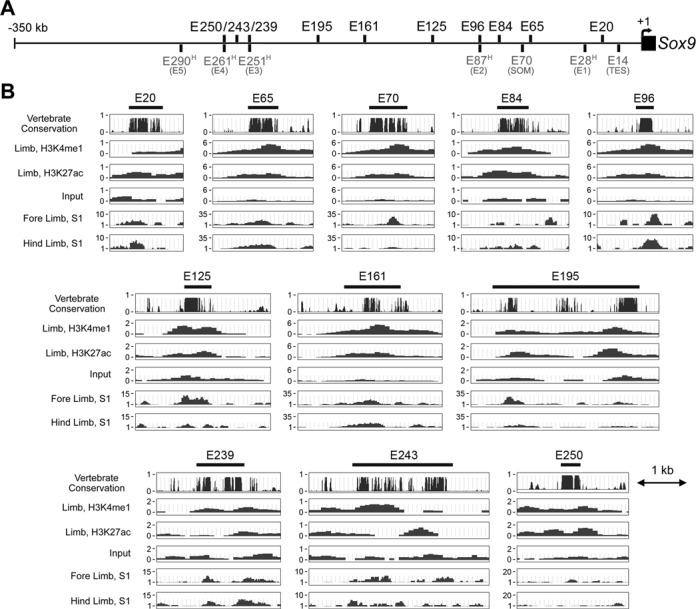
Identification of ten putative Sox9 enhancers. (A) Schematic showing the position of newly identified enhancers (black, top) and previously identified enhancers (gray, bottom) in the –350-kb region (horizontal line) located directly upstream of the mouse Sox9 gene (black box with arrow marking the transcription start site). Enhancers identified in the human genome are presented with an ‘H’ in superscript and number corresponding to their position relative to the human SOX9 gene. Names previously given to enhancers are indicated in parentheses. (B) Specific features of E70 and ten most promising enhancers. The black bar underneath each element acronym indicates the sequence tested in reporter genes. The top graph depicts sequence conservation in 30 vertebrate genomes. The next three schematics are profiles of histone marks for active/poised enhancers (H3K4me1) and active enhancers (H3K27ac) and input background detected in E14.5 mouse embryo limbs through ChIP-seq performed by the ENCODE/LICR project. The bottom two profiles show DNase I hypersensitive sites (S1) detected in E11.5 mouse limb buds by the ENCODE/University of Washington project. See Supplementary Figure S1 for related data.
E84 and E195 potently activate a Sox9 reporter in differentiated chondrocytes in vitro
Histone modifications and DNase I hypersensitivity are well-accepted signatures of gene regulatory regions, but they do not provide functional information on enhancers. To obtain such information, we constructed reporter plasmids using either pLER (19) or pWHERE (Figure 2A) as templates. Both types of plasmids contained up to four copies of a prospective enhancer cloned upstream of the Sox9 promoter and the E. coli β-galactosidase gene (lacZ). We transiently transfected the plasmids in four different cell types: rat chondrosarcoma derived RCS cells, which express a faithful phenotype of growth plate chondrocytes at the proliferating/early prehypertrophic stage (31,32; Liu, C.-F., Lefebvre, V., submitted for publication); primary chondrocytes freshly isolated from neonatal mouse rib cages and prone to dedifferentiation in culture; mouse embryo mesenchymal C3H10T1/2 cells, which are prochondrocytic and weakly express Sox9 (30,33); and human embryo kidney epithelial HEK-293 cells, which are nonchondrocytic and weakly express SOX9 (34,35). As previously seen for Sox9 promoter-pLER (19), Sox9 promoter-pWHERE was minimally active in all cells and was robustly activated (230-fold) in RCS cells by E70 (Figure 2B). Four copies of E84 and two copies of E195 activated the Sox9 promoter powerfully in RCS cells (∼80-fold), but other enhancers were inactive or hardly active in these cells. E195 increased the Sox9 promoter activity almost 30-fold in primary chondrocytes, but other enhancers were inactive in these cells. E195 was modestly active in C3H10T1/2 cells, as were E20 and E125. E250 was mildly active in HEK-293 cells (three- to nine-fold). E70, E84 and E195 were active as a single copy in RCS cells, but E195 was an order of magnitude more potent than E70 and E84 (Figure 2C). We concluded that the newly identified E84 and E195 enhancers might participate in Sox9 expression in chondrocytes in vivo and several of the other elements might have this function in mesenchymal cells or other types of cells.
Figure 2.
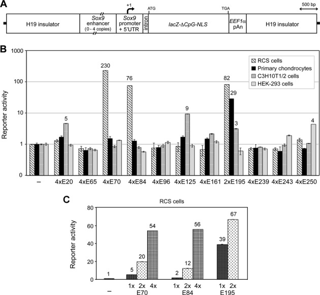
Schematic and activity of Sox9 enhancers in cultured cells. (A) Schematic of Sox9 enhancer-promoter-pWHERE reporters. Mouse H19 insulator sequences flank up to four copies of a Sox9 enhancer, the Sox9 307-bp proximal promoter and 364-bp 5′ untranslated region, a small intron, a modified lacZ coding sequence and the human EEF1α polyadenylation signal (pAn). The variable size of enhancers is shown with broken lines. The start of transcription (+1, angled arrow) and the start and end of translation are indicated. (B) Activities of Sox9 enhancer-promoter-pWHERE reporters in RCS cells, primary chondrocytes, C3H10T1/2 cells and HEK-293 cells. Reporters contained no enhancer (–), four tandem copies (4×) of most Sox9 enhancers, or two copies of E195. Only two copies of E195 were tested because this element is very large (3022 bp) compared to others (365–2064 bp) and more active than others. Reporter activities were normalized for transfection efficiency and calculated relative to that of the Sox9 promoter-only reporter in each cell type. Data are presented as the mean with standard deviation for technical triplicates in an experiment representative of several independent others. Activation folds ≥ 3 are indicated. (C) Activities of Sox9 enhancer-promoter-pLER reporters harboring up to four copies of E70, E84 or E195 in RCS cells.
E84, E195 and E250 activate a Sox9 reporter in the chondrocyte lineage in vivo
We assayed the activity of all new Sox9 enhancer-promoter-pWHERE reporters in transgenic mice. At least two independent transgenic mouse lines were generated per enhancer and found to yield comparable data. We therefore show results obtained for only one line per enhancer. We first collected embryos at E14.5 and stained them with the β-galactosidase substrate X-gal. The Sox9 promoter-only reporter showed no significant activity in any tissue (data not shown), as reported for the equivalent construct in pLER (19). Three enhancers showed activity in the chondrocyte lineage: E84, E195 and E250 (Figure 3). This was readily visible for E250 and E84 in whole-mount embryo staining, whereas strong staining in peripheral nerves and melanocytes blocked the view on internal tissues for E195 (Figure 3A). E250 and E84 showed complementary domains of activity in cartilage, E250 being most active in cartilage regions still containing prechondrocytes, such as the tip of digits, ribs and tail, and E84 in cartilage regions containing overtly differentiated chondrocytes, such as the middle segment of ribs, proximal tibia and lumbar vertebrae. Analysis of histology sections confirmed these findings (Figure 3B and C). In addition, it revealed that E195, like E84, was predominantly active in overtly developed cartilage. E195, however, was mostly active in columnar/proliferating chondrocytes of cartilage growth plates, whereas E84 was equally active in presumptive joint-lining, epiphyseal and columnar chondrocytes. Like Sox9, both enhancers were inactive by the time chondrocytes reached hypertrophic differentiation. Beside cartilage, all three enhancers showed activity in few other tissues, such as neocortex for E250 and E84, heart valves for E84, and peripheral nerves, kidney ureters and dorsal root ganglia for E195 (Supplementary Figure S2A). All these tissues express Sox9, except dorsal root ganglia, which express Sox10, a close relative of Sox9. E20, E65, E161 and E239 did not show activity in cartilage, but showed activity in several other tissues (Supplementary Figure S2B to F). Some sites were ectopic with regards to Sox9 expression, such as mesenchyme between limb skeletal elements and non-Sertoli cells (likely germ cells) in testis, but others, namely olfactory epithelium, upper lip, tooth mesenchyme and palatal shelf, were derivatives of cranial neural crest that expresses Sox9 (36,37). E96 could not activate the Sox9-promoter reporter, and E125 and E243 did not show reproducible activity (data not shown). Thus, in addition to E70, the –350-kb region upstream of Sox9 harbors multiple enhancers active in Sox9 expression domains. At least four of them direct transgene expression in cranial neural crest-derived cells and three of them in chondrocytes. We focused the rest of the study on the latter.
Figure 3.
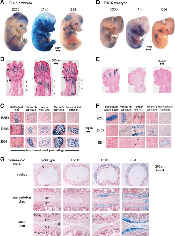
Activities of E250, E195 and E84 in transgenic mice. (A) E14.5 transgenic embryos stained with X-gal. The transgenes were carrying four copies of E250, two copies of E195, or four copies of E84 upstream of the Sox9 promoter in the pWHERE reporter. Tissues expressing the reporters are seen in blue. (B) Longitudinal sections through paws and forearms. J, phalangeal joint. E, C and H stand for the epiphyseal, columnar and hypertrophic zones of cartilage growth plates, respectively. (C) High-magnification pictures of sections through various types of cartilage at different stages of development. C, cartilage. J, presumptive joint, still filled with Sox9-expressing mesenchyme. T, joint tendinous capsule. (D) E12.5 transgenic embryos stained with X-gal. (E) Paw sections showing precartilaginous digital condensations (arrow). (F) Sections through distinct cartilage tissues. C, cartilage. G, root ganglia. (G) X-gal-stained sections through distinct cartilage types from 3-week-old pups. AC, articular cartilage. B, bone. C, cartilage. GP, growth plate cartilage. M, meniscal cartilage. NP, nucleus pulposus. Bone and nucleus pulposus are not Sox9-expressing tissues. See Supplementary Figures S2 and S3 for related data.
We analyzed E12.5 embryos to explore the activity of E250, E195 and E84 at the onset of chondrogenesis. E250 was active in all precartilaginous condensations, as illustrated for paws and ribs (Figure 3D–F). As seen at E14.5, it was no longer active in developing and developed cartilage, such as vertebral, Meckel's and basioccipital cartilage. E195 and E84 were silent in precartilaginous condensations, but E195 was active in developing and overtly developed cartilage, and E84 was active in the most advanced cartilage elements. Each enhancer showed activity in few noncartilaginous sites, as seen at E14.5 (Supplementary Figure S3A). At E11.5, E250 was active in already formed precartilaginous condensations, such as that of the humerus anlagen, but it was silent in the mesenchymal precursors of condensations, such as those filling limb buds (Supplementary Figure S3B and C). Cartilage structures are fully developed by the time pups can be weaned (21 days of age). At that time, E250 was virtually mute in all cartilage (Figure 3G). E195 activity was readily detectable in most chondrocytes of vertebral growth plates and the trachea, but only in few articular chondrocytes, whereas E84 was well active in all cartilage types.
Taken together, these data indicated that E84, E195 and E250 likely contribute to activate Sox9 in the chondrocyte lineage. E250 may render expression in condensing prechondrocytes, E195 in proliferating chondrocytes, and E84 in fully differentiated, nonhypertrophic chondrocytes.
SOX9 differentially regulates E250, E195, E84 and E70
SOX9 has been proposed to autoregulate its own expression, as do many cell-fate determining transcription factors. ChIP-seq assays in RCS cells revealed that the Sox9 –350-kb region was displaying strong marks of active enhancers at the levels of E195, E84 and E70 and that these enhancers were bound by SOX9 and SOX6 (Figure 4A). SOX6 and its close relative SOX5 are co-expressed with SOX9 in chondrocytes, but belong to a different SOX group than SOX9. They act redundantly to boost the activity of SOX9 on many cartilage enhancers and thereby potentiate the chondrogenic activity of SOX9 (30,38). The three proteins are therefore often referred to as the chondrogenic SOX trio. We hence tested whether SOX9 and SOX5/SOX6 co-activated the Sox9 cartilage enhancers. Co-transfection of HEK-293 cells with Sox9 reporters and SOX expression plasmids showed that SOX9 could powerfully activate E70 (22.5-fold), as previously described (19), but that SOX5 and SOX6 interfered with SOX9's activity (Figure 4B). In the absence of SOX protein overexpression, both E84 and E195 mediated downregulation of the Sox9 promoter by two-fold. Expression of either SOX5/SOX6 or SOX9 had little if any effect, but co-expression of SOX9 with SOX5 and/or SOX6 resulted in weak activation of E84 (2.2-fold) and robust activation of E195 (14.2- to 20.6-fold). E250 was weakly active in HEK-293 cells both without and upon SOX protein overexpression (2.7- to 4.2-fold). Similar results were obtained in C3H10T1/2 and COS-7 cells (data not shown). The relative effects of the SOX proteins on the four enhancers correlated well with ChIP-seq and reporter assay data obtained in RCS cells (Figures 2B and 4A and C). We concluded that these four Sox9 enhancers rely on distinct regulatory mechanisms, some of which involve SOX9 and SOX5/SOX6.
Figure 4.
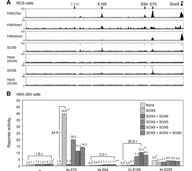
Binding and activation of Sox9 enhancers by SOX9 and SOX5/SOX6. (A) ChIP-seq data obtained for histone modifications, SOX9 and SOX6, and input material for each SOX protein in the –350-kb region upstream of Sox9 in RCS cells. Vertical arrows, location of the four cartilage enhancers. Vertical bars, summits of SOX peaks identified using MACS software. (B) Activities of Sox9 enhancers in HEK-293 cells co-transfected with Sox9 reporters and expression plasmids for no protein (–) or SOX proteins, as indicated. Reporter activities were normalized for transfection efficiency and calculated relative to that of the Sox9 promoter-only reporter. Data are presented as the mean with standard deviation for technical triplicates in an experiment representative of several independent others. Δ, P <0.01 in a t-test for the difference in Sox9 promoter activity caused by an enhancer in HEK-293 cells not forced to express SOX proteins. * and **, P <0.01 and <0.001, respectively, in a t-test for the difference in a reporter activity caused by SOX protein overexpression.
The chondrogenic SOX trio directly contributes to E195 activity
ChIP-seq and reporter data indicated that SOX9 and SOX6 are bound to E195 and capable of activating this enhancer, but they did not prove that the proteins directly bind to the enhancer. We therefore performed new experiments to test this possibility. ChIP-seq data revealed that the SOX proteins were bound to the 3′ third of E195 (Figure 5A). Accordingly, the conserved segments located in the E195 5′ third (E195A) and middle third (E195B) had no enhancer activity in RCS cells, whereas the 3′ third (E195C) and its conserved core (E195D) were as potent as the full-length enhancer (Figure 5A and B). Further truncations demonstrated that the 3′ fourth of E195D facilitated enhancer activity, but was not essential, whereas the 5′ fourth and third fourth (from the 5′ end) were critically needed (Figure 5C and D). Sequence analysis of E195D revealed four segments harboring putative binding sites for SOX9 and SOX5/SOX6 (Figure 5E). These segments indeed contained several sequences resembling the SOX consensus binding site (CT/ATTGT/AT/A). These sequences were presenting as inverted pairs separated by four nucleotides (A/TA/TCAAA/TGNNNNCT/ATTGT/AT/A) or as tandem repeats separated by no or few nucleotides (CT/ATTGT/AT/A[Ns]CT/ATTGT/AT/A). These two distinct configurations of SOX site pairs correspond to the motifs most often bound by SOX9 and SOX5/SOX6, respectively, in the RCS genome (Liu, C.-F., Lefebvre, V., submitted for publication). EMSAs with extracts from COS-7 cells forced to express SOX proteins showed that SOX5/SOX6 and SOX9 were able to bind to all four segments, except segment 3 (Figure 5F; Supplementary Figure S4A). SOX5/SOX6 preferred segment 1 to segment 2, whereas SOX9 preferred segment 2 to segment 1. Binding of the SOX proteins to segment 4 was apparent only upon antibody supershifting because other protein(s) formed a complex with this segment that co-migrated with the SOX/DNA complexes (Figure 5G). Mutations introduced in the reporters to prevent SOX protein binding revealed that the SOX-like motifs in segments 1 and 2 were necessary for enhancer activity in RCS cells (Figure 5H and Supplementary Figure S4B) and for induction of enhancer activity by SOX9 and SOX6 in HEK-293 cells (Figure 5I). Those present in segments 3 and 4 were not essential, but significantly contributed to full enhancer activity. These data thus consolidated evidence that the SOX trio directly participates in E195 activation in chondrocytes.
Figure 5.
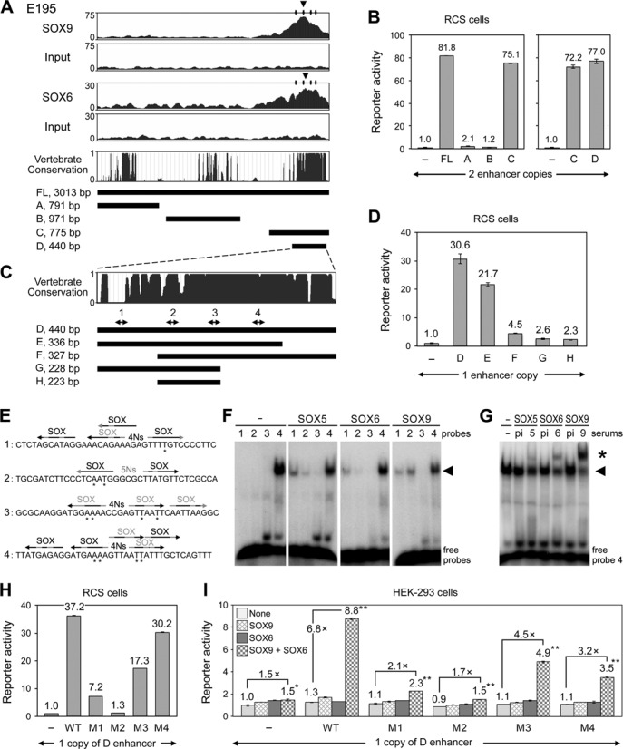
E195 is directly activated by the SOX trio. (A) ChIP-seq data, vertebrate conservation and schematics of the full-length E195 enhancer (FL) and segments A to D. The top four graphs show ChIP-seq profiles obtained in RCS cells for SOX9 and SOX6 and for respective input material at the level of E195. Large arrowheads, summits of SOX peaks identified by MACS software. Small arrows, SOX-binding regions (see panel E). The vertebrate conservation graph was obtained by comparing 30 genomes, from lamprey to human, in the UCSC genome browser. (B) Activity of Sox9 promoter reporters harboring two enhancer copies in RCS cells. (C) Conservation and schematics of E195D to H segments. Double arrows and numbers indicate putative the location of putative SOX-binding sites (see panel E). (D) Activity of reporters harboring one copy of E195D to E195H in RCS cells. (E) Probe sequences used for EMSA. Sequences are shown for the upper strand only. Each arrow represents a putative SOX-binding site. The black and gray shades highlight nucleotides that match and do not match the SOX domain-binding consensus CT/ATTGT/AT/A, respectively. SOX is written in black for sequences presenting at least five consensus nucleotides, and in gray for others. Inverted sites separated by four nucleotides (strong SOX9 site) or five nucleotides (weak SOX9 site) are indicated. Asterisks mark nucleotides mutated in reporters used in panels H and I. (F) EMSA with probes 1–4 and nuclear extracts from COS-7 cells forced to express no protein, SOX5, SOX6 or SOX9 (Supplementary Figure S4A shows that the extracts contained similar amounts of SOX protein). Note that probe 4 forms a complex with a nonspecific protein migrating at the same level as SOX/DNA complexes (arrowhead). (G) EMSA of extracts incubated with probe 4 and preimmune serum (pi) or antiserums against SOX5 (5), SOX6 (6) or SOX9 (9). Star, antibody supershifts. (H) Reporter activities generated by one copy of wild-type and mutant E195D enhancers in RCS cells. The M1 to M4 enhancers featured point mutations, as shown in panel E and Supplementary Table S2. (I) Same experiment as in panel G, but using HEK-293 cells co-transfected with SOX expression plasmids. Reporter activities are presented as described in Figures 2B and 4B. See Supplementary Figure S4A and B for related data.
The SOX trio is directly necessary but not sufficient for E84 robust activity
ChIP-seq data showed that SOX6 and SOX9 were binding to the 5′ half of E84 in RCS cells (Figure 6A). Accordingly, reporters containing only the 5′ half of the enhancer (E84A) or just its most conserved region (E84C) had strong activity, whereas a reporter containing only the 3′ half (E84B) was virtually inactive (Figure 6A and B). Truncations of E84C indicated that a 120-bp segment (E84E) was sufficient to generate vigorous enhancer activity (Figure 6C). Its 5′ and 3′ ends were both necessary to generate this activity, as deletion of either end (E84F and E84G) resulted in complete loss of activity. Sequence analysis of E84E revealed two segments harboring putative SOX5/SOX6 and SOX9 binding sites (Figure 6D). EMSA showed that both segments could bind SOX5/SOX6, but preferred SOX9 (Figure 6E). Mutations introduced in either segment to prevent SOX protein binding abrogated the activity of E84E in RCS cells (Supplementary Figure S4C and Figure 6F). Forced expression of SOX9 and SOX6 in HEK-293 cells resulted in weak activation of E84E, as seen for the full-length enhancer, and this effect was dependent upon the SOX binding sites (Figure 6G and Supplementary Figure S4C). Altogether, these data support the notion that E84 is a direct target of the SOX trio, but that additional mechanisms likely exist that allow the SOX trio to forcefully activate the enhancer in chondrocytes.
Figure 6.
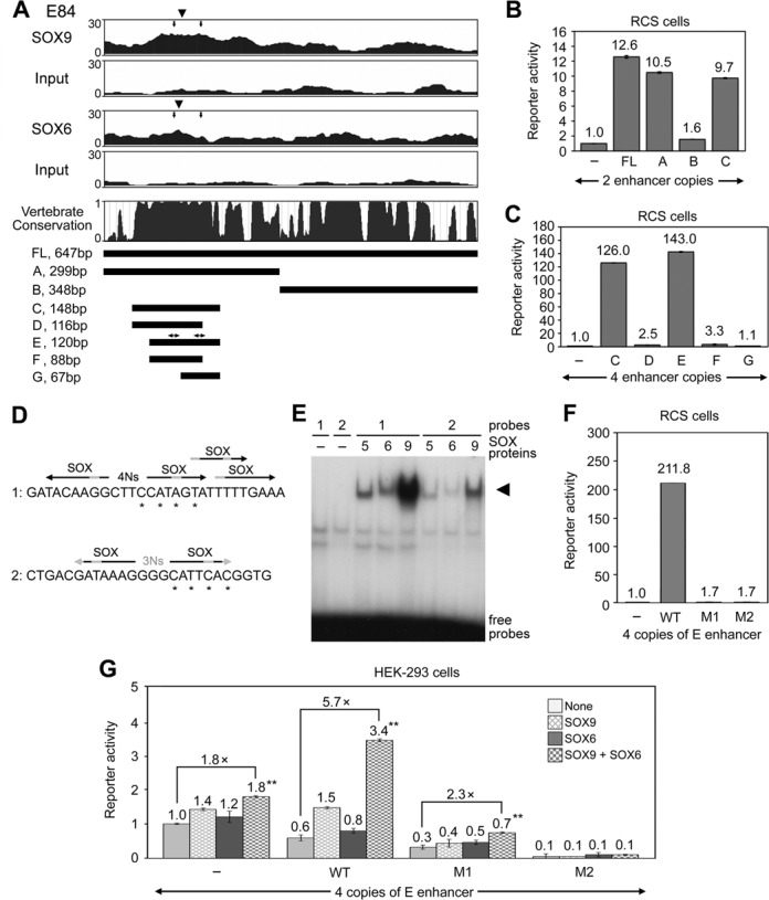
E84 activity involves the SOX trio. (A) ChIP-seq data, vertebrate conservation and schematics of the full-length E84 enhancer (FL) and segments A to G. The top four graphs show ChIP-seq profiles obtained in RCS cells for SOX9 and SOX6 and for respective input material at the level of E84. Large arrowheads, summits of SOX peaks identified using MACS software. Small arrows, SOX-binding regions (see panel D). The vertebrate conservation graph was obtained by comparing 30 genomes, from lamprey to human, in the UCSC genome browser. Double arrows above the E segment schematic indicate the positions of SOX-binding regions. (B) Activities of reporters harboring two copies of E84FL, A, B and C in RCS cells. (C) Activities of reporters harboring four copies of E84C to E84G in RCS cells. (D) EMSA probe sequences. Only upper strands are shown. See Figure 5E for labeling explanations. Asterisks mark nucleotides mutated in reporters tested in panels F and G. (E) EMSA with SOX protein-containing extracts from COS-7 cells and probes described in panel D. Arrowhead, SOX protein/DNA complexes. (F) Reporter activities generated by four copies of wild-type and mutant E84E enhancers in RCS cells. Mutations were as described in panel D. (G) Same experiment as in panel F, but in HEK-293 cells co-transfected with SOX expression plasmids. Reporter activities were calculated and are presented as described in Figures 2B and 4B. See Supplementary Figure S4A and C for related data.
Sox9 enhancers synergize with each other
Key cell fate determining transcription factors, such as the embryonic stem cell master regulators SOX2, OCT4, and KLF4, have been shown to regulate their own expression and that of other pivotal regulatory genes through super-enhancers, i.e. clusters of synergizing enhancers (39,40). To test whether the newly identified Sox9 enhancers might likewise synergize with each other, we cloned them in tandems in reporters. We did not find evidence of synergy between E84 and E195 in RCS cells (Figure 7A). Interestingly, however, each of E84 and E195 was able to synergize with E70, leading to activation of the Sox9 promoter 2.3- to 3.4-fold more efficiently than expected from the sum of individual enhancer activities. Such a synergy was not observed in SOX6/SOX9-expressing HEK-293 cells (Figure 7B), suggesting that chondrocytes may possess non-SOX proteins that help enhancer interactions. We thus concluded that E70, E84 and E195 might be engaged in a super-enhancer complex that generates strong expression of Sox9 in chondrocytes.
Figure 7.
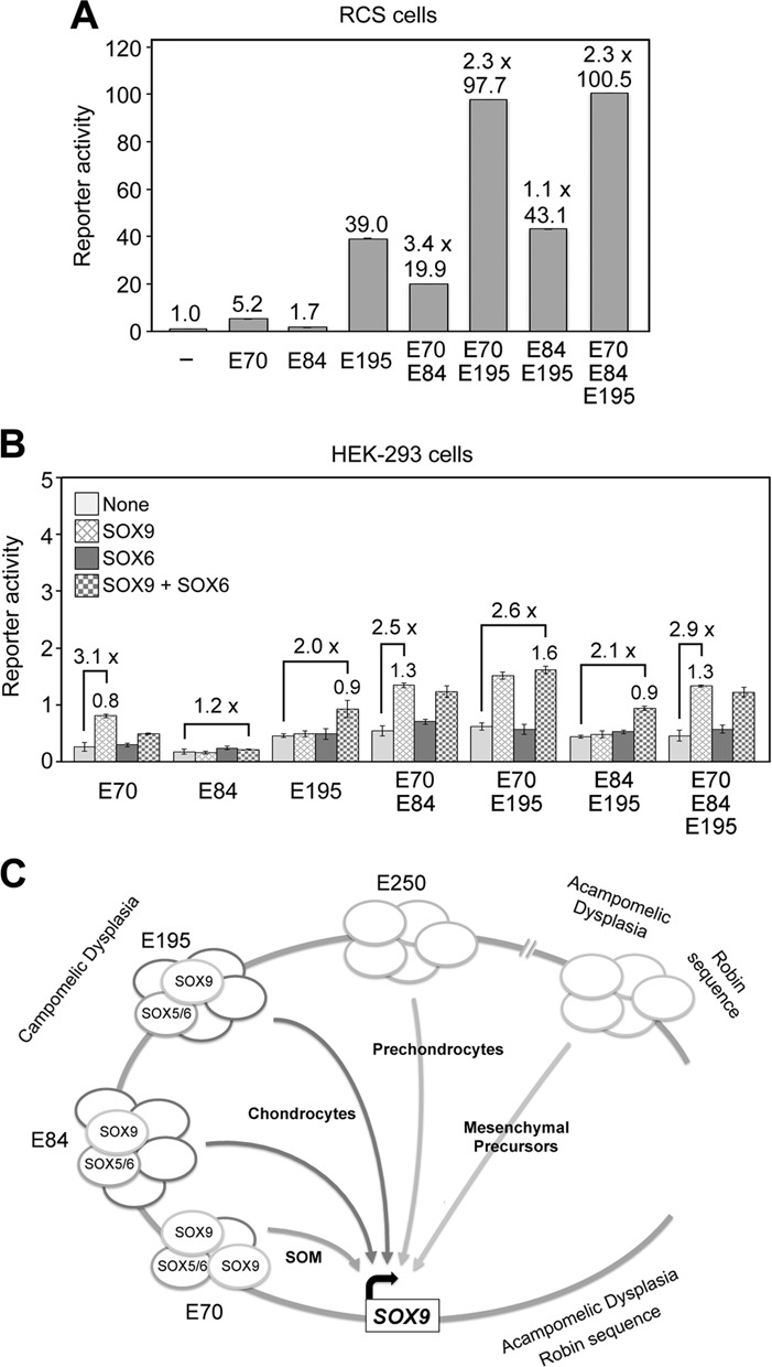
Sox9 enhancer synergy and model for the activation of SOX9 in chondrocytes. (A) Activities of reporters carrying one copy of E70, E84 or E195, or tandem combinations of one copy of these enhancers in RCS cells. Synergy between enhancers (1.1 to 3.4×) was calculated as the fold activation of the Sox9 promoter obtained relative to the sum of the activities of individual enhancers. (B) Activities of the same reporters as in panel A in HEK-293 cells co-transfected with SOX expression plasmids. The best activation fold of the Sox9 promoter achieved by SOX proteins (SOX9 alone or together with SOX6) is indicated for each reporter. (C) Model. See the first paragraph of Discussion for explanations.
DISCUSSION
This study moves forward understanding of mechanisms that drive SOX9 expression in the chondrocyte lineage and that are likely disrupted in campomelic dysplasia. It allows a major update of the previously proposed model that SOX9 expression is brought up by numerous enhancers spread over large distances (27,19; Figure 7C). This study has focused on the –350-kb region located directly upstream of SOX9. This region was formerly shown to home E70, a SOX9-driven enhancer active in most Sox9-expressing somatic tissues (19). The present study has revealed that three distinct enhancers with prominent activity in chondrocytes also reside in this region. The most distal one, E250, is specific to condensing prechondrocytes. More proximally, E195 has distinct activity in proliferating chondrocytes, and even more proximally, E84 indistinguishably targets all types of differentiated chondrocytes. E84 and E195 directly depend on SOX9 for activation, as does E70, but unlike E70, they also require SOX5/SOX6 and other proteins to generate potent, tissue-specific transactivation. E84 and E195 are able to synergize with E70 in growth plate chondrocytes. Their dissociation from SOX9 by chromosomal translocations occurring in the –350-kb region may thus explain the severe cartilage growth deficiency observed in campomelic dysplasia. Previous and new data indicate that several other enhancers, active in mesenchymal progenitors and in other SOX9-expressing cells, are located in the –350-kb region, farther upstream of this region, and downstream of SOX9. Removal of such enhancers by translocations in the –350-kb region may contribute to many of the dysmorphic features of campomelic dysplasia patients. Removal by translocations occurring further upstream of the –350-kb region or downstream of SOX9 likely explains the features of acampomelic dysplasia and Robin sequence.
E250 is uniquely active in chondrogenic mesenchymal cells condensing to form precartilaginous anlagen. Various signaling pathways are implicated in this important step in the generation and patterning of skeletal elements. They are elicited by Notch, transforming growth factor-beta (TGFβ) and bone morphogenetic protein ligands, but beside SOX9, other cell-specific transacting factors remain unknown (41–44). E250 is, to our knowledge, the first enhancer shown to have specific activity in prechondrocytes. We thus anticipate that it will be an instrumental tool to identify these factors. E250 is not bound by SOX9 in RCS cells and is not activated upon forced expression of SOX9 in mesenchymal cells. These results suggest that SOX9 is not implicated in the activity of E250. However, we cannot fully rule out this possibility. We have indeed observed that RCS cells, which model growth plate chondrocytes at the proliferating/early prehypertrophic stage, do not allow SOX9 to bind targets specific of other chondrocyte differentiation stages (Liu, C.-F., Lefebvre, V., submitted for publication). It thus remains possible that E250 is dependent on SOX9 and on factors that permit SOX9 binding to the enhancer in prechondrocytes. The Scherer group previously tested the human ortholog of E250, which they named E4, but they did not detect enhancer activity in transgenic mouse embryos (27). A possible explanation to account for the differences between the two studies is that the Scherer group cloned only one copy of E4 in their reporter, whereas we empowered our reporter by inserting four tandem copies of E250. In addition, unlike the Bagheri-Fam reporter, the pWHERE reporter that we used here included insulator sequences to protect transgene activity from site-of-integration effects, and it also featured a lacZ coding sequence optimized for expression and protein detection. We believe that these pWHERE features are significant because we observed in preliminary work that pWHERE was superior to our previously generated pLER reporter to generate strong and reproducible transgene activities.
E195, E84 and E70 overlap in activity in differentiated chondrocytes and all are dependent upon SOX9. There are, however, notable differences between these enhancers. E70 is active in most somatic tissues expressing Sox9, including all Sox9-expressing chondrocytic stages, and it is less active in cartilage than in other tissues. E195 is very active in proliferating chondrocytes, and also shows activity in a limited set of Sox9-expressing cells. E84 is the most specific of the three enhancers for chondrocytes, and it displays strong activity in all overtly developed cartilage types. E195 may upregulate Sox9 expression in growth plates. E84 may ensure maintenance of Sox9 expression in all chondrocytes. E70 may serve in chondrocytes as well as in other cell types to boost the activity of cell type-specific enhancers. Qualitative and quantitative differences in activities between the three enhancers were explained by showing that SOX5/SOX6 boost activation of E195 and E84 by SOX9, but have a repressor effect on E70. This result fits with evidence that SOX5/SOX6 are necessary for chondrocyte overt differentiation through stimulating activation of numerous cartilage-specific genes by SOX9, whereas they repress gene activation by SOX9 in several other cell lineages (30,38,45,46). This finding does not just identify additional chondrocyte enhancers controlled by the SOX trio, but it also uncovers that SOX5/SOX6 likely elevate Sox9 expression in differentiated chondrocytes. This role of the proteins had not been revealed before. Cartilage primordia were shown to form and express Sox9 at a seemingly normal level in Sox5/Sox6 double-null embryos, proving that SOX5/SOX6 are not necessary for Sox9 expression in early chondrocytes (36). However, the virtual inability of mutant primordia to further develop did not allow testing the contribution of SOX5/SOX6 to Sox9 expression in fully differentiated chondrocytes. The almost exclusive confinement of E84 activity to chondrocytes is likely related to the fact that the SOX trio, although necessary for enhancer activity, is not sufficient. The additional proteins that permit the trio to activate this enhancer only in chondrocytes remain to be identified. They may include inhibitory proteins in nonchondrocytic cells expressing SOX9, and permissive proteins in chondrocytes.
Besides E250, E195, E84 and E70, the –350-kb region comprises several regions evolutionarily conserved and displaying enhancer marks in developing limbs. In vitro and in vivo reporter assays ascertained that at least four of these regions are able to function as enhancers. One of them is E239. Named E3, it was previously shown by the Scherer group to be active in E8.5 to E10.5 embryos in the cranial neural crest and inner ear, important sites of Sox9 expression (27). Its activity was shown to be enhanced when fused with E250/E4 and a third element, called E5 and located 30 kb further upstream. In line with the first findings, we found strong activity of E239 in cranial neural crest-derived cells, namely in the olfactory epithelium, developing tongue, teeth and palate. The ability of E4/E5 to enhance E3 activity is intriguing with respect to our finding that E250/E4 is active primarily in prechondrocytic condensations. It is thus possible that E5, whose individual activity has not been tested, was responsible for enhancing the activity of E239/E3 rather than E250/E4. E20, E65 and E161 were found to have enhancer activity in transgenic embryos in sites where Sox9 expression is undetectable. Several of these sites, such as mesenchyme located between cartilage primordia, derived from Sox9-expressing cells. In the testis, E239 and E65 were inactive in Sertoli cells, which robustly express endogenous Sox9, and in mesenchyme, which derives from Sox9-expressing cells. In contrast, they were active in cells that likely correspond to germ cells. These cells do not express endogenous Sox9 and do not derive from Sox9-expressing cells (47). These enhancers are thus unlikely to contribute to the testis differentiating activity of Sox9. It is possible that the cloning of multimers just upstream of the Sox9 promoter allowed these enhancers to exhibit prolonged activity or an activity level that in the endogenous Sox9 locus escapes detection.
This study focused on the –350-kb region upstream of SOX9 based on evidence that most chromosomal translocations causing campomelic dysplasia occur within this region and based on evidence that a YAC transgene encompassing this region was sufficient to drive expression of a reporter gene in most of the Sox9 expression domain. The enhancers that we have identified in this region between –70kb and –250-kb do not explain, however, why a campomelic dysplasia patient who had a chromosomal breakpoint at 288–319 kb upstream of SOX9 presented with the full spectrum of skeletal features characteristic of the disease (48) and why another patient with a chromosomal breakpoint at –375kb was a classical case of mild campomelic dysplasia (23). Based on these two cases and our lack of identification of chondrocyte enhancers between 250 and 350 kb upstream of SOX9, we anticipate that one or several additional enhancers with important roles in the chondrocyte lineage may be located more than 375 kb upstream of SOX9. These enhancers may overlap in activity and even synergize with the enhancers identified in the present study or they may be active in chondrogenic mesenchymal cells before precartilaginous condensation.
The presence of many enhancers around SOX9 is not a surprise as more and more genes, especially those involved in pivotal regulatory functions, are associated with multiple enhancers, collectively called super-enhancers. This has been shown, for instance, in embryonic stem cells (39,40). This enhancer multiplicity may serve several purposes, besides synergistic activation of gene expression. In the case of enhancers controlling SOX9 expression in the chondrocyte lineage, it may significantly contribute to the great diversity in skeleton patterning existing among and within vertebrate species. For instance, polymorphism in E195 could generate variable levels of SOX9 expression in the growth plate and thereby differential growth rates. Polymorphism in E250, which is active in condensing prechondrocytes, could result in variations in the number, size and shape of cartilage structures. Polymorphism in E84, which is active in all types of differentiated chondrocytes, could account for variation in both size and strength of temporary and adult cartilage tissues. While polymorphisms in enhancer sequences may underlie variations within what is considered a normal range, mutations drastically affecting the activity of enhancers may underlie many types of yet unexplained genetically inherited diseases. Severe diseases could include campomelic dysplasia and acampomelic dysplasia. Milder diseases could comprise minor skeletal dysplasias leading to early-onset osteoarthritis. As most SOX9 enhancers are active in several cell types, polymorphisms and disease-causing mutations are likely, as in campomelic dysplasia, to affect both skeletal and nonskeletal processes.
In conclusion, this study greatly illuminates current understanding of the molecular control of SOX9 expression in chondrocytes and other cells. It has shed new light on possible disease mechanisms, including but not limited to campomelic dysplasia. Furthermore, it has provided new tools to further dissect mechanisms underlying normal and pathological processes and to design novel strategies to prevent and treat skeletal and other types of diseases.
SUPPLEMENTARY DATA
Supplementary Data are available at NAR Online.
Acknowledgments
We thank Peter Dy, Abhilash Gadi, Jocelyn Ohlemacher, Jamie Shah and Dan Coleman for scientific and technical help. We thank the Case Western Reserve University Gene Targeting and Transgenic Facility for generating transgenic founder mice.
FUNDING
National Institute of Arthritis and Musculoskeletal and Skin Diseases (NIAMS) [AR46249 and AR60016 to V.L.]; F32 postdoctoral fellowship (AR063548 to T.J.M.); Morgenthaler postdoctoral fellowships (to C.F.L. and T.J.M.); Arthritis National Research Foundation (ANRF) (1405PB to P.B.). Funding for open access charge: Cleveland Clinic.
Conflict of interest statement. None declared.
REFERENCES
- 1.Foster J.W., Dominguez-Steglich M.A., Guioli S., Kwok C., Weller P.A., Stevanovic M., Weissenbach J., Mansour S., Young I.D., Goodfellow P.N., et al. Campomelic dysplasia and autosomal sex reversal caused by mutations in an SRY-related gene. Nature. 1994;372:525–530. doi: 10.1038/372525a0. [DOI] [PubMed] [Google Scholar]
- 2.Wagner T., Wirth J., Meyer J., Zabel B., Held M., Zimmer J., Pasantes J., Bricarelli F.D., Keutel J., Hustert E., et al. Autosomal sex reversal and campomelic dysplasia are caused by mutations in and around the SRY-related gene SOX9. Cell. 1994;79:1111–1120. doi: 10.1016/0092-8674(94)90041-8. [DOI] [PubMed] [Google Scholar]
- 3.Unger S., Scherer G., Superti-Furga A. GeneReviews. Seattle: University of Washington; 2013. Campomelic dysplasia. [PubMed] [Google Scholar]
- 4.Akiyama H., Chaboissier M. C., Behringer R. R., Rowitch D. H., Schedl A., Epstein J. A., de Crombrugghe B. Essential role of Sox9 in the pathway that controls formation of cardiac valves and septa. Proc. Natl. Acad. Sci. U.S.A. 2004;101:6502–6507. doi: 10.1073/pnas.0401711101. [DOI] [PMC free article] [PubMed] [Google Scholar]
- 5.Stolt C.C., Wegner M. SoxE function in vertebrate nervous system development. Int. J. Biochem. Cell Biol. 2010;42:437–440. doi: 10.1016/j.biocel.2009.07.014. [DOI] [PubMed] [Google Scholar]
- 6.Akiyama H., Lefebvre V. Unraveling the transcriptional regulatory machinery in chondrogenesis. J. Bone. Miner. Metab. 2011;29:390–395. doi: 10.1007/s00774-011-0273-9. [DOI] [PMC free article] [PubMed] [Google Scholar]
- 7.Reginensi A., Clarkson M., Neirijnck Y., Lu B., Ohyama T., Groves A.K., Sock E., Wegner M., Costantini F., Chaboissier M.C., et al. SOX9 controls epithelial branching by activating RET effector genes during kidney development. Hum. Mol. Genet. 2011;20:1143–1153. doi: 10.1093/hmg/ddq558. [DOI] [PMC free article] [PubMed] [Google Scholar]
- 8.Kawaguchi Y. Sox9 and programming of liver and pancreatic progenitors. J. Clin. Invest. 2013;123:1881–1886. doi: 10.1172/JCI66022. [DOI] [PMC free article] [PubMed] [Google Scholar]
- 9.Svingen T., Koopman P. Building the mammalian testis: origins, differentiation, and assembly of the component cell populations. Genes Dev. 2013;27:2409–2426. doi: 10.1101/gad.228080.113. [DOI] [PMC free article] [PubMed] [Google Scholar]
- 10.Bi W., Deng J. M., Zhang Z., Behringer R. R., de Crombrugghe B. Sox9 is required for cartilage formation. Nat. Genet. 1999;22:85–89. doi: 10.1038/8792. [DOI] [PubMed] [Google Scholar]
- 11.Akiyama H., Chaboissier M.C., Martin J.F., Schedl A., de Crombrugghe B. The transcription factor Sox9 has essential roles in successive steps of the chondrocyte differentiation pathway and is required for expression of Sox5 and Sox6. Genes Dev. 2002;16:2813–2828. doi: 10.1101/gad.1017802. [DOI] [PMC free article] [PubMed] [Google Scholar]
- 12.Dy P., Wang W., Bhattaram P., Wang Q., Wang L., Ballock R.T., Lefebvre V. Sox9 directs hypertrophic maturation and blocks osteoblast differentiation of growth plate chondrocytes. Dev. Cell. 2012;22:597–609. doi: 10.1016/j.devcel.2011.12.024. [DOI] [PMC free article] [PubMed] [Google Scholar]
- 13.Henry S.P., Liang S., Akdemir K.C., de Crombrugghe B. The postnatal role of Sox9 in cartilage. J. Bone Miner. Res. 2012;27:2511–2525. doi: 10.1002/jbmr.1696. [DOI] [PMC free article] [PubMed] [Google Scholar]
- 14.Fukui N., Ikeda Y., Ohnuki T., Tanaka N., Hikita A., Mitomi H., Mori T., Juji T., Katsuragawa Y., Yamamoto S., et al. Regional differences in chondrocyte metabolism in osteoarthritis: a detailed analysis by laser capture microdissection. Arthritis Rheum. 2008;58:154–163. doi: 10.1002/art.23175. [DOI] [PubMed] [Google Scholar]
- 15.Haag J., Gebhard P.M., Aigner T. SOX gene expression in human osteoarthritic cartilage. Pathobiology. 2008;75:195–199. doi: 10.1159/000124980. [DOI] [PubMed] [Google Scholar]
- 16.Zhou Z.Q., Ota S., Deng C., Akiyama H., Hurlin P.J. Mutant activated FGFR3 impairs endochondral bone growth by preventing SOX9 downregulation in differentiating chondrocytes. Hum. Mol. Genet. 2014;24:1764–1773. doi: 10.1093/hmg/ddu594. [DOI] [PMC free article] [PubMed] [Google Scholar]
- 17.Matheu A., Collado M., Wise C., Manterola L., Cekaite L., Tye A.J., Canamero M., Bujanda L., Schedl A., Cheah K.S., et al. Oncogenicity of the developmental transcription factor Sox9. Cancer Res. 2012;72:1301–1315. doi: 10.1158/0008-5472.CAN-11-3660. [DOI] [PMC free article] [PubMed] [Google Scholar]
- 18.Kumar D., Lassar A.B. The transcriptional activity of Sox9 in chondrocytes is regulated by RhoA signaling and actin polymerization. Mol. Cell. Biol. 2009;29:4262–4273. doi: 10.1128/MCB.01779-08. [DOI] [PMC free article] [PubMed] [Google Scholar]
- 19.Mead T.J., Wang Q., Bhattaram P., Dy P., Afelik S., Jensen J., Lefebvre V. A far-upstream (-70 kb) enhancer mediates Sox9 auto-regulation in somatic tissues during development and adult regeneration. Nucleic Acids Res. 2013;41:4459–4469. doi: 10.1093/nar/gkt140. [DOI] [PMC free article] [PubMed] [Google Scholar]
- 20.Bi W., Huang W., Whitworth D.J., Deng J.M., Zhang Z., Behringer R.R., de Crombrugghe B. Haploinsufficiency of Sox9 results in defective cartilage primordia and premature skeletal mineralization. Proc. Natl. Acad. Sci. U. S. A. 2001;98:6698–6703. doi: 10.1073/pnas.111092198. [DOI] [PMC free article] [PubMed] [Google Scholar]
- 21.Meyer J., Südbeck P., Held M., Wagner T., Schmitz M.L., Bricarelli F.D., Eggermont E., Friedrich U., Haas O.A., Kobelt A., et al. Mutational analysis of the SOX9 gene in campomelic dysplasia and autosomal sex reversal: lack of genotype/phenotype correlations. Hum. Mol. Genet. 1997;6:91–98. doi: 10.1093/hmg/6.1.91. [DOI] [PubMed] [Google Scholar]
- 22.Velagaleti G.V., Bien-Willner G.A., Northup J.K., Lockhart L.H., Hawkins J.C., Jalal S.M., Withers M., Lupski J.R., Stankiewicz P. Position effects due to chromosome breakpoints that map approximately 900 Kb upstream and approximately 1.3 Mb downstream of SOX9 in two patients with campomelic dysplasia. Am. J. Hum. Genet. 2005;76:652–662. doi: 10.1086/429252. [DOI] [PMC free article] [PubMed] [Google Scholar]
- 23.Leipoldt M., Erdel M., Bien-Willner G.A., Smyk M., Theurl M., Yatsenko S.A., Lupski J.R., Lane A.H., Shanske A.L., Stankiewicz P., et al. Two novel translocation breakpoints upstream of SOX9 define borders of the proximal and distal breakpoint cluster region in campomelic dysplasia. Clin. Genet. 2007;71:67–75. doi: 10.1111/j.1399-0004.2007.00736.x. [DOI] [PubMed] [Google Scholar]
- 24.Gordon C.T., Tan T.Y., Benko S., Fitzpatrick D., Lyonnet S., Farlie P.G. Long-range regulation at the SOX9 locus in development and disease. J. Med. Genet. 2009;46:649–656. doi: 10.1136/jmg.2009.068361. [DOI] [PubMed] [Google Scholar]
- 25.Benko S., Fantes J.A., Amiel J., Kleinjan D.J., Thomas S., Ramsay J., Jamshidi N., Essafi A., Heaney S., Gordon C.T., et al. Highly conserved non-coding elements on either side of SOX9 associated with Pierre Robin sequence. Nat. Genet. 2009;41:359–364. doi: 10.1038/ng.329. [DOI] [PubMed] [Google Scholar]
- 26.Wunderle V.M., Critcher R., Hastie N., Goodfellow P.N., Schedl A. Deletion of long-range regulatory elements upstream of SOX9 causes campomelic dysplasia. Proc. Natl. Acad. Sci. U. S. A. 1998;95:10649–10654. doi: 10.1073/pnas.95.18.10649. [DOI] [PMC free article] [PubMed] [Google Scholar]
- 27.Bagheri-Fam S., Barrionuevo F., Dohrmann U., Gunther T., Schule R., Kemler R., Mallo M., Kanzler B., Scherer G. Long-range upstream and downstream enhancers control distinct subsets of the complex spatiotemporal Sox9 expression pattern. Dev. Biol. 2006;291:382–397. doi: 10.1016/j.ydbio.2005.11.013. [DOI] [PubMed] [Google Scholar]
- 28.Sekido R., Lovell-Badge R. Sex determination involves synergistic action of SRY and SF1 on a specific Sox9 enhancer. Nature. 2008;453:930–934. doi: 10.1038/nature06944. [DOI] [PubMed] [Google Scholar]
- 29.Han Y., Lefebvre V. L-Sox5 and Sox6 drive expression of the aggrecan gene in cartilage by securing binding of Sox9 to a far-upstream enhancer. Mol. Cell. Biol. 2008;8:4999–5013. doi: 10.1128/MCB.00695-08. [DOI] [PMC free article] [PubMed] [Google Scholar]
- 30.Lefebvre V., Li P., de Crombrugghe B. A new long form of Sox5 (L-Sox5), Sox6 and Sox9 are coexpressed in chondrogenesis and cooperatively activate the type II collagen gene. EMBO J. 1998;17:5718–5733. doi: 10.1093/emboj/17.19.5718. [DOI] [PMC free article] [PubMed] [Google Scholar]
- 31.King K.B., Kimura J.H. The establishment and characterization of an immortal cell line with a stable chondrocytic phenotype. J. Cell. Biochem. 2003;89:992–1004. doi: 10.1002/jcb.10571. [DOI] [PubMed] [Google Scholar]
- 32.Mukhopadhyay K., Lefebvre V., Zhou G., Garofalo S., Kimura J.H., de Crombrugghe B. Use of a new rat chondrosarcoma cell line to delineate a 119-base pair chondrocyte-specific enhancer element and to define active promoter segments in the mouse pro-alpha 1(II) collagen gene. J. Biol. Chem. 1995;270:27711–27719. doi: 10.1074/jbc.270.46.27711. [DOI] [PubMed] [Google Scholar]
- 33.Taylor S.M., Jones P.A. Multiple new phenotypes induced in C3H10T1/2 and 3T3 cells treated with 5-azacytidine. Cell. 1979;17:771–779. doi: 10.1016/0092-8674(79)90317-9. [DOI] [PubMed] [Google Scholar]
- 34.Graham F.L., Smiley J., Russell W.C., Nairn R. Characteristics of a human cell line transformed by DNA from human adenovirus type 5. J. Gen. Virol. 1977;36:59–74. doi: 10.1099/0022-1317-36-1-59. [DOI] [PubMed] [Google Scholar]
- 35.Bhattaram P., Penzo-Méndez A., Kato K., Bandyopadhyay K., Gadi A., Taketo M.M., Lefebvre V. SOXC proteins amplify canonical WNT signaling to secure nonchondrocytic fates in skeletogenesis. J. Cell Biol. 2014;207:657–671. doi: 10.1083/jcb.201405098. [DOI] [PMC free article] [PubMed] [Google Scholar]
- 36.Yamashiro T., Wang X.P., Li Z., Oya S., Aberg T., Fukunaga T., Kamioka H., Speck N.A., Takano-Yamamoto T., Thesleff I. Possible roles of Runx1 and Sox9 in incipient intramembranous ossification. J. Bone Miner. Res. 2004;19:1671–1677. doi: 10.1359/JBMR.040801. [DOI] [PubMed] [Google Scholar]
- 37.Forni P.E., Wray S. Neural crest and olfactory system: new prospective. Mol. Neurobiol. 2012;46:349–360. doi: 10.1007/s12035-012-8286-5. [DOI] [PMC free article] [PubMed] [Google Scholar]
- 38.Smits P., Li P., Mandel J., Zhang Z., Deng J.M., Behringer R.R., de Crombrugghe B., Lefebvre V. The transcription factors L-Sox5 and Sox6 are essential for cartilage formation. Dev. Cell. 2001;1:277–290. doi: 10.1016/s1534-5807(01)00003-x. [DOI] [PubMed] [Google Scholar]
- 39.Whyte W.A., Orlando D.A., Hnisz D., Abraham B.J., Lin C.Y., Kagey M.H., Rahl P.B., Lee T.I., Young R.A. Master transcription factors and mediator establish super-enhancers at key cell identity genes. Cell. 2013;153:307–319. doi: 10.1016/j.cell.2013.03.035. [DOI] [PMC free article] [PubMed] [Google Scholar]
- 40.Hnisz D., Abraham B.J., Lee T.I., Lau A., Saint-André V., Sigova A.A., Hoke H.A., Young R.A. Super-enhancers in the control of cell identity and disease. Cell. 2013;155:934–947. doi: 10.1016/j.cell.2013.09.053. [DOI] [PMC free article] [PubMed] [Google Scholar]
- 41.Karlsson C., Lindahl A. Notch signaling in chondrogenesis. Int. Rev. Cell Mol. Biol. 2009;275:65–88. doi: 10.1016/S1937-6448(09)75003-8. [DOI] [PubMed] [Google Scholar]
- 42.Wang W., Rigueur D., Lyons K.M. TGF-beta signaling in cartilage development and maintenance. Birth Defects Res., Part C. 2014;102:37–51. doi: 10.1002/bdrc.21058. [DOI] [PMC free article] [PubMed] [Google Scholar]
- 43.Kozhemyakina E., Lassar A.B., Zelzer E. A pathway to bone: signaling molecules and transcription factors involved in chondrocyte development and maturation. Development. 2015;142:817–831. doi: 10.1242/dev.105536. [DOI] [PMC free article] [PubMed] [Google Scholar]
- 44.Lim J., Tu X., Choi K., Akiyama H., Mishina Y., Long F. BMP-Smad4 signaling is required for precartilaginous mesenchymal condensation independent of Sox9 in the mouse. Dev. Biol. 2015;400:132–138. doi: 10.1016/j.ydbio.2015.01.022. [DOI] [PMC free article] [PubMed] [Google Scholar]
- 45.Lefebvre V. The SoxD transcription factors–Sox5, Sox6, and Sox13–are key cell fate modulators. Int. J. Biochem. Cell. Biol. 2010;42:429–432. doi: 10.1016/j.biocel.2009.07.016. [DOI] [PMC free article] [PubMed] [Google Scholar]
- 46.Reiprich S., Wegner M. From CNS stem cells to neurons and glia: Sox for everyone. Cell Tissue Res. 2015;359:111–124. doi: 10.1007/s00441-014-1909-6. [DOI] [PubMed] [Google Scholar]
- 47.Akiyama H., Kim J.E., Nakashima K., Balmes G., Iwai N., Deng J.M., Zhang Z., Martin J.F., Behringer R.R., Nakamura T., et al. Osteo-chondroprogenitor cells are derived from Sox9 expressing precursors. Proc. Natl. Acad. Sci. U.S.A. 2005;102:14665–14670. doi: 10.1073/pnas.0504750102. [DOI] [PMC free article] [PubMed] [Google Scholar]
- 48.Pfeifer D., Kist R., Dewar K., Devon K., Lander E.S., Birren B., Korniszewski L., Back E., Scherer G. Campomelic dysplasia translocation breakpoints are scattered over 1 Mb proximal to SOX9: evidence for an extended control region. Am. J. Hum. Genet. 1999;65:111–124. doi: 10.1086/302455. [DOI] [PMC free article] [PubMed] [Google Scholar]
Associated Data
This section collects any data citations, data availability statements, or supplementary materials included in this article.