

Abstract
Background
HIV-1 gp120/gp41 is heavily modified by n-linked carbohydrates that play important roles either in correct folding or in shielding vulnerable viral protein surfaces from antibody recognition.
Methods
In our previous work, 25 potential N-linked glycosylation sites (PNGS) of a CRF07_BC isolate of HIV-1 were individually mutated, and the resulting effects on infectivity and antibody-mediated neutralization were evaluated. In order to further understand the functional role of these PNGS, we generated double and multiple mutants from selected individual PNGS mutants. The effects were then evaluated by examining infectivity and sensitivity to antibody-mediated neutralization by neutralizing monoclonal antibodies (nMAbs) and serum antibodies from HIV-1 positive donors.
Results
Infectivity results showed that, among the twelve combined PNGS mutants, only 197M.1 (N197D/N301Q) lost infectivity completely, while all others (except for 197M.6) showed reduced viral infectivity. In terms of neutralization sensitivity to known nMAbs, we found that adding N463Q mutation to all the gp120 mutants containing N197D significantly increased neutralization sensitivity to VRC01 and VRC03, suggesting N197 and N463 have a strong synergistic effect in regulating the neutralizing sensitivity of HIV-1 to the anti-CD4bs nMAbs VRC01/VRC03. Structural analysis based on the available structures of gp120 alone and in complex with CD4 and various nMAbs elucidates a molecular rationale for this experimental observation.
Conclusions
The data indicate that N463 plays an important role in regulating the CD4bs MAbs VRC01/VRC03 sensitivity in the genetic background of N197D mutation of gp120, which should provide valuable information for a better understanding of the interplay between HIV-1 and VRC01/03.
Keywords: HIV, N-linked glycosylation site, Pseudovirus, Change of Neutralization Sensitivity, Structural Modeling and Analysis
Introduction
Induction of an antibody response capable of neutralizing diverse human immunodeficiency virus type 1 (HIV-1) isolates is a critical step for an effective vaccine that can protect against HIV-1 infection. Although vaccines have thus far failed to induce broadly neutralizing antibody responses, it is reported that approximately 1 to 30% of HIV-1-infected subjects eventually develop potent and broadly reactive neutralizing antibodies1–3. Most broadly neutralizing antibodies in chronic HIV-1 sera have been shown to target the region around the CD4 binding site, the CD4 induced site, the glycan-dependent or quaternary epitopes on the gp120, and the conserved elements of V3 region4–8. In recent years, based on new neutralizing antibody screening systems, many potent and broadly MAbs have been isolated9–11.
The CD4-binding site (CD4bs) on gp120 is essential for viral entry, and is highly conserved compared with other envelope regions. The CD4bs contains the key epitopes recognized by several broadly neutralizing antibodies obtained so far. MAb b12, a CD4bs-MAb, was isolated from a phage display library and can neutralize about 40% of known HIV-1 isolates12, 13. Recently, a battery of anti-CD4bs MAbs, including VRC01/VRC0311, 3BNC11714, PGV0415, and NIH45-4616 show significantly higher potency and wider breadth compared with b12. VRC01 can neutralize ~90% of HIV-1 isolates at a low inhibitory concentration (IC50) and structural studies show that it achieves this neutralization by precisely recognizing the initial site of CD4 attachment on HIV-1 gp120. Mutagenesis studies indicated that VRC01 contacts within the gp120 loop D, the CD4 binding loop, and the V5 region were necessary for optimal VRC01 neutralization17, 18.
N-linked glycans are important for processing of gp120 that ultimately influences envelop conformation, oligomerization, receptor binding, membrane fusion and viral entry, infectivity, and antibody neutralization of HIV virus19–23. In our previous work, we individually mutated the 25 PNGS of a CRF07_BC isolate to study the effect on viral infectivity and antibody-mediated neutralization finding that certain N-linked glycosylation sites are critical for virus to infect target cells and protect the virus from neutralizing by monoclonal antibodies24. The partial deglycosylated HIV envelope protein was previously shown to enhance the immunogenicity of some epitopes17, 25, 26. Some studies reported that combined PNGS elimination at specific sites in clade B HIV virus can increase the neutralizing sensitivity to certain nMAbs27, 28. For example, mutating N186 and N197 simultaneously on gp120 of 3 CRF01_AE clones increased sensitivity to b12, whereas the mutating N186 or N197 individually was not sufficient to increase b12 sensitivity29. Also the combined N460 and N463 mutation on V5 region of 08_B'C viruses resulted in higher VRC01 sensitivity than each single mutation alone30. The conserved PNGS in gp41 were also eliminated individually and in combination to test the viral replication31. Clade 07_BC viruses, which have been one of the most predominantly circulated HIV-1 strains in China, have a somewhat different glycan arrangement than the B/AE and other clade viruses, and the elimination of combined PNGS located on different regions of the gp120 of the predominant Clade of HIV viruses in China has not been evaluated before.
In order to further understand the biological functions of the gp120 PNGS of the Clade 07_BC HIV-1 strain predominantly circulating in China, we constructed combined PNGS mutants based on previous single PNGS mutants to evaluate their infectivity and neutralizing sensitivity to nMAbs. We identified certain combined PNGS mutants that become highly sensitive to certain nMAbs, suggesting that the PNGS sites on these mutants likely play a critical role for shielding the virus from being recognized by these nMAbs and targeting/recognition by the host humoral responses.
Methods
Cells and Plasmid
TZM-bl cells, Env-deficient HIV-1 backbone (pSG3ΔEnv) were obtained from the US National Institutes of Health (NIH) AIDS Research and Reference Reagent Program (ARRRP), as contributed by John C. Kappes, Xiaoyun Wu, and Tranzyme (Birmingham, AL). The 293FT cells were obtained from Invitrogen (Carlsbad, CA).
MAbs & Positive Serum
MAbs 2F5, 4E10, 2G12 and b12 were obtained from the International AIDS Vaccine Initiative (IAVI, New York, NY); MAbs 2F5 and 4E10 were contributed by Hermann Katinger (Institute of Applied Microbiology, Vienna, Austria) and b12 was contributed by Dennis Burton and Carlos Barbas (The Scripps Research Institute, La Jolla, CA); MAbs VRC01, VRC03, PG9, PG16 were obtained from the ARRRP (NIH).
For use in the study, 20 subtype BC HIV-1 infected positive sera were obtained from chronically HIV-1-infected blood donors in main HIV-1 epidemic regions of China. Individual samples were coded based on the region of China. These samples were collected from Beijing (bj20, bj22), Sichuan (sc59r), Guangdong (gd64), Yunnan (yn99r, yn148r), Guangxi (gx66, gx75, gx76, gx77, gx78, gx79, gx82, gx85, gx87, gx93, gx94, gx95) province and Xinjiang autonomous region (xj16, xj50). Samples were stored at −70 °C and freezing/thawing cycles were avoided. All serum samples were heat-inactivated at 56 °C for 1 h before use.
Elimination of PNGS by Mutagenesis
The motif for an N-linked glycosylation site is Asn-X-Thr/Ser, where X can be any amino acid except proline32. Elimination of PNGS was performed using site-directed mutagenesis, by changing an asparagine (N) to a glutamine (Q) or aspartate (D). The wt env gene was inserted into pcDNA 3.1D/V5-His-TOPO (Invitrogen) as a template for mutagenesis. Mutagenesis was performed as described previously24. Standard PCR and cloning procedure were used to obtain the mutant clones. The entire env gene of each mutant was sequenced to confirm mutation.
Pseudovirus Preparation, Infectivity, Titration and Neutralization Assays
Pseudoviruses were produced by co-transfection of 293FT cells (>90% confluency in a 25 cm2 rectangular canted neck cell culture flask, Corning, USA) with 5.3 µg pSG3ΔEnv plasmid and 2.7 µg Env-expressing plasmids using the Lipofectamine 2000 reagent (Invitrogen). Supernatants were harvested 48 hr after transfection, filtered (0.45-µm pore size), and stored at −80 °C. The concentration of HIV-1 Gag p24 antigen in viral supernatants was measured by enzyme-linked immunosorbent assay (ELISA) (Vironostika HIV-1 antigen micro-ELISA system; bioMérieux, Boxtel, The Netherlands).
A fixed amount of pseudovirus (equivalent to 1.0 ng p24 antigen) was added to TZM-bl cells at 70−80% confluency in a 96-well plate in the presence of 15 µg/ mL DEAE-dextran, in a total volume of 200 µL. 48 hr after infection, the luciferase activity in infected cells was measured using the Bright-Glo™ luciferase assay system (Promega, Madison, WI). Relative infectivity was calculated by dividing the Log10 (RLU of mutant) by Log10 (RLU of wt).
The 50% tissue culture infectious dose (TCID50) of a single infectious pseudovirus batch was determined in TZM-bl cells, as described previously33.
Neutralization was measured as a reduction in luciferase expression after a single-round infection of TZM-bl cells with pseudoviruses according to previously published method34.
Structural Modeling
The full-length HIV FE gp120 was generated using the homology modeling software Modeller 9.1335. The gp120 pdb structures 4nco, 2ny7, 3ngb, and 3se8 were used as templates for modeling and glycans were modeled from 4nco. The interfacial residues that make up the defined epitope/paratope for the gp120 and protein ligands where calculated using PDBe PISA v1.48 server 'Protein interfaces, surfaces and assemblies' service PISA at the European Bioinformatics Institute (http://www.ebi.ac.uk/pdbe/prot_int/pistart.html)36.
Results
Construction of the Combined PNGS Mutants and Viral Infectivity
In the previous study, the asparagine residue in all 25 PNGS on the wild-type gp120/41 of the HIV strain FE were mutated individually to glutamine or aspartate at the following positions: 88 (on C1 of gp120); 133, 142, 156, 160 (on V1); 181 (on V2); 197, 234, 241, 262, 289 (on C2); 301 (on V3); 339, 355 (on C3); 392, 408, 411 (on V4 loop); 442, 448 (on C4); 463, 466 in V5; 611, 616, 625, 637 (on gp41) (residue positions on gp120/41 are based on HXB2 numbering, Supplemental Fig. 1).
The effects of these individual PNGS mutants on nMAbs-mediated neutralization have been previously examined24. Here, we generated twelve combined PNGS mutants that contain different combinations of the selected PNGS point mutations to evaluate their influence on infectivity and neutralization of the resulting mutant viruses. The twelve combined PNGS mutants constructed in this study were shown in Supplemental Table 1. Eleven of the twelve mutants (except for M46) contain the N197D mutation, and eight of them contain N197D/N463Q mutations. All mutants were confirmed by sequencing.
Among all the combined mutants studied here, only 197M.1 (N197D/N301Q) has completely lost infectivity. Other multiple mutants showed no significant reduction of viral infectivity when compared to the two single point mutants, N197D or N301Q (Fig. 1).
Figure 1. Infectivity of the wt HIV strain and the PNGS mutants (see Supplemental Table 1 for the listed mutants).

Infectivity is shown as relative luminescence units (RLU) in a logarithmic scale. Relative infectivity was calculated by dividing the Log10 (RLU of mutant) by Log10 (RLU of wt). The data represent the means of three independent experiments, and the error bars indicate the standard deviations from the means.
Effect of Combined PNGS Mutations on Neutralization by nMAbs
The non-infectious mutant 197M.1 (N197D/N301Q) was excluded from the further neutralization assay study. All other mutant viruses on Supplemental Table 1, together with some single mutants, were examined for neutralization by nMAbs. In previous work, the single N197D mutant showed an obvious increase in susceptibility to neutralization by b12 (~17-fold increase) and VRC03 (~37-fold increase), as well as a modest 2-fold increase to VRC01 when compared to wild-type24. However, the neutralization result for the eight combined PNGS mutants in this study showed a much more dramatic increase in susceptibility to neutralization by VRC01/VRC03 compared to the single N197D or to wild-type (Table 1, Fig. 2). For example, mutants 197M.3 (N197D/N463Q), 197M.4 (N197D/N463Q/N442Q), 197M.5 (N197D/N463Q/N625Q), 197M.8 (N197D/N463Q/N625Q/N442Q/N339Q/N448) and 197M.9 (N197D/N463Q/N625Q/N442Q/N339Q) showed a 867-fold increase in susceptibility to neutralization by VRC03 when compared with wild-type, whereas mutants 197M.10 (N197D/N463Q/N625Q/N442Q/N339Q/N466Q) and 197M.11 (N197D/N463Q/N625Q/N442Q/N339Q) showed a ~1300-fold increase, and 197M.2 (N197D/N625Q), 197M.6 (N197D/N625Q/N442Q) and 197M.7 (N197D/N625Q/N442Q/N339Q) showed a ~100-fold increase (Table 1, Fig. 2). As for the neutralization by another nMAb VRC01, mutants 197M.3, 197M.4, 197M.5, 197M.8, 197M.9, 197M.10 and 197M.11 showed a ~275–420 fold increase, whereas 197M.2, 197M.6 and 197M.7 had only ~2-fold increase in neutralizing sensitivity compared to wild-type. M46 (N625Q/N463Q) showed no effect on VRC01/VRC03 mediated neutralization. However, all other combined PNGS mutants containing N197D mutation showed little changes of sensitivity to b12 or other nMAbs (PG9, PG16, 2F5, 4E10 and 2G12) when compared to the N197D single point mutant (Table 1, Fig. 2).
Table 1.
Neutralization of Env glycosylation mutants by nMAbs
| nMAbs | |||||||||
|---|---|---|---|---|---|---|---|---|---|
| Env | Region | 2F5 | 4E10 | b12 | VRC03 | VRC01 | PG9 | PG16 | 3869 |
| FE(WT) | - | 2.224 (100) | 2.986 (100) | 0.088 (100) | 2.601 (100) | 5.500 (100) | 1.415 (100) | 7.600 (100) | >25 (100) |
| N197D | V1/V2 | 0.396 (562) | 0.477 (626) | 0.005 (1760) | 0.070 (3716) | 2.355 (234) | 16.123 (9) | >25 (<30) | 5.540 (>451) |
| N301Q | V3 | 0.210 (1059) | 0.585 (510) | 0.040 (220) | 1.235 (211) | 2.330 (236) | 0.875 (162) | 14.884 (51) | ND |
| N339Q | C3 | 1.127 (197) | 1.579 (189) | 0.083 (106) | 1.799 (145) | 4.899 (112) | 1.664 (85) | 14.644 (52) | ND |
| N442Q | C4 | 0.781 (285) | 0.934 (320) | 0.045 (196) | 0.791 (329) | 4.120 (133) | 1.650 (86) | 16.111 (47) | ND |
| N448Q | C4 | 0.635 (350) | 0.694 (430) | 0.057 (154) | 2.917 (89) | 4.987 (110) | 0.878 (161) | 13.386 (57) | ND |
| N463Q | V5 | 0.763 (291) | 0.895 (334) | 0.056 (157) | 2.170 (120) | 3.450 (159) | 2.544 (56) | 16.333 (47) | ND |
| N466Q | V5 | 1.339 (166) | 1.397 (214) | 0.087 (101) | 2.139 (122) | 3.204 (172) | 3.070 (46) | 16.378 (46) | ND |
| N611Q | gp41 | 1.836 (121) | 0.704 (424) | 0.116 (76) | 3.103 (84) | 5.431 (101) | 1.797 (79) | 15.830 (48) | ND |
| N625Q | gp41 | 0.400 (556) | 0.577 (518) | 0.041 (215) | 3.329 (78) | 5.398 (102) | 1.069 (132) | 16.450 (46) | ND |
| M46 | combination | 0.435 (511) | 0.628 (475) | 0.149 (59) | 1.734 (150) | 3.683 (149) | 2.160 (66) | 16.560 (46) | ND |
| 197M.2 | combination | 0.237 (938) | 0.329 (908) | 0.008 (1100) | 0.031 (8390) | 2.685 (205) | 12.694 (11) | >25 (<30) | ND |
| 197M.3 | combination | 0.363 (613) | 0.389 (768) | 0.004 (2200) | 0.003 (86700) | 0.020 (27500) | 16.105 (9) | >25 (<30) | ND |
| 197M.4 | combination | 0.486 (458) | 0.706 (423) | 0.004 (2200) | 0.003 (86700) | 0.018 (30556) | 15.755 (9) | >25 (<30) | ND |
| 197M.5 | combination | 0.372 (598) | 0.585 (510) | 0.005 (1760) | 0.003 (86700) | 0.020 (27500) | 16.360 (9) | >25 (<30) | ND |
| 197M.6 | combination | 0.284 (783) | 0.523 (571) | 0.006 (1467) | 0.043 (6049) | 2.043 (269) | 16.230 (9) | >25 (<30) | ND |
| 197M.7 | combination | 0.253 (879) | 0.399 (748) | 0.005 (1760) | 0.023 (11309) | 1.564 (352) | 16.378 (9) | >25 (<30) | ND |
| 197M.8 | combination | 0.284 (783) | 0.463 (645) | 0.004 (2200) | 0.003 (86700) | 0.017 (32353) | 15.611 (9) | >25 (<30) | ND |
| 197M.9 | combination | 0.457 (487) | 0.700 (427) | 0.005 (1760) | 0.003 (86700) | 0.014 (39286) | 16.580 (9) | >25 (<30) | ND |
| 197M.10 | combination | 0.464 (479) | 0.886 (337) | 0.004 (2200) | 0.002 (130050) | 0.017 (32353) | 16.177 (9) | >25 (<30) | ND |
| 197M.11 | combination | 0.435 (511) | 0.536 (557) | 0.003 (2933) | 0.002 (130050) | 0.013 (42308) | 16.325 (9) | >25 (<30) | ND |
Both IC50(µg/mL) and Percentage of neutralization sensitivity relative to WT (%) were showed in each box.
Percentage of neutralization sensitivity relative to WT (%) = IC50 WT / IC50 mutant.
ND: not detected
Figure 2. Fold difference of neutralizing sensitivity of PNGS mutants to the three available Anti-CD4bs neutralizing nMAbs.

Fold difference was calculated by dividing the mean IC50 (wt) by mean IC50 (mutant), and was expressed in logarithm. The mean IC50 of three independent experiments was shown in Table 1.
The results shown above indicate that whenever a combined mutant of the HIV FE strain (a BC clade virus) contains N197Q together with N463Q, a dramatic increase of the neutralizing sensitivity to VRC01/VRC03 is observed. To investigate whether the combination of N197Q and N463Q mutation has the same effect in other clade viruses to these CD4bs MAbs neutralization, a clade B virus B05 and a clade AE virus GX74.20 were tested. The N197Q mutation alone in the two viruses showed a ~2–4 fold increase in susceptibility to neutralization by VRC01/VRC03, whereas the N463Q mutation alone showed no effect (Supplemental Fig. 2). The combination of N197Q and N463Q showed a less dramatic, but still significant increase in susceptibility to neutralization by VRC01/VRC03, ranging between 8 to 20 fold increase. For the sensitivity to b12, similar to the behavior of the FE strain mutants, the combined N197Q and N463Q mutations in these two clade viruses showed no obvious changes when compared with the N197Q single mutant (Supplemental Fig. 2).
Effect of PNGS Mutations on Neutralization by Serum Antibodies
Here we used serum collected from 20 subjects infected with subtype BC isolates to test the single and combined PNGS mutants for neutralization phenotype. The ID50 for neutralization is shown in Table 2. Among the single PNGS mutants, N197D had a 2−3-fold ID50 increase in the seven sera of the serum panel; N301Q showed a 2−5-fold ID50 increase in eight of the serum panel; N442Q showed a 2−5-fold ID50 increase in seven of the serum panel; and the N625Q made a 2−4-fold ID50 increase in seven of the serum panel. In addition, the N611Q mutant was neutralized by serum gx66, gx76 with a titer increase of over three-fold compared to the wild-type virus, and the N637Q mutant was neutralized by serum yn148r with a five-fold titer increase compared to the wild-type virus. A few PNGS mutations also decrease the neutralizing sensitivity to HIV-1 positive serum. The N197D mutation resulted a 2−fold reduction in serum xj50 neutralization, this neutralization reduction also happened in the N611Q mutation to serum yn99r, N289D to serum bj22 and gx75, N448Q to serum sc59r and N625Q to serum yn99r. These data suggest that the presence of the PNGS in the C2 (N197), V3 (N301), C4 (N442) regions and gp41 (N625) protect FE from antibody-mediated neutralization by the serum panel.
Table 2.
Neutralization of Env glycosylation mutants by serum antibodies.
| Serum/ID50 | |||||||||||||||||||||
|---|---|---|---|---|---|---|---|---|---|---|---|---|---|---|---|---|---|---|---|---|---|
| Env | region | xj16 | bj20 | bj22 | xj50 | sc59r | gd64 | gx66 | gx75 | gx76 | yn148r | gx77 | gx78 | gx79 | gx82 | gx85 | gx87 | gx93 | gx94 | gx95 | yn99r |
| FE(WT) | - | 1494 | 75 | 239 | 800 | 302 | 205 | 156 | 90 | 246 | 1309 | 402 | 188 | 624 | 528 | 150 | 62 | 232 | 543 | 201 | 125 |
| N87D | C1 | 1893 | 54 | 251 | 930 | 253 | 200 | 206 | 119 | 197 | 1152 | 559 | 354 | 555 | 319 | 122 | 62 | 152 | 514 | 202 | 137 |
| N133Q | V1 | 2734 | 82 | 234 | 1130 | 531 | 216 | 191 | 90 | 217 | 1284 | 452 | 188 | 734 | 485 | 84 | 62 | 186 | 451 | 191 | 83 |
| N142Q | V1 | 1961 | 109 | 445 | 1287 | 372 | 280 | 279 | 225 | 406 | 1989 | 730 | 222 | 1557 | 886 | 177 | 76 | 243 | 841 | 318 | 207 |
| N197D | C2 | 1226 | 275 | 284 | 316 | 396 | 164 | 214 | 273 | 377 | 1498 | 540 | 249 | 1262 | 1214 | 223 | 85 | 145 | 1654 | 410 | 291 |
| N289D | C2 | 1508 | 99 | 103 | 824 | 290 | 213 | 181 | 39 | 283 | 1244 | 492 | 183 | 832 | 561 | 122 | 62 | 140 | 527 | 222 | 131 |
| N301Q | V3 | 3215 | 235 | 375 | 950 | 327 | 307 | 232 | 131 | 229 | 1510 | 1187 | 162 | 799 | 694 | 171 | 137 | 1182 | 1681 | 601 | 270 |
| N339Q | C3 | 1611 | 98 | 250 | 976 | 263 | 241 | 207 | 152 | 333 | 1607 | 641 | 244 | 809 | 549 | 161 | 73 | 256 | 704 | 236 | 150 |
| N355Q | C3 | 2253 | 59 | 208 | 1584 | 175 | 183 | 112 | 102 | 186 | 707 | 670 | 197 | 1005 | 986 | 115 | 58 | 248 | 1059 | 185 | 84 |
| N392Q | V4 | 2058 | 67 | 200 | 1387 | 285 | 209 | 180 | 107 | 235 | 1344 | 461 | 174 | 628 | 414 | 126 | 70 | 294 | 636 | 216 | 111 |
| N408Q | V4 | 1478 | 80 | 268 | 1129 | 261 | 225 | 183 | 157 | 309 | 1266 | 502 | 216 | 690 | 572 | 169 | 72 | 252 | 611 | 240 | 157 |
| N411Q | V4 | 2051 | 116 | 317 | 1181 | 420 | 308 | 195 | 182 | 324 | 1096 | 415 | 217 | 613 | 487 | 124 | 63 | 220 | 600 | 203 | 109 |
| N442Q | C4 | 2636 | 292 | 488 | 1228 | 563 | 366 | 278 | 481 | 303 | 1329 | 685 | 323 | 1240 | 1135 | 253 | 77 | 395 | 1017 | 414 | 174 |
| N448Q | C4 | 1075 | 82 | 224 | 1092 | 151 | 204 | 180 | 69 | 248 | 1507 | 396 | 168 | 590 | 493 | 83 | 59 | 206 | 1164 | 221 | 127 |
| N463Q | V5 | 1868 | 119 | 338 | 1492 | 347 | 249 | 208 | 130 | 325 | 1376 | 634 | 254 | 2250 | 1146 | 129 | 75 | 270 | 1836 | 192 | 196 |
| N466Q | V5 | 1811 | 72 | 224 | 1222 | 382 | 197 | 154 | 116 | 337 | 1587 | 517 | 259 | 1121 | 741 | 187 | 77 | 211 | 790 | 261 | 144 |
| N611Q | gp41 | 2322 | 68 | 223 | 1350 | 206 | 195 | 766 | 172 | 750 | 1108 | 368 | 200 | 1089 | 762 | 99 | 44 | 173 | 898 | 134 | 63 |
| N625Q | gp41 | 2293 | 199 | 396 | 1506 | 413 | 242 | 242 | 181 | 260 | 1544 | 904 | 376 | 1034 | 2136 | 286 | 87 | 350 | 790 | 409 | 227 |
| N637Q | gp41 | 1979 | 82 | 374 | 1402 | 267 | 180 | 160 | 117 | 249 | 6714 | 539 | 210 | 719 | 532 | 133 | 63 | 231 | 1090 | 219 | 137 |
| M46 | combination | 2537 | 110 | 240 | 1008 | 386 | 215 | 128 | 190 | 320 | 1238 | 487 | 380 | 685 | 1580 | 165 | 75 | 225 | 1128 | 586 | 152 |
| 197M.2 | combination | 1904 | 563 | 311 | 544 | 403 | 320 | 394 | 237 | 375 | 1401 | 574 | 438 | 1404 | 2480 | 287 | 106 | 288 | 1850 | 594 | 292 |
| 197M.3 | Combination | 1860 | 483 | 365 | 525 | 413 | 332 | 251 | 865 | 423 | 2533 | 612 | 305 | 8210 | 8005 | 290 | 110 | 235 | 8350 | 581 | 525 |
| 197M.4 | combination | 2018 | 350 | 320 | 480 | 475 | 230 | 305 | 580 | 420 | 1580 | 690 | 428 | 1350 | 1860 | 235 | 80 | 230 | 1523 | 625 | 538 |
| 197M.5 | combination | 2033 | 375 | 335 | 456 | 660 | 256 | 330 | 883 | 415 | 1695 | 699 | 465 | 8335 | 6853 | 440 | 83 | 260 | 7895 | 674 | 550 |
| 197M.6 | combination | 2943 | 625 | 333 | 557 | 814 | 344 | 386 | 987 | 470 | 1671 | 1053 | 418 | 1437 | 2386 | 430 | 101 | 184 | 1592 | 613 | 425 |
| 197M.7 | combination | 2493 | 727 | 325 | 563 | 866 | 325 | 363 | 1996 | 654 | 2236 | 1904 | 553 | 2403 | 2987 | 447 | 93 | 212 | 3043 | 652 | 404 |
| 197M.8 | combination | 1223 | 567 | 291 | 476 | 345 | 341 | 440 | 446 | 471 | 3075 | 983 | 510 | 8737 | 7083 | 301 | 89 | 1329 | 12346 | 579 | 666 |
| 197M.9 | combination | 2774 | 620 | 305 | 486 | 789 | 390 | 400 | 1143 | 438 | 2702 | 944 | 636 | 9166 | 7481 | 442 | 392 | 1252 | 9477 | 643 | 755 |
| 197M.10 | combination | 2062 | 617 | 241 | 413 | 1072 | 367 | 382 | 1071 | 695 | 4135 | 1493 | 434 | 16294 | 7786 | 353 | 109 | 3313 | 11378 | 582 | 658 |
| 197M.11 | combination | 2557 | 640 | 303 | 402 | 1038 | 358 | 3281 | 1085 | 3018 | 4742 | 1560 | 403 | 21096 | 7546 | 454 | 79 | 3452 | 11078 | 621 | 660 |
Boxes are color coded as follows: yellow, 2–10 fold increase of ID50; orange, > 10-fold increase of ID50; gray, > 2-fold decrease of ID50.
Interestingly, compared with the single point mutants, most of the combined PNGS mutants displayed higher neutralizing sensitivity to the serum. All the eleven combined PNGS mutants (except for M46) showed a 2−34-fold ID50 increase in 7−15 of the serum panels, with the mutant 197M.11 (N197D/N463Q/N625Q/N442Q/N339Q) showing 2–34 fold increase in 15 tested serum samples compare to the respective single PNGS mutants and wild-type virus. 197M.11 also showed a ~9-fold and ~4-fold increase in neutralization sensitivity to serum gx66 and gx76, compared to the other mutants (Table 2). Another interesting observation is that, while some the single PNGS mutants showed no effect on neutralizing sensitivity by serum, combining such single PNGS mutants somehow displayed increased neutralizing sensitivity to serum antibodies, as shown by the sensitivity increase of mutants 197M.6 (N197D/N625Q/N442Q) and 197M.7 (N197D/N625Q/N442Q/N339Q) to serum sc59R, and mutants 197M.8 (N197D/N463Q/N625Q/N442Q/N339Q/N448), 197M.9 (N197D/N463Q/N625Q/N442Q/N339Q) and 197M.10 (N197D/N463Q/N625Q/N442Q/N339Q/N466Q) to serum gx93 (Table 2).
Structural Modeling and Rationalization
In order to understand the molecular basis for the observed increase of sensitivity to VRC01/VRC03 when N463Q mutation is added on top of N197D, we analyzed the available structural information for gp120 structures alone and its complexes with CD4 and various nMAbs18, 30, 37–40. In doing so, the antibody bound gp120 structures available to date were aligned to the FE gp120 model, and the structural basis by which specific glycosylation positions influence neutralization were examined. Of the 25 potential PNGS sites, both N197 and N463 map to the CD4 binding “face” of gp120 (Fig. 3A, 3B). However, N197 and N463 are not directly on top of the CD4 binding interface, neither N197 nor N463 appears to occlude CD4 binding (Fig. 3A, 3B), suggesting that they should not directly interfere with or participate in CD4 binding for infection. This is consistent with the observed viral infectivity for the mutants containing N197/N463 residues (Fig. 1). As for the antibody neutralization, nMAb b12 relies solely on heavy chain contacts that partially overlap with the CD4 binding site40, and unlike the VRC antibodies, neutralization of HIV FE by b12 is not as dramatically affected by FE gp120 N197D/N463Q mutation (Fig. 2, Table 1). Based on the structural modeling, N197 and N463 of FE gp120 should not present steric hindrance to b12 antibody binding to the CD4 binding site (Fig. 3C, 3D). Compared to the binding interfaces for b12 and CD4, the N197 and N463 residues of FE gp120 are located directly adjacent to or within theVRC01 epitope/paratope (Fig. 3E, 3F), and these two residues are located within the VRC03 epitope/paratope (Fig. 3G, 3H). The degree of steric occlusion on the structural model appears to be directly related to the level of our observed sensitivity changes of neutralization by VRC01 and VRC03, where VRC03 binding to gp120 should be more hindered by glycosylation at N197/N463 compared to VRC01 binding, which is consistent with the observation that mutation at N197/N463 has even more marked increase of neutralizing sensitivity to VRC03 than VRC01. Therefore, the effects of FE gp120 glycosylation at N197 and N463 on neutralization sensitivity can be directly correlated to the structural properties that rationalize nMAb recognition and binding.
Figure 3. Structural basis for effects of N197D/N463Q mutations of HIV FE gp120 on neutralization of by different CD4bs nMAbs.
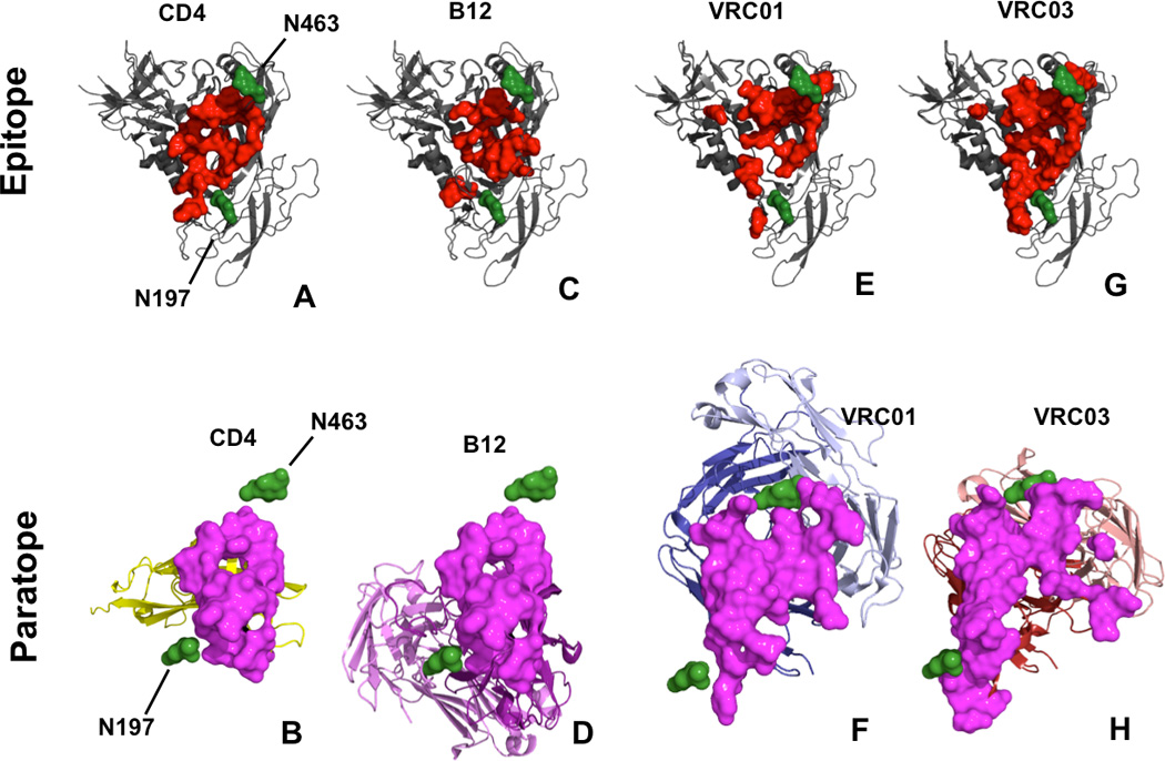
Shown is a FE gp120 structural model aligned to the structures of gp120 binding to CD4, b12, VRC01, or VRC03. (Top panels) The respective gp120 epitopes (red) are mapped with respect to the N197 and N463 glycans (green). (Bottom panels) The CD4, b12, VRC01, and VRC03 paratopes (magenta) for binding FE gp120 are shown with respect to N197 and N463 glycans of FE gp120. Combined mutations of N197D and N463Q did not affect viral infectivity and minimally affect b12-mediated neutralization. In contrast, the N197D and N463Q drastically increased neutralization sensitivity to VRC01 and VRC03.
Discussion
Among the twelve combined PNGS mutants of the HIV-1 gp120/gp41, eleven of them had no significant loss of infectivity, suggesting that these PNGS are not important for gp120/gp41 folding, maturation, viral assembly, and cell entry during infection. Surprisingly, one of the twelve mutants, the double mutant 197M.1 (N197D and N301Q) resulted in a complete loss of viral infectivity, which is rather surprising considering that as all the other combined mutants contain the N197D mutation had infectivity, and some of them having additional 3 to 7 point mutations on top of N197D. Our previous study indicated that N197D or N301Q mutation alone did not have severe impact on the infectivity24, which is also confirmed here (Fig. 1). However, the combination of the two point mutations proved to be deadly for the virus, which suggests that such double mutant somehow abolish certain essential function(s) required for viral infection.
For the eleven combined PNGS mutants that showed viral infectivity, we examined their sensitivity to seven broadly neutralizing MAbs41, 42. The neutralization ability of CD4bs MAbs VRC03 showed a marked increase for combined PNGS mutants that contained the both N197D and N463Q mutations, with a remarkable ~800–1300 fold increase over wt FE. These mutants also showed a ~300–400 fold increase in sensitivity to VRC01. Compared with the combined PNGS mutants, only the N197D and the N442Q among the single mutants showed relatively modest increase in neutralizing sensitivity to VRC03, with N197D showing 37-fold increase and N442Q showing 3-fold to VRC03. N197D also showed a 2-fold increase sensitivity to VRC01. However, N463Q single mutant, together with most other single mutants alone, showed little effect on neutralization by VRC01/VRC03, b12 or other nMAbs (Table 1).
Two pairs of combined mutants 197M.2/197M.5, and 197M.7/197M.9 (Supplemental Table 1), which differ only in containing N463Q mutation or not, showed significant difference in VRC01/VRC03 sensitivity, with the mutants containing N463Q displaying much higher VRC01/03 sensitivity (Fig. 2, Table 1). In fact, all the seven mutants that contain both N197D and N463Q in them showed the same dramatic increase of sensitivity to VRC01/VRC03, whereas those combined mutants with N197D but without N463Q (mutants 197M.2, 197M.6, and 197M.7) showed little effect (Supplemental Table 1 & Table 1). Interestingly, further mutations of the PNGS up to five sites on top of the N197D/N463Q mutation appear to have very minor additional effect on the sensitivity level to the two VRC nMAbs. Thus, N463Q mutation seems to play an important role in determining the neutralizing sensitivity to nMAbs VRC01/VRC03 in the N197D mutant background. In other words, even though the effect of N463Q mutation alone did little in altering the sensitivity to nMAbs VRC01/VRC03 (with ~1.5 fold increase), combining N463Q mutation with N197D has an unexpected synergistic effect in increasing the sensitivity to VRC01/VRC03 by a remarkable 300–1300 fold (Fig. 2, Table 1), and a less pronounced, but still significant change was also observed in clade B/AE virus in our experiment (Supplemental Fig. 2). The fact that neither single mutant of N197 and N463 exhibits a drastic change in neutralization sensitivity may be a bit confusing/confounding but in considering the structural properties of the N-linked sugars (being rather bulky and unstructured), it seems not surprising that changes neutralization sensitivity do not appear until both N197 and N463 are mutated. Firstly, N197 and N463 are both located on flexible loops within gp120 and so their positioning within gp120 may be quite dynamic. This property has direct implications when considering interactions with CD4bs antibodies. Secondly, and in addition to the structural properties of N197/463, the structural properties of the sugars (mainly mannose 5 or 9 with regards to gp120) linked to glycosylated N residues should be taken into account. Such synergistic effect could be the result of the absence of glycosylation on the PNGS mutant, as suggested by the structural modeling. Alternatively, the PNGS mutation could also affect the Env conformation to increase the sensitivity to some of the nMAbs.
Very interestingly, the remarkable increase of sensitivity of mutants containing N463Q/N197D was only observed for nMAbs VRC01/VRC03, and not for other tested nMAbs (Table 1). This is rather intriguing especially considering b12 belongs to the class of CD4bs nMAbs as VCR01/03. The results further confirm that b12 neutralizes HIV-1 with mechanisms that are not identical to those used by VCR01/03, despite the fact that they all bind to the general area of CD4bs15, 17, 18, 39, 43–45.
The neutralization phenotype of combined mutants using the serum antibodies showed a much less profound impact on sensitivity to the serum antibody than to the nMAbs, with ~20 fold as the highest increase of sensitivity for mutant 10 and 11 (197M.10 and 197M.11). Another interesting observation is that, while the combined mutants containing N197D/N463Q did not show continued increase of neutralizing sensitivity to nMAbs with additional PNGS mutations, these combined mutants displayed a general trend of cumulative mutational effect on the increase of neutralizing sensitivity to the serum antibodies (Table 2). These combined mutants also showed a broader sensitivity increase to HIV-1 positive serum than the single PNGS mutants (Table 2). However, there appears to be a general trend that the observed cumulative effect to the serum antibodies become stronger when the combined mutants contain at least the N197D and N463Q mutations, which further suggests that the neutralizing antibodies in HIV-1 positive serum targeting the CD4bs and the conserved elements of V3 region may play a dominant role in neutralizing the virus for those patient serum in our assays.
Through analyzing HIV sequences, we found that only ~30% of 4,829 sequences of HIV Env retrieved from LANL HIV Databases have N463 site. ~70% of them have Asn-X-Thr/Ser PNGS motif and others lost it by amino acid substitution at Thr/Ser. Thus, the glycosylation at N463 is present in small part of HIV isolates and they maybe involved in viral evolution to escape from neutralization.
In conclusion, our results indicate that N463 plays an important role in regulating the CD4bs MAbs VRC01/VRC03 sensitivity in the genetic background of N197D mutation of gp120. We also found that combined PNGS mutations that include N197D/N463Q mutations of gp120 made the virus more readily to be neutralized by serum polyclonal antibodies from patients. We have provided a structural basis to rationalize the observed synergistic effects of this combined mutations through molecular modeling. In the future, it remains to be examined regarding the issue of whether the modified gp120 Env bearing such combined mutations can induce a stronger immune response as a more potent vaccine candidate source.
Supplementary Material
Acknowledgements
We thank all who provided MAbs and TZM-bl cells for these studies.
This study was supported by the key project on infectious diseases such as AIDS, Hepatitis, Tuberculosis (grant: 2012ZX10004701-001) from the Ministry of Science and Technology, China and in part by NIH GM087986.
Abbreviations
- HIV
human immunodeficiency virus
- Env
envelope glycoprotein
- PNGS
potential N-linked glycosylation site
- CRFs
The circulating recombinant forms
- TCID50
the 50% tissue culture infectious dose
- nMAb
neutralizing monoclonal antibody
- MPER
membrane proximal external region.
Footnotes
Conflict of interest: The authors declare that they have no conflict of interest.
REFERENCE
- 1.Doria-Rose NA, Klein RM, Manion MM, et al. Frequency and phenotype of human immunodeficiency virus envelope-specific B cells from patients with broadly cross-neutralizing antibodies. J Virol. 2009 Jan;83(1):188–199. doi: 10.1128/JVI.01583-08. [DOI] [PMC free article] [PubMed] [Google Scholar]
- 2.Sather DN, Armann J, Ching LK, et al. Factors associated with the development of cross-reactive neutralizing antibodies during human immunodeficiency virus type 1 infection. J Virol. 2009 Jan;83(2):757–769. doi: 10.1128/JVI.02036-08. [DOI] [PMC free article] [PubMed] [Google Scholar]
- 3.Simek MD, Rida W, Priddy FH, et al. Human immunodeficiency virus type 1 elite neutralizers: individuals with broad and potent neutralizing activity identified by using a high-throughput neutralization assay together with an analytical selection algorithm. J Virol. 2009 Jul;83(14):7337–7348. doi: 10.1128/JVI.00110-09. [DOI] [PMC free article] [PubMed] [Google Scholar]
- 4.Dhillon AK, Donners H, Pantophlet R, et al. Dissecting the neutralizing antibody specificities of broadly neutralizing sera from human immunodeficiency virus type 1-infected donors. J Virol. 2007 Jun;81(12):6548–6562. doi: 10.1128/JVI.02749-06. [DOI] [PMC free article] [PubMed] [Google Scholar]
- 5.Lavine CL, Lao S, Montefiori DC, Haynes BF, Sodroski JG, Yang X. High-mannose glycan-dependent epitopes are frequently targeted in broad neutralizing antibody responses during human immunodeficiency virus type 1 infection. J Virol. 2012 Feb;86(4):2153–2164. doi: 10.1128/JVI.06201-11. [DOI] [PMC free article] [PubMed] [Google Scholar]
- 6.Li Y, Migueles SA, Welcher B, et al. Broad HIV-1 neutralization mediated by CD4-binding site antibodies. Nat Med. 2007 Sep;13(9):1032–1034. doi: 10.1038/nm1624. [DOI] [PMC free article] [PubMed] [Google Scholar]
- 7.Li Y, Svehla K, Louder MK, et al. Analysis of neutralization specificities in polyclonal sera derived from human immunodeficiency virus type 1-infected individuals. J Virol. 2009 Jan;83(2):1045–1059. doi: 10.1128/JVI.01992-08. [DOI] [PMC free article] [PubMed] [Google Scholar]
- 8.Stamatatos L, Morris L, Burton DR, Mascola JR. Neutralizing antibodies generated during natural HIV-1 infection: good news for an HIV-1 vaccine? Nat Med. 2009 Aug;15(8):866–870. doi: 10.1038/nm.1949. [DOI] [PubMed] [Google Scholar]
- 9.Mouquet H, Scharf L, Euler Z, et al. Complex-type N-glycan recognition by potent broadly neutralizing HIV antibodies. Proc Natl Acad Sci U S A. 2012 Nov 20;109(47):E3268–E3277. doi: 10.1073/pnas.1217207109. [DOI] [PMC free article] [PubMed] [Google Scholar]
- 10.Walker LM, Phogat SK, Chan-Hui PY, et al. Broad and potent neutralizing antibodies from an African donor reveal a new HIV-1 vaccine target. Science. 2009 Oct 9;326(5950):285–289. doi: 10.1126/science.1178746. [DOI] [PMC free article] [PubMed] [Google Scholar]
- 11.Wu X, Yang ZY, Li Y, et al. Rational design of envelope identifies broadly neutralizing human monoclonal antibodies to HIV-1. Science. 2010 Aug 13;329(5993):856–861. doi: 10.1126/science.1187659. [DOI] [PMC free article] [PubMed] [Google Scholar]
- 12.Binley JM, Wrin T, Korber B, et al. Comprehensive cross-clade neutralization analysis of a panel of anti-human immunodeficiency virus type 1 monoclonal antibodies. J Virol. 2004 Dec;78(23):13232–13252. doi: 10.1128/JVI.78.23.13232-13252.2004. [DOI] [PMC free article] [PubMed] [Google Scholar]
- 13.Burton DR, Pyati J, Koduri R, et al. Efficient neutralization of primary isolates of HIV-1 by a recombinant human monoclonal antibody. Science. 1994 Nov 11;266(5187):1024–1027. doi: 10.1126/science.7973652. [DOI] [PubMed] [Google Scholar]
- 14.Scheid JF, Mouquet H, Ueberheide B, et al. Sequence and structural convergence of broad and potent HIV antibodies that mimic CD4 binding. Science. 2011 Sep 16;333(6049):1633–1637. doi: 10.1126/science.1207227. [DOI] [PMC free article] [PubMed] [Google Scholar]
- 15.Falkowska E, Ramos A, Feng Y, et al. PGV04, an HIV-1 gp120 CD4 binding site antibody, is broad and potent in neutralization but does not induce conformational changes characteristic of CD4. J Virol. 2012 Apr;86(8):4394–4403. doi: 10.1128/JVI.06973-11. [DOI] [PMC free article] [PubMed] [Google Scholar]
- 16.Diskin R, Scheid JF, Marcovecchio PM, et al. Increasing the potency and breadth of an HIV antibody by using structure-based rational design. Science. 2011 Dec 2;334(6060):1289–1293. doi: 10.1126/science.1213782. [DOI] [PMC free article] [PubMed] [Google Scholar]
- 17.Li Y, O'Dell S, Walker LM, et al. Mechanism of neutralization by the broadly neutralizing HIV-1 monoclonal antibody VRC01. J Virol. 2011 Sep;85(17):8954–8967. doi: 10.1128/JVI.00754-11. [DOI] [PMC free article] [PubMed] [Google Scholar]
- 18.Zhou T, Georgiev I, Wu X, et al. Structural basis for broad and potent neutralization of HIV-1 by antibody VRC01. Science. 2010 Aug 13;329(5993):811–817. doi: 10.1126/science.1192819. [DOI] [PMC free article] [PubMed] [Google Scholar]
- 19.Pollakis G, Kang S, Kliphuis A, Chalaby MI, Goudsmit J, Paxton WA. N-linked glycosylation of the HIV type-1 gp120 envelope glycoprotein as a major determinant of CCR5 and CXCR4 coreceptor utilization. J Biol Chem. 2001 Apr 20;276(16):13433–13441. doi: 10.1074/jbc.M009779200. [DOI] [PubMed] [Google Scholar]
- 20.Huang X, Jin W, Hu K, et al. Highly conserved HIV-1 gp120 glycans proximal to CD4-binding region affect viral infectivity and neutralizing antibody induction. Virology. 2012 Feb 5;423(1):97–106. doi: 10.1016/j.virol.2011.11.023. [DOI] [PubMed] [Google Scholar]
- 21.Kumar R, Tuen M, Li H, Tse DB, Hioe CE. Improving immunogenicity of HIV-1 envelope gp120 by glycan removal and immune complex formation. Vaccine. 2011 Nov 8;29(48):9064–9074. doi: 10.1016/j.vaccine.2011.09.057. [DOI] [PMC free article] [PubMed] [Google Scholar]
- 22.McLellan JS, Pancera M, Carrico C, et al. Structure of HIV-1 gp120 V1/V2 domain with broadly neutralizing antibody PG9. Nature. 2011 Dec 15;480(7377):336–343. doi: 10.1038/nature10696. [DOI] [PMC free article] [PubMed] [Google Scholar]
- 23.Scanlan CN, Pantophlet R, Wormald MR, et al. The broadly neutralizing anti-human immunodeficiency virus type 1 antibody 2G12 recognizes a cluster of alpha1-->2 mannose residues on the outer face of gp120. J Virol. 2002 Jul;76(14):7306–7321. doi: 10.1128/JVI.76.14.7306-7321.2002. [DOI] [PMC free article] [PubMed] [Google Scholar]
- 24.Wang W, Nie J, Prochnow C, et al. A systematic study of the N-glycosylation sites of HIV-1 envelope protein on infectivity and antibody-mediated neutralization. Retrovirology. 2013;10:14. doi: 10.1186/1742-4690-10-14. [DOI] [PMC free article] [PubMed] [Google Scholar]
- 25.Bolmstedt A, Hinkula J, Rowcliffe E, Biller M, Wahren B, Olofsson S. Enhanced immunogenicity of a human immunodeficiency virus type 1 env DNA vaccine by manipulating N-glycosylation signals. Effects of elimination of the V3 N306 glycan. Vaccine. 2001 Nov 12;20(3–4):397–405. doi: 10.1016/s0264-410x(01)00358-9. [DOI] [PubMed] [Google Scholar]
- 26.Li Y, Cleveland B, Klots I, et al. Removal of a single N-linked glycan in human immunodeficiency virus type 1 gp120 results in an enhanced ability to induce neutralizing antibody responses. J Virol. 2008 Jan;82(2):638–651. doi: 10.1128/JVI.01691-07. [DOI] [PMC free article] [PubMed] [Google Scholar]
- 27.Koch M, Pancera M, Kwong PD, et al. Structure-based, targeted deglycosylation of HIV-1 gp120 and effects on neutralization sensitivity and antibody recognition. Virology. 2003 Sep 1;313(2):387–400. doi: 10.1016/s0042-6822(03)00294-0. [DOI] [PubMed] [Google Scholar]
- 28.Reynard F, Fatmi A, Verrier B, Bedin F. HIV-1 acute infection env glycomutants designed from 3D model: effects on processing, antigenicity, and neutralization sensitivity. Virology. 2004 Jun 20;324(1):90–102. doi: 10.1016/j.virol.2004.03.022. [DOI] [PubMed] [Google Scholar]
- 29.Utachee P, Nakamura S, Isarangkura-Na-Ayuthaya P, et al. Two N-linked glycosylation sites in the V2 and C2 regions of human immunodeficiency virus type 1 CRF01_AE envelope glycoprotein gp120 regulate viral neutralization susceptibility to the human monoclonal antibody specific for the CD4 binding domain. J Virol. 2010 May;84(9):4311–4320. doi: 10.1128/JVI.02619-09. [DOI] [PMC free article] [PubMed] [Google Scholar]
- 30.Guo D, Shi X, Arledge KC, et al. A single residue within the V5 region of HIV-1 envelope facilitates viral escape from the broadly neutralizing monoclonal antibody VRC01. J Biol Chem. 2012 Dec 14;287(51):43170–43179. doi: 10.1074/jbc.M112.399402. [DOI] [PMC free article] [PubMed] [Google Scholar]
- 31.Johnson WE, Sauvron JM, Desrosiers RC. Conserved, N-linked carbohydrates of human immunodeficiency virus type 1 gp41 are largely dispensable for viral replication. J Virol. 2001 Dec;75(23):11426–11436. doi: 10.1128/JVI.75.23.11426-11436.2001. [DOI] [PMC free article] [PubMed] [Google Scholar]
- 32.Marshall RD. Glycoproteins. Annu Rev Biochem. 1972;41:673–702. doi: 10.1146/annurev.bi.41.070172.003325. [DOI] [PubMed] [Google Scholar]
- 33.Li M, Gao F, Mascola JR, et al. Human immunodeficiency virus type 1 env clones from acute and early subtype B infections for standardized assessments of vaccine-elicited neutralizing antibodies. J Virol. 2005 Aug;79(16):10108–10125. doi: 10.1128/JVI.79.16.10108-10125.2005. [DOI] [PMC free article] [PubMed] [Google Scholar]
- 34.Chong H, Hong K, Zhang C, et al. Genetic and neutralization properties of HIV-1 env clones from subtype B/BC/AE infections in China. J Acquir Immune Defic Syndr. 2008 Apr 15;47(5):535–543. doi: 10.1097/QAI.0b013e3181663967. [DOI] [PubMed] [Google Scholar]
- 35.Eswar N, John B, Mirkovic N, et al. Tools for comparative protein structure modeling and analysis. Nucleic Acids Res. 2003 Jul 1;31(13):3375–3380. doi: 10.1093/nar/gkg543. [DOI] [PMC free article] [PubMed] [Google Scholar]
- 36.Krissinel E, Henrick K. Inference of macromolecular assemblies from crystalline state. J Mol Biol. 2007 Sep 21;372(3):774–797. doi: 10.1016/j.jmb.2007.05.022. [DOI] [PubMed] [Google Scholar]
- 37.Julien JP, Cupo A, Sok D, et al. Crystal structure of a soluble cleaved HIV-1 envelope trimer. Science. 2013 Dec 20;342(6165):1477–1483. doi: 10.1126/science.1245625. [DOI] [PMC free article] [PubMed] [Google Scholar]
- 38.Kwong PD, Wyatt R, Robinson J, Sweet RW, Sodroski J, Hendrickson WA. Structure of an HIV gp120 envelope glycoprotein in complex with the CD4 receptor and a neutralizing human antibody. Nature. 1998 Jun 18;393(6686):648–659. doi: 10.1038/31405. [DOI] [PMC free article] [PubMed] [Google Scholar]
- 39.Wu X, Zhou T, Zhu J, et al. Focused evolution of HIV-1 neutralizing antibodies revealed by structures and deep sequencing. Science. 2011 Sep 16;333(6049):1593–1602. doi: 10.1126/science.1207532. [DOI] [PMC free article] [PubMed] [Google Scholar]
- 40.Zhou T, Xu L, Dey B, et al. Structural definition of a conserved neutralization epitope on HIV-1 gp120. Nature. 2007 Feb 15;445(7129):732–737. doi: 10.1038/nature05580. [DOI] [PMC free article] [PubMed] [Google Scholar]
- 41.Pejchal R, Wilson IA. Structure-based vaccine design in HIV: blind men and the elephant? Curr Pharm Des. 2010;16(33):3744–3753. doi: 10.2174/138161210794079173. [DOI] [PMC free article] [PubMed] [Google Scholar]
- 42.Saphire EO, Parren PW, Pantophlet R, et al. Crystal structure of a neutralizing human IGG against HIV-1: a template for vaccine design. Science. 2001 Aug 10;293(5532):1155–1159. doi: 10.1126/science.1061692. [DOI] [PubMed] [Google Scholar]
- 43.Duenas-Decamp MJ, O'Connell OJ, Corti D, Zolla-Pazner S, Clapham PR. The W100 pocket on HIV-1 gp120 penetrated by b12 is not a target for other CD4bs monoclonal antibodies. Retrovirology. 2012;9:9. doi: 10.1186/1742-4690-9-9. [DOI] [PMC free article] [PubMed] [Google Scholar]
- 44.Tran EE, Borgnia MJ, Kuybeda O, et al. Structural mechanism of trimeric HIV-1 envelope glycoprotein activation. PLoS Pathog. 2012;8(7):e1002797. doi: 10.1371/journal.ppat.1002797. [DOI] [PMC free article] [PubMed] [Google Scholar]
- 45.Watkins JD, Diaz-Rodriguez J, Siddappa NB, Corti D, Ruprecht RM. Efficiency of neutralizing antibodies targeting the CD4-binding site: influence of conformational masking by the V2 loop in R5-tropic clade C simian-human immunodeficiency virus. J Virol. 2011 Dec;85(23):12811–12814. doi: 10.1128/JVI.05994-11. [DOI] [PMC free article] [PubMed] [Google Scholar]
Associated Data
This section collects any data citations, data availability statements, or supplementary materials included in this article.