Abstract
Background
Mechanisms underlying the transition from commensalism to virulence in Enterococcus faecalis are not fully understood. We previously identified the enterococcal leucine-rich protein A (ElrA) as a virulence factor of E. faecalis. The elrA gene is part of an operon that comprises four other ORFs encoding putative surface proteins of unknown function.
Results
In this work, we compared the susceptibility to phagocytosis of three E. faecalis strains, including a wild-type (WT), a ΔelrA strain, and a strain overexpressing the whole elr operon in order to understand the role of this operon in E. faecalis virulence. While both WT and ΔelrA strains were efficiently phagocytized by RAW 264.7 mouse macrophages, the elr operon-overexpressing strain showed a decreased capability to be internalized by the phagocytic cells. Consistently, the strain overexpressing elr operon was less adherent to macrophages than the WT strain, suggesting that overexpression of the elr operon could confer E. faecalis with additional anti-adhesion properties. In addition, increased virulence of the elr operon-overexpressing strain was shown in a mouse peritonitis model.
Conclusions
Altogether, our results indicate that overexpression of the elr operon facilitates the E. faecalis escape from host immune defenses.
Electronic supplementary material
The online version of this article (doi:10.1186/s12866-015-0448-y) contains supplementary material, which is available to authorized users.
Keywords: Enterococcus faecalis, Macrophage, elrA, elr operon
Background
As a natural inhabitant of the oral cavity, gastrointestinal tract, and female vaginal tract in humans, Enterococcus faecalis is normally considered a nonpathogenic microorganism. However, it is a common opportunistic pathogen in immunocompromised patients, causing nosocomial infections. While our current understanding of the mechanisms that lead to the lifestyle shift from commensalism to virulence in enterococci remains an emerging area of research, the pathogenesis of E. faecalis is clearly nonetheless a complex multifactorial process that currently remains poorly understood. In this regard, we have previously identified the enterococcal leucine-rich protein A (ElrA), a protein that possesses a leucine-rich repeat (LRR) domain and a carboxy-terminal WxL domain, which promotes non-covalent association to the bacterial surface [1]. ElrA is encoded by the elr operon , which encodes two other WxL surface proteins, a small LPXTG-motif protein and a putative transmembrane protein proposed to form cell surface complexes [1–4]. Expression of the elr operon is under the control of the positive regulator elrR [4, 5]. The elrA gene is poorly expressed in vitro, but it can be induced by complex biological milieu such as serum or urine, which suggests the tightly regulated control of elrA expression in response to in vivo signals [5, 6]. Previously, we showed that inactivation of the elrA gene resulted in significantly reduced virulence in a mouse model of peritonitis [4]. We also observed reduced secretion of interleukin-6 (IL-6, a pro-inflammatory cytokine) upon in vivo infection with the ΔelrA mutant strain and we hypothesized that ElrA may be involved in this modulation, by stimulating host immune cells to counteract E. faecalis infection [4].
Macrophages are potent antigen presenting cells that play a key role in initiating an immune response against invading bacteria. In turn, some pathogens have evolved strategies in order to circumvent macrophage functions [7]. Previous studies have shown that E. faecalis can survive in peritoneal macrophages better than other non-pathogenic bacteria [8, 9]. In addition, it possesses mechanisms permitting escape from murine or human macrophages [8, 10, 11]. E. faecalis cell wall glycopolymers play a key role in the resistance to phagocytosis. In particular, capsular polysaccharide serotypes C and D contribute to complement evasion [12, 13] and rhamnopolysaccharide Epa protects from phagocytic killing [13, 14], most likely by preventing uptake by macrophages as we recently showed in zebrafish model [15].
In the present study, we sought to evaluate whether the expression of elrA alone or that of the entire elr operon most influences the capability of E. faecalis to be phagocytized by the RAW 264.7 mouse macrophages in vitro. To circumvent the aforementioned low level of elrA expression in vitro, a genetically modified E. faecalis strain harboring a constitutive promoter upstream of the elr operon (P+-elrA-E) was constructed. The ability of this elr-overexpressing strain to be internalized was compared with a wild-type strain of E. faecalis, and with different isogenic-elr mutant strains, obtained by genetic manipulation of the E. faecalis P+-elrA-E strain.
Results and discussion
Production of ElrA requires other gene(s) of the elr operon
As previously discussed, E. faecalis ElrA protein is poorly expressed in vitro, but induced in vivo and is particularly important for E. faecalis virulence [4, 5]. Moreover, this protein could not be detected by Western blot experiments in total protein extracts prepared from E. faecalis wild-type strain OG1RF (WT) [4, 5]. Located immediately downstream of elrA in the five-gene elr operon (see Materials and Methods and Fig. 1), there is a gene encoding a small protein with an LPXTG anchor motif (ElrB), followed by two further proteins each possessing a carboxy-terminal WxL anchor motif (ElrC and ElrD), and finally a putative transmembrane protein (ElrE), a member of the DUF916 protein family [4]. A BLAST (Basic Local Alignment Search Tool) analysis performed on the sequence of elrABCDE using the NCBI non-redundant protein sequence (nr) database revealed novel orthologs for the five proteins (ElrA-ElrE). The corresponding best matches for ElrA were observed with the hypothetical protein WP_022792020.1 of Weissella halotolerans (34 % identity and 49 % homology between residues 85 to 718 of ElrA and residues 8 to 680 of WP_022792020.1) and the hypothetical protein UC3_01347 of Enterococcus phoeniculicola (36 % identity and 54 % homology between residues 1 to 471 of ElrA and residues 1 to 467 of UC3_01347), followed by InlA of L. monocytogenes as initially reported [4]. Orthologs of proteins ElrB to ElrE were detected in various species with the respective best matches for Enterococcus pallens (ElrB), Enterococcus phoeniculicola (ElrC), Lactococcus garvieae and Enterococcus avium (ElrD), Carnobacterium divergens and Carnobacterium maltaromaticum (ElrE). Proteins possessing WxL domains, cognate putative transmembrane and LPXTG proteins have been proposed to form multicomponent complexes on the bacterial surface [2]. This hypothesis is supported by the recent work of Galloway-Pena et al. who showed interaction between E. faecium locus A-encoded WxL proteins and the cognate transmembrane protein in vitro [3]. In this context, the organization of the elr operon suggests that the four proteins (ElrB-ElrE) have a function related to ElrA. To address the role of ElrA in vitro while maintaining Elr protein stoichiometry, we engineered an E. faecalis strain overexpressing the whole elr operon. This strain, E. faecalis P+-elrA-E, was generated by replacement of the elr operon promoter region by the E. faecalis PaphA3 constitutive promoter (P+ strain, see Material and Methods) [4, 16] (Fig. 1). Subsequently, to explore the role of ElrB-ElrE in ElrA function, the P+-elrA-E strain was used to generate: i) a strain expressing the elr operon but without elrA: E. faecalis P+-ΔelrA, ii) a strain overexpressing only elrA: E. faecalis P+-elrA-ΔelrB-E strain, and iii) a strain where the entire elr operon was inactivated: E. faecalis P+-ΔelrA-E (Fig. 1). Growth of the three strains was comparable (data not shown), indicating that neither expression of elr operon nor parts of it impacted bacterial growth under the conditions tested.
Fig. 1.
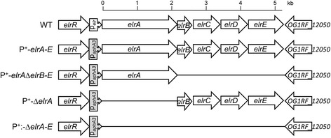
Schematic representation of the elrA operon in strains used. In E. faecalis WT elrA is followed by four genes encoding proteins of unknown function (OG1RF_12054 or elrB to OG1RF_12051 or elrE). The gene products are all predicted secreted proteins with an amino-terminal signal peptide. ElrA, ElrC and ElrD display a C-terminal WxL domain. ElrB possess a carboxy-terminal LPxTG anchor. ElrE belongs to the DUF916 family protein and has a predicted C-terminal transmembrane anchor. In P+-elrA-E the natural elrA promoter was replaced by the constitutive promoter of the kanamycin resistance gene (PaphA3). The P+-elrA-E strain was used as recipient for all mutant constructions
We first tested ElrA production in each of the different strains by Western blot analysis. As expected, ElrA was not detected in protein extracts prepared from WT, P+-ΔelrA, or P+-ΔelrA-E culture. Strikingly, protein extracts prepared from P+-elrA-E strain revealed a band of the expected size for ElrA (80-kDa, Fig. 2A), confirming that the endogenous PelrA promoter is inactive in vitro, and that its replacement by a constitutive promoter allows expression of elrA in vitro. No ElrA was detected in protein extracts prepared from the P+-elrA-ΔelrB-E strain (Fig. 2A), suggesting an important role for at least one of the four other proteins present in the elr operon in either the production or stability of ElrA. To corroborate this hypothesis and study elrA transcription in the P+-elrA-ΔelrB-E strain, elrA transcripts were analyzed by Northern blotting hybridization using total RNA prepared from the WT, P+-elrA-E and P+-elrA-ΔelrB-E strains. As expected, elrA transcript was not detected in the WT strain by Northern blotting under laboratory growth conditions, confirming our previous results [4, 5]. Similar analyses in respect of the P+-elrA-E and P+-elrA-ΔelrB-E overexpression strains resulted in strong hybridization signals corresponding to transcripts, of sizes of ~5 and 2.4 kb, corresponding to the predicted full-length, and the elrBCDE-deleted elr operon transcripts, respectively (Fig. 2B). Detection of elrA transcripts in P+-elrA-ΔelrB-E strain strongly indicates a post-transcriptional control of ElrA expression, confirming the important role of at least one of the four other elr operon genes for either ElrA production or stabilization.
Fig. 2.
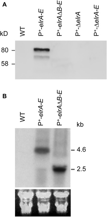
Detection of ElrA protein and elr transcript. a) Western blot analysis of total protein extracts from WT and mutant strains of E. faecalis, that was performed using a 12 % SDS-PAGE and polyclonal rat anti-ElrA antibodies, is shown. Band at ~80kD corresponds to the predicted size of ElrA, whereas the additional band represents a degradation product. b) Northern blot analysis of elr operon performed with ~40 μg of total RNA which was extracted from exponentially growing cells. Names of strains analyzed are indicated at the top of each lane. Probes used were elrA-specific oligonucleotide probes. The estimated length of transcripts that agrees with their predicted sizes is shown on the right. Below, ribosomal RNAs were used as loading controls
Overexpression of elr operon impairs phagocytosis
We then explored the effect of elr operon overexpression on the E. faecalis interaction with macrophages by monitoring phagocytosis of GFP-labeled E. faecalis strains by RAW cells using flow cytometry analysis. Firstly, we evaluated the phagocytosis dynamics of the E. faecalis WT strain at different multiplicities of infection (MOI, data not show) and decided to use a MOI of 1:100 in which approximately 58 % ±11 (mean ± SEM after three independent experiments each one in triplicate) of macrophages were GFP positive after 30 min of interaction. This value was used as reference (nominal 100 %) in order to estimate the phagocytosis index (PI) of the different E. faecalis mutant strains (see Materials and Methods). Operon inactivation (P+-ΔelrA-E) did not affect bacterial uptake when compared to the WT strain (105 ± 6.7 %, Fig. 3A). In contrast, a significant reduction of phagocytosis was observed with P+-elrA-E strain (PI = 39 % ± 4.4; P <0.0001, Fig. 3A). Although constitutive expression of the four other operon proteins (P+-ΔelrA strain) appeared to reduce phagocytosis (PI = 81.3 % ± 4.4), this reduction was not statistically significant (Fig. 3A). As expected, given that the P+-elrA-ΔelrB-E strain does not appear to produce ElrA (Western blot results, Fig. 2A), no difference in phagocytosis was observed when this strain was compared to the WT strain (PI = 98.2 % ± 5.8) (Fig. 3A). Lower levels of uptake by phagocytosis of the strain P+-elrA-E compared to the WT was confirmed by double-labeling fluorescence microscopy analysis (data not shown). These results support a link between the expression of elr operon and the uptake of E. faecalis.
Fig. 3.
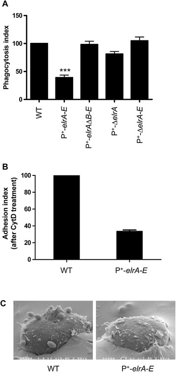
Phagocytosis of isogenic strains overexpressing full-length or partially deleted elr operon by RAW macrophages. a) For all E. faecalis strains tested, the phagocytosis index (PI) was calculated as average ± SEM from three independent experiments. Statistical significance was measured by ANOVA and Dunnett's multiple comparison test, ***P< 0.001. b) Adhesion index (AI) of E. faecalis strains after treatment with cytochalasin D. AI was calculated as follows: AI = % of GFP-labeled macrophages after infection with the mutant strain X 100/% of GFP-labeled macrophages after infection with the WT strain. Shown is the mean ± SEM from two independent experiments performed in duplicate. c) Scanning electronic microscopy (SEM) showing E. faecalis adhesion. Micrographs of macrophages infected for 30 min with E. faecalis strains observed by electron scanning microscopy. The micrographs are representative of two independent experiments
Overexpression of elr operon modifies bacterial adhesion
Phagocytosis is initiated with the recognition of ligands on bacterial cell surfaces by receptors including scavenger receptors, glucan receptors, and integrins present on the membrane of macrophages, which leads to bacteria engulfment via an actin-dependent mechanism. To test whether the impairment of phagocytosis seen for the P+-elrA-E strain correlated with reduced adhesion of the bacterium to macrophage cells, we used cytochalasin D (CytD), which inhibits phagocytosis, but does not prevent the initial step of bacterial adhesion [17, 18]. Macrophages were therefore infected (as described above for the phagocytosis test) with either WT or P+-elrA-E strains in the presence or absence of CytD, and the percentages of GFP+ macrophages were measured by flow cytometry analysis (uninfected macrophages were used as negative control). Comparison of forward scatter (FSC) and side scatter (SSC) values from uninfected cells (CytD treated or untreated) confirmed that macrophages were not altered by CytD (Additional file 1: Figure S1). As shown in Fig. 3B, P+-elrA-E strain was 60 % less adherent to macrophages than the WT strain. These results are in agreement with scanning microscopy observations of infected macrophages, that showed a sharp contrast between adhesion of WT and P+-elrA-E strains (Fig. 3C).
Because proteins encoded by the elr operon demonstrate characteristics of surface proteins (WxL, and LPXTG motifs) and could form a surface complex, we hypothesized that overexpression of elr operon could result in the formation of surface structures, which in turn resulted in the inhibition of phagocytosis as observed in vitro. Analyses of bacterial strains using transmission electron microscopy (thin sections and negative staining) and scanning electron microscopy revealed no differences at the surface structure level between the WT and the elr operon-overexpressing strain (data not shown). This indicated that no major structural modification was detected under the tested conditions. Since high expression levels of surface proteins can modify physicochemical properties of bacterial cell surface such as charge or hydrophobicity [19, 20], we compared the affinity of bacterial cells of WT and P+-elrA-E strains to the solvents using a MATS test as described by Bellon-Fontaine et al. [21]. Both strains exhibited similar affinity for the apolar solvents decane (~40 %) and hexadecane (~30 %) and for the acidic solvent chloroform (~70 %), indicating no major changes of the surface hydrophobicity upon elr overexpression. In turn, the affinity of the strain P+-elrA-E for the basic solvent ethyl acetate (~35 %) increased significantly compared to the WT (<1 %), indicating that expression of elr operon enhances the negative charge of the bacterial cells. Thus we hypothesize that poor adhesion of strain P+-elrA-E may result from repulsive forces between the negatively charged macrophage membrane and bacterial surface, which is loaded with Elr proteins.
Overexpression of elr operon increases E. faecalis virulence
Inactivation of ElrA reduces virulence in a mouse model of peritonitis [4] and we show that overexpression of elr operon impairs phagocytosis in vitro. We hypothesized that overexpression of elr may enhance dissemination, and thus E. faecalis virulence. To test this hypothesis, we assessed the survival of mice following peritoneal infection with WT, P+-elrA-E, or ΔelrA strains. Mice were injected with three different doses of WT or mutant strains, and the mortality rates were compared. No differences in mortality levels were found when mice were infected with 109 CFU of WT or mutant strains (data not shown). Interestingly, mortality was significantly increased for mice infected with 3 x 10⁸ and 1 x 10⁸ CFU of P+-elrA-E strain compared to WT and ΔelrA strains at 72 h post-infection (Fig. 4). Eighty five percent of the mice infected with 3 x 10⁸ CFU of P+-elrA-E strain died, whereas 45 % and 10 % mice died when infected with the WT and ΔelrA strains, respectively (P = 0.049 and P < 0.0001) (Fig. 4A). Similarly, 65, 30, and 5 % of the mice infected with 1 x 10⁸ CFU of P+-elrA-E, WT, and ΔelrA strains died, respectively (P = 0.044 and P < 0.0001) (Fig. 4B). These results show that overexpression of elr operon increases E. faecalis virulence. We also compared the dissemination of the WT, P+-elrA-E, or ΔelrA strains in organs of mice at 24 h postinfection by determining bacterial loads (Fig. 5A). A 0.70- and 1.79-log10 increase in the bacterial counts in the liver and spleen, respectively, were observed for the P+-elrA-E compared to the WT and ΔelrA strains, respectively, when mice were challenged with inocula of 1x10⁸ CFU. Similar trends were observed with inocula of 3x10⁸ CFU (1.05- and 0.93-log10, respectively), although to a somewhat lesser extent (Fig. 5B). These results indicate that the virulence phenotype correlates with higher dissemination of the strain P+-elrA-E. The correlation between increased virulence and avoidance of phagocytosis observed in vitro corroborates our hypothesis that elr operon may be involved in the evasion of the immune response by E. faecalis. We previously linked the attenuated virulence of an elrA deficient strain with the decreased organ burden and survival in peritoneal macrophages using an in vivo–in vitro infection model [4]. These new data suggest that expression of elrA and/or elr operon contributes to the escape of E. faecalis from phagocytosis in vivo, promoting dissemination and enhancing virulence of the pathogen.
Fig. 4.
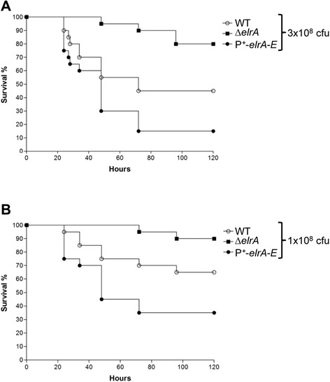
Effect of overexpression of elr operon on E. faecalis virulence. Kaplan-Meier survival analysis in a mouse peritonitis model with the E. faecalis WT strain (open circles), the ΔelrA strain (squares), and the P+-elrA-E strain (closed circles). A total of 10 mice were infected intraperitoneally with ~3 x 10⁸ (a) or ~1 x 10⁸ (b) CFU of each strain. For pairwise comparisons of P+-elrA-E / WT and P+-elrA-E / WT, P values were < 0.05 for each inoculum
Fig. 5.
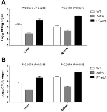
Overexpression of elr operon in E. faecalis increases bacterial dissemination in mice. E. faecalis organ burden in 10 mice were infected intraperitoneally with ~3 x 10⁸ (a) or ~1 x 10⁸ (b) CFU of each strain. The results represent the means and standard deviations of the number of bacteria able to colonize the spleen and liver at 24 h postinfection
Discussion
Previous studies have shown that E. faecalis survives into peritoneal macrophages better than non-pathogenic bacteria [8]. Since then, E. faecalis virulence factors able to interfere with uptake and survival in macrophages have been described [22]. We previously linked the attenuated virulence of an E. faecalis strain deleted for elrA with decreased organ burden and survival in peritoneal macrophages [4]. In this study, we show that overexpression of elr operon by E. faecalis confers resistance to phagocytosis by interfering with bacterial adhesion to macrophages. We also correlated E. faecalis avoidance of phagocytosis observed in vitro with increased virulence and dissemination in a mouse peritonitis model. These data contrast with our previous report that WT and ΔelrA strains were evenly phagocytosed [4]. Nevertheless, these studies are difficult to compare since macrophage infections were performed differently (i.e. in vivo versus in vitro infection and duration of infection). Moreover, the expression level of Elr proteins in vivo is unknown. The tight control of expression of the elr operon suggests that the operon may be required in specific conditions that remain to be identified [4, 5]. The 162-fold increased level of erlA transcript in E. faecalis strain MMH594 grown in urine [6], supports that expression of Elr proteins may vary in response to host-derived cues. We assume that elr operon may enhance E. faecalis virulence by promoting initial dissemination in the host after escape of bacteria from phagocytosis, but also by contributing to E. faecalis survival within infected macrophages depending on the tissues or cell types encountered by E. faecalis. From this study we propose that high-level expression of elr operon may, in some circumstances, occur in vivo and promotes escape of E. faecalis from phagocytosis.
The present study also revealed that ElrA requires at least one other elr gene to be expressed at a detectable level and confirmed that elrA gene is cotranscribed with the other elr genes [4]. The elr operon is a typical gene cluster of WxL surface proteins that associate non-covalently to the peptidoglycan of low-GC gram-positive bacteria. The operonic organization of the elr operon and the need of at least one other protein encoded by elr operon for ElrA production in vitro further support the hypothesis that cell-surface proteins, encoded by the elr operon, may participate in the formation of a multicomponent complex at the surface as it has been previously proposed [1, 2, 4]. Based on recent work by Galloway-Pena et al. who showed that WxL and DUF916 proteins interact in vitro [3], we believe that ElrA may be protected from degradation by interacting with at least another elr-encoded protein. If neither surface appendages nor modification could be observed upon overexpression of elr operon, other experiments are needed to establish if elr operon drives the formation of a surface complex in E. faecalis. Nevertheless, overexpression of Elr proteins seems to increase the negative charge of the bacterial surface, suggesting that E. faecalis evasion of phagocytosis by immune cells is driven by electrostatic repulsion. Even if elr overexpression emphasizes the steric or charge hindrance by Elr proteins in vitro, one cannot exclude that similar physicochemical changes occur in vivo in response to environmental cues [6], and confers to the E. faecalis cells anti-adhesion properties that promote escape from phagocytosis. These in vitro findings are reminiscent of the acidic LRR protein Slr from Streptococcus pyogenes that is involved in phagocytosis evasion [23], probably by enhancing the anti-adhesive properties of streptococcal cells. Another possibility would be that high level of Elr proteins sterically hinders E. faecalis-associated molecular patterns important for recognition by scavenger receptors. Altogether, this work shows that expression of elr operon contributes to the escape of E. faecalis from phagocytosis, promoting dissemination and enhancing virulence of the pathogen. Further investigations will focus on characterizing the precise role of each of the Elr proteins.
Conclusions
In summary, this work shows that high-level expression of elr operon by E. faecalis increases virulence and confers resistance to phagocytosis, probably through charge repulsion. Consistently, the strain expressing elr operon displays stabilization of ElrA, further supporting that Elr proteins form an extracellular protein complex as part of the virulence process. Structural and functional characterization of the Elr proteins will help to understand E. faecalis pathogenesis and provide clues on WxL- and associated proteins of low-GC Gram-positive bacteria.
Methods
Reagents
All reagents were obtained from Sigma-Aldrich (St. Louis, MO), unless otherwise stated.
Bacterial strains and plasmids
Bacterial strains and plasmids used in this work are listed in Table 1. E. faecalis strains were grown in M17 medium supplemented with 0.5 % glucose (GM17) at 37 °C without aeration. Escherichia coli strains were grown aerobically in Luria-Bertani medium at 37 °C. Plasmid constructions were first established in E. coli TG1 strain and then transferred into E. faecalis by electrotransformation using a Bio-Rad Gene Pulser Electroporator (Bio-Rad Laboratories). E. faecalis strains expressing green-fluorescent protein (GFP) were obtained by electroporation with pMV158-GFP plasmid [24]. Recombinant bacteria were selected by the addition of antibiotics as follows: for E. faecalis chloramphenicol 4 μg/ml, tetracycline 4 μg/ml and erythromycin (Ery) 30 μg/ml; for E. coli, chloramphenicol 10 μg/ml and ampicillin 100 μg/ml. DNA manipulations were performed as previously described [25].
Table 1.
Strains and plasmids used in this work
| Strain | Designation relevant characteristics | Source or Reference |
|---|---|---|
| E. faecalis | ||
| WT | Fusr Rifr; plasmid-free wild-type strain | [35] |
| ΔelrA | OG1RF ΔelrA | [4] |
| P+-elrA-E | OG1RF PaphA3::elrA-E | This work |
| P+-ΔelrA | OG1RF PaphA3::ΔelrA | This work |
| P+-elrA-ΔelrB-E | OG1RF PaphA3::elrA-ΔelrB-E | This work |
| P+ -ΔelrA-E | OG1RF PaphA3::ΔelrA-E | This work |
| E. coli | ||
| TG1 | supE hsdD5 thi (Δlac-proAB) F’ (traD36 proAB-lacZΔM15) | [36] |
| BL21(λDE3) | F− ompT gal dcm hsdSB(rB−mB−) λDE3 | [37] |
| Plasmids | ||
| pACYC177 | Ampr, Kanr, ori p15A | [38] |
| pET2817 | Ampr, ori colE1, T7 promoter, His-Tag coding sequence | [39] |
| pGEM-T easy | Ampr, ori ColE1, linearized with 3’ T overhangs | Promega |
| pGhost9 | Ermr, ori pWV01, repA(Ts) | [27] |
| pMV158-GFP | pMV158 with the gene encoding the green fluorescent protein | [24] |
| pTCV-lac(PaphA3) | Tetr, ori ColE1, ori pAMβ1, lacZ harboring PaphA3 promoter | [16] |
| pVE14009 | Ermr, ori pWV01, repA(Ts), with elrA deletion | [4] |
| pVE14047 | Ampr, pET2817 with 6x His::ElrA | This work |
| pVE14142 | Ampr, Kanr, ori p15A, ‘elrR-elrA’ region | This work |
| pVE14145 | Ampr, Kanr, ori p15A, ‘elrR-PaphA3::elrA’ region | This work |
| pVE14146 | Ermr, ori pWV01, repA(Ts), ‘elrR-PaphA3::elrA’ region | This work |
| pVE14178 | Ampr, ori colE1, with elrA-E deletion | This work |
| pVE14179 | Ampr, ori colE1, with elrB-E deletion | This work |
| pVE14450 | Ermr, ori pWV01, repA(Ts), with elrB-E deletion | This work |
| pVE14455 | Ampr, Kanr, ori p15A, with PaphA3 ::elrA-E deletion | This work |
| pVE14456 | Ermr, ori pWV01, repA(Ts), with PaphA3 ::elrA-E deletion | This work |
| pVE14457 | Ermr, ori pWV01, repA(Ts), with PaphA3::elrA deletion | This work |
Cell line and culture conditions
The RAW 264.7 mouse macrophage cell line (ATCC®−TIB-71) was maintained in DMEM supplemented with 10 % heat-inactivated fetal bovine serum (FBS) and 2 mM l-glutamine [26]. For phagocytosis assays, cells were seeded at 0.5 × 10⁶/well into 12-well tissue culture plates (TPP, Domique Dutscher, Brumath, France) and incubated overnight at 37 °C under 6 % CO2. For microscopy experiments, cells were cultured in tissue culture plates containing poly-L-lysine pretreated coverslips for microscopy or on Lab-tek chamber slides (Nunc, Domique Dutscher). Comparative analysis of phagocytosis using either heat-inactivated serum or serum-free media (Macrophage-SFM, GIBCO, Invitrogen) did not show differences (data not shown). Thus, for practical reasons we decided to use heat-inactivated serum in all experiments of this work.
Generation of anti-ElrA rat polyclonal antibodies
Recombinant ElrA was purified to produce polyclonal rat anti-ElrA antibodies by Proteogenix (Oberhausbergen, France). Briefly, a DNA fragment encoding elrA was PCR-amplified from E. faecalis chromosomal DNA using OEF275 and OEF276 primers (Table 2). The PCR product was digested with BamHI/PciI and cloned into purified BamHI/NcoI-digested pET2817 vector backbone, resulting in plasmid VE14047 which was transformed into E. coli BL21(λDE3). For ElrA production and purification, the resulting recombinant strain was cultured at 25 °C and induced with 1 mM of IPTG (isopropyl β-D-1-thiogalactopyranoside) for 5 hrs. Recombinant 6xHis::ElrA protein was purified under denaturing conditions on Ni-NTA columns using the QIAexpress kit (Qiagen, Courtaboeuf, France).
Table 2.
Primers used in this study
| Name | Sequence 5'-3' | Source or Reference |
|---|---|---|
| OEF9 | TTGACCATCACGAGATACC | This work |
| OEF13 | CTATCTTGGTCAAAAGAGCG | This work |
| OEF15 | TATTCGATGTTGGCGTTGG | [4] |
| OEF18 | GGAGGATGCGATTGTTTCG | [4] |
| OEF212 | CTCTTCTGCCGATGAAGTTTCTGG | [4] |
| OEF275 | CAAACATGTTAGAAACGACCGAAACAATCGC | This work |
| OEF276 | TTGGATCCACTCACCCCCTATTTTGC | This work |
| OEF343 | GCGAATTCGAAGATCTGAGAAAATATCAGGAGGTGAAG | This work |
| OEF344 | ATGGATCCAGACGGAGTAGGTTATTTGC | This work |
| OEF345 | TTCTCAGATCTTCGAATTCGCTGAATATCAACTGAAAATGGG | This work |
| OEF346 | ATCTCGAGTTGCGTATTTCGGATTTAGCC | This work |
| OEF49 | CACGCTGTACGATCAGCAAC | This work |
| OEF595 | CAATCCTAATAGCAATACACC | This work |
| OEF596 | GGTGTATTGCTATTAGGATTGTGCCTGTTCATCATTTTACG | This work |
| OEF598 | CGAAACAATCGCATCCTCCTGCCTGTTCATCATTTTACG | This work |
| Vlac1 | GTTGAATAACACTTATTCCTATC | [16] |
| Vlac2 | CTTCCACAGTAGTTCACCACC | [16] |
Construction of mutant and over-expressing strains of E. faecalis
The E. faecalis elrA gene is part of a five-gene operon elrA (OG1RF_12055), elrB (OG1RF_12054), elrC (OG1RF_12053), elrD (OG1RF_12052), and elrE (OG1RF_12051) (Fig. 1), encoding putative surface proteins of unknown function. To circumvent the lack of ElrA production in vitro, we constructed a genetically modified E. faecalis strain harboring the constitutive promoter PaphA3 (hereafter named P+), instead of the native promoter, PelrA, upstream of the whole elr operon (i.e., elrA-E, Fig. 1). This genetically modified strain (called P+-elrA-E), was constructed by a double cross-over event using the pGhost9 plasmid [27]. Briefly, two overlapping fragments were PCR-amplified from E. faecalis OG1RF chromosomal DNA with primers OEF343/OEF344 and OEF345/OEF346 (Table 2). The two PCR products were then fused by PCR using the external primers OEF344/OEF346, and the resulting product was cloned into purified XhoI-BamHI-digested pACYC177 vector, resulting in plasmid pVE14142. The PaphA3 promoter was PCR-amplified with primers Vlac1 and Vlac2 from pTCV-lac(PaphA3) plasmid [16]. An EcoRI-BamHI fragment, containing the PaphA3 promoter, was then cloned into EcoRI-BglII-digested pVE14142 vector to obtain plasmid pVE14145. Then, a 2.3 kb XhoI-EaeI fragment from pVE14145 plasmid (containing the promoter and the targeted region) was cloned into pGhost9 vector to generate the final vector pVE14146. This plasmid was established in E. faecalis OG1RF strain and a markerless insertion of PaphA3 upstream of the elrA-E operon was performed as previously described [1]. Correct integration of PaphA3 into the chromosomal locus was confirmed by sequencing. All the following mutant constructs were performed using P+−elrA-E strain as a recipient in order to have the same genetic background (Table 1). For the construction of a strain expressing only elrA under the control of PaphA3 promoter, a fused DNA fragment using primers OEF13/OEF595 and OEF596/OEF49 amplified from OG1RF strain DNA was cloned into pGEM-T easy vector (Promega) to generate pVE14179. A 4.5 kb PstI fragment was then cloned into PstI-digested pGhost9 to obtain plasmid pVE14450 and established in P+-elrA-E strain to obtain the P+-elrA-ΔelrB-E strain. For the construction of a strain expressing elr operon lacking elrA, a 6.6 kb Bst/ApeI fragment from pVE14009 was cloned into Bst/ApeI-digested pVE14146 vector, resulting in pVE14457. This plasmid was established in P+-elrA-E strain to obtain the P+-ΔelrA strain. To inactivate the whole elr operon (i.e., elrA-E), we first generated an in-frame deletion of the whole operon by PCR. For this, we used OEF15/OEF18 and OEF598/OEF49 primers described for the first PCR. The two PCR products were fused by PCR using external primers OEF49/OEF15, and the resulting product was cloned into pGEM-T, resulting in plasmid pVE14178. A BstAPI/AatII 890bp DNA fragment from pVE14178 was then cloned into pVE14145 to generate pVE14455. The final plasmid was generated by cloning a 4.5 kb XhoI DNA fragment from pVE14455 vector into XhoI-digested pGhost9 to obtain pVE14456. This plasmid was established in P+-elrA-E strain and the resulting strain was named P+-ΔelrA-E. All expected modifications or deletions were confirmed by sequencing.
Preparation of protein extracts, SDS gel electrophoresis, and immunoblot analysis
Total protein extraction from bacteria, SDS-PAGE, and Western blot immunodetection were carried out using standard methods (24) with some modifications. Strains were grown at 37 °C overnight and then diluted 100-fold and grown under the same conditions to an OD600~1. Protein crude extract was obtained by trichloroacetic acid (TCA) precipitation by mixing 800 μl of bacterial culture with 200 μl of ice cold TCA solution (100 % w/v). The protein pellet was then obtained by centrifugation and recovered directly into SDS sample buffer. Anti-ElrA antibody was used at a dilution of 1:500 for Western blot immunodetection.
RNA isolation and Northern blotting
Total RNA was extracted as previously described [28]. Northern blots were performed on 40 μg of total RNA separated on a 0.9 % denaturing agarose gel as previously described [29]. Specific oligonucleotides OEF9 and OEF212 were used to detect elrA transcripts. Oligonucleotides were labelled with [γ-32P]-ATP and T4 polynucleotide kinase (NEB Biolabs) according to the recommendations of the manufacturer (NEB Biolabs). Analysis was performed from RNA extracted from two independent experiments.
Phagocytosis assay with RAW macrophages
Fluorescent E. faecalis were grown on GM17 plates containing erythromycin (GM17-Ery), with a single colony subsequently being selected and grown overnight in GM17-Ery broth. A 100 μl aliquot was then transferred into 10 ml of fresh GM17-Ery and incubated until cultures reached an OD600 ~1. Bacteria were then pelleted by centrifugation, washed three times with PBS, and adjusted to a concentration of 1 × 10⁹ CFU/ml in supplemented DMEM. The number of bacteria present in each suspension was confirmed by plating onto solid GM17-Ery.
For phagocytosis experiments, adherent RAW cells were infected with fluorescent E. faecalis at a multiplicity of infection (MOI) of 100:1 (bacterium/cell ratio). After 30 min of interaction, cells were washed twice with PBS, recovered with cell dissociation buffer (GIBCO, Invitrogen), washed again, and finally fixed in 3 % paraformaldehyde (PFA) solution. Fluorescence of RAW cells due to infecting bacteria was detected by a flow cytometer in the FL-1 channel. The phagocytosis index (PI) was calculated using the percent of fluorescent macrophages after E. faecalis wild-type (WT) strain infection and applying the following formula: PI = (percent of fluorescent macrophages after infection X 100 /percent of fluorescent macrophages after WT infection) [30, 31]. Results are expressed as the mean ± SEM from three independent experiments usually performed in duplicate or triplicate.
Bacterial adhesion assay
To separate adhesion from subsequent steps of phagocytosis, cells were pretreated 30 min with 1 μg/ml of cytochalasin D (CytD), an actin polymerization inhibitor, as described [17]. A CytD (1000X) stock solution in DMSO was prepared according to manufacturer's recommendations and stored at −20 °C. DMEM supplemented medium (see above) was used to dilute stock solution. RAW cells were seeded at 1 × 10⁶/well into 6-well tissue culture plates (TPP, Dominique Dutscher) and incubated O/N at 37 °C under 6 % CO2. Macrophages pre-treated with CytD were first washed twice with fresh medium and then infected at a MOI of 100:1, similar to phagocytosis analysis above; CytD-untreated and uninfected macrophages were used as negative controls. Fluorescence in RAW cells due to infecting bacteria was detected by flow cytometry. Adhesion Index (AI) = (percent of GFP+ macrophages pre-treated with CytD, after infection by the E. faecalis mutant strain X 100/percent of GFP+ macrophages pre-treated with CytD after WT infection).
Fluorescence and electron microscopy
Raw macrophages were seeded in 12-well cell culture plates on a glass slide and infected with GFP-labeled E. faecalis wild-type (WT) or P+-elrA-E strains at a MOI of 1:100, with uninfected macrophages serving as negative control. After 30 min of interaction, macrophages were washed twice with PBS, fixated and immunolabeled with Streptococcus group D antiserum (BD Diagnostics, Le Pont de Claix, France) as previously described [4]. Fluorescence was examined using a Carl Zeiss microscope (Axiovert 200 M, in the ApoTome mode) at MIMA2 platform (INRA, Jouy en Josas). Images were processed with Axiovision version 4.6 (Carl Zeiss).
Imaging of bacterial-cells interaction was performed using a Hitachi S-4500 scanning electron microscope (SEM) at the MIMA2 imaging platform. Macrophages were seeded in 12-well cell culture plates and infected with either E. faecalis wild-type (WT) or P+-elrA-E strains at a MOI of 1:100, with uninfected macrophages serving as negative control. After 30 min of interaction, macrophages were washed twice with PBS, recovered with cell dissociation buffer (GIBCO, Invitrogen), washed again, and suspended in a fixative solution and treated as previously described [32].
Preparation of bacterial samples for transmission and scanning electron microscopy was performed as previously described [32, 33]. Thin-sections and negative-stains were observed with a Zeiss EM902 electron microscope operated at 80 kV (MIMA2 - UR 1196 Génomique et Physiologie de la Lactation, INRA, plateau de Microscopie Electronique, 78352 Jouy-en-Josas, France). Microphotographies were acquired using MegaView III CCD camera and analyzed with the ITEM software (Eloise SARL, Roissy CDG, France).
Microbial adhesion to solvents
Microbial adhesion to solvents (MATS) analysis was carried out as described previously by Bellon-Fontaine and collaborators [21]. In brief, a single colony of each of the E. faecalis strains studied was subcultured four times in BHI and harvested at stationary phase. Bacterial cells were centrifuged at 5000 ×g for 8 min and washed twice in 0.15 M NaCl and re-suspended to a final OD400 ~0.8. Bacterial suspensions (2.4 ml) were vortexed for 1 min with 0.4 ml of highest purity grade chloroform (Sigma-Aldrich), hexadecane (Sigma-Aldrich), ethyl acetate (Merck), or decane (Merck). The emulsion was left to stand for 20 min to allow complete phase separation, and the OD400 of 1 ml from the aqueous phase was measured. Affinity of the cells for each solvent (% affinity) = ((ODf-ODi)/ ODi)x100 where ODi is the initial optical density of the bacterial suspension before mixing with the solvent, and ODf the final absorbance after mixing and phase separation. Analysis was performed twice in triplicate.
Mouse peritonitis model
The mouse experiments were approved by the Institutional Animal Use and Care Committee at the Università Cattolica del Sacro Cuore, Rome, Italy (permit number Z21, 1 November 2010), and authorized by the Italian Ministry of Health, according to the Legislative Decree 116/92, which implemented the European Directive 86/609/EEC on laboratory animal protection in Italy. Animal welfare was routinely checked by veterinarians of the Service for Animal Welfare.
Virulence of strains OG1RF, ΔelrA, and P+-elrA-E was tested as described previously [4]. The inoculum size was confirmed by determining the number of CFU on brain heart infusion agar. Each inoculum was 10-fold diluted in 25 % sterile rat fecal extract prepared from a single batch as previously described [34]. Groups of 10 ICR outbred mice (Harlan Italy Srl, San Pietro al Natisone, Italy) were challenged intraperitoneally with 1 ml of each bacterial inoculum, housed five per cage, and fed ad libitum. A control group of mice was injected with 25 % sterile rat fecal extract only. Survival was monitored every 3 to 6 h. In another set of experiments, groups of mice were killed 24 h postinfection, and livers and spleens were removed, weighed, homogenized, and serially diluted in saline solution for colony counts.
Statistical analysis
Statistics were performed using GraphPad Prism (Version 4.00 for Windows, GraphPad Software, San Diego California, USA). One-way analysis of variance (ANOVA) was followed by Dunnett's multiple-comparison test when comparing multiple groups for one factor. For animal experiments, survival estimates were constructed by the Kaplan-Meier method and compared by log rank analysis, and comparisons with P values of <0.05 were considered to be significant.
Acknowledgements
We thank A. Navickas and F. Wessner for technical support on MATS assays and RNA extractions, and P. Adenot and R. Fleurot of the Platform MIMA2 for access to the Apotome microscope. We also thank P. Lee, A. Gruss, D. Lereclus, C. Archambaud and S. Aymerich for critical revision of the manuscript. We are thankful to M. Mangan for careful reading of the manuscript for the English editing. This work was supported by the Institut National de la Recherche Agronomique. R.D. was supported by a fellowship from the Région Ile-de-France in the framework of the Dim MalinF.
Additional file
Bacterial adhesion assay. RAW macrophages were pretreated with cytochalasin D (CytD) or not (-) before infection with E. faecalis strains WT and P+-elrA-E expressing GFP. After 30 min of interaction, cells were washed twice with PBS, recovered with cell dissociation buffer. GFP positives macrophages were detected by flow cytometry. Graphs represent green fluorescence intensity. Results are representative of two independent experiments. In the absence of CytD pretreatment, the population of GFP-labeled macrophages infected with the P+-elrA-E strain (55.5 %) is decreased compared to the macrophages infected with the WT strain (86.6 %). CytD is known to inhibit actin rearrangement and blocks bacterial entry. To establish whether differential GFPlabeling of macrophages resulted from adhesion or entry defect of strain P+-elrA-E, similar experiment was performed on macrophages pre-treated with CytD. When infected with the WT strain, the population of GFP-labeled pre-treated macrophages (77.9 %) slightly decreased compared to untreated ones. This observation indicates that the majority of the GFP-labeled macrophages detected harbor adherent GFP-bacteria at their surface. In contrast, the intensity and the population of GFP-labeled pre-treated macrophages infected with the P+-elrA-E strain (28.4 %) decreased drastically compared to untreated ones, indicating that P+-elrA-E bacteria adhered less efficiently to macrophages.
Footnotes
Competing interests
The authors declare that they have no competing interests.
Authors’ contributions
NGCP designed, performed, and interpreted in vitro experiments, and contributed to write the manuscript. RD and SG constructed the bacterial strains. CL and RM carried out RNA experiments, KP performed Western Blot experiments, SC and TM performed electron microscopy. FB performed mouse experiments and BP, MS and LRG designed animal experiments and helped writing the manuscript. PL critically revised the manuscript for intellectual content. PS designed and coordinated the study and wrote the manuscript. All authors read and approved the final manuscript.
Contributor Information
Naima G. Cortes-Perez, Email: naima.cortes-perez@jouy.inra.fr
Romain Dumoulin, Email: rom.dumoulin@gmail.com.
Stéphane Gaubert, Email: stephane.gaubert@jouy.inra.fr.
Caroline Lacoux, Email: caroline.lacoux@jouy.inra.fr.
Francesca Bugli, Email: francesca.bugli@rm.unicatt.it.
Rebeca Martin, Email: rebeca.martinrosique@jouy.inra.fr.
Sophie Chat, Email: sophie.chat@jouy.inra.fr.
Kevin Piquand, Email: kevin.piquand@gmail.com.
Thierry Meylheuc, Email: thierry.meylheuc@jouy.inra.fr.
Philippe Langella, Email: philippe.langella@jouy.inra.fr.
Maurizio Sanguinetti, Email: msanguinetti@rm.unicatt.it.
Brunella Posteraro, Email: bposteraro@rm.unicatt.it.
Lionel Rigottier-Gois, Email: lionel.rigottier-gois@jouy.inra.fr.
Pascale Serror, Email: pascale.serror@jouy.inra.fr.
References
- 1.Brinster S, Furlan S, Serror P. C-terminal WxL domain mediates cell wall binding in Enterococcus faecalis and other gram-positive bacteria. J Bacteriol. 2007;189(4):1244–1253. doi: 10.1128/JB.00773-06. [DOI] [PMC free article] [PubMed] [Google Scholar]
- 2.Siezen R, Boekhorst J, Muscariello L, Molenaar D, Renckens B, Kleerebezem M. Lactobacillus plantarum gene clusters encoding putative cell-surface protein complexes for carbohydrate utilization are conserved in specific gram-positive bacteria. BMC Genomics. 2006;7:126. doi: 10.1186/1471-2164-7-126. [DOI] [PMC free article] [PubMed] [Google Scholar]
- 3.Galloway-Pena JR, Liang X, Singh KV, Yadav P, Chang C, La Rosa SL, et al. The identification and functional characterization of WxL proteins from Enterococcus faecium reveal surface proteins involved in extracellular matrix interactions. J Bacteriol. 2015;197(5):882–892. doi: 10.1128/JB.02288-14. [DOI] [PMC free article] [PubMed] [Google Scholar]
- 4.Brinster S, Posteraro B, Bierne H, Alberti A, Makhzami S, Sanguinetti M, et al. Enterococcal Leucine-Rich Repeat-Containing Protein Involved in Virulence and Host Inflammatory Response. Infect Immun. 2007;75(9):4463–4471. doi: 10.1128/IAI.00279-07. [DOI] [PMC free article] [PubMed] [Google Scholar]
- 5.Dumoulin R, Cortes-Perez N, Gaubert S, Duhutrel P, Brinster S, Torelli R, et al. Enterococcal Rgg-Like Regulator ElrR Activates Expression of the elrA Operon. J Bacteriol. 2013;195(13):3073–3083. doi: 10.1128/JB.00121-13. [DOI] [PMC free article] [PubMed] [Google Scholar]
- 6.Shepard BD, Gilmore MS. Differential expression of virulence-related genes in Enterococcus faecalis in response to biological cues in serum and urine. Infect Immun. 2002;70(8):4344–4352. doi: 10.1128/IAI.70.8.4344-4352.2002. [DOI] [PMC free article] [PubMed] [Google Scholar]
- 7.Knodler LA, Celli J, Finlay BB. Pathogenic trickery: deception of host cell processes. Nat Rev Mol Cell Biol. 2001;2(8):578–588. doi: 10.1038/35085062. [DOI] [PubMed] [Google Scholar]
- 8.Gentry-Weeks CR, Karkhoff-Schweizer R, Pikis A, Estay M, Keith JM. Survival of Enterococcus faecalis in Mouse Peritoneal Macrophages. Infect Immun. 1999;67(5):2160–2165. doi: 10.1128/iai.67.5.2160-2165.1999. [DOI] [PMC free article] [PubMed] [Google Scholar]
- 9.Verneuil N, Sanguinetti M, Le Breton Y, Posteraro B, Fadda G, Auffray Y, et al. Effects of the Enterococcus faecalis hypR gene encoding a new transcriptional regulator on oxidative stress response and intracellular survival within macrophages. Infect Immun. 2004;72(8):4424–4431. doi: 10.1128/IAI.72.8.4424-4431.2004. [DOI] [PMC free article] [PubMed] [Google Scholar]
- 10.Baldassarri L, Bertuccini L, Creti R, Filippini P, Ammendolia MG, Koch S, et al. Glycosaminoglycans mediate invasion and survival of Enterococcus faecalis into macrophages. J Infect Dis. 2005;191(8):1253–1262. doi: 10.1086/428778. [DOI] [PubMed] [Google Scholar]
- 11.Coburn PS, Baghdayan AS, Dolan GT, Shankar N. An AraC-type transcriptional regulator encoded on the Enterococcus faecalis pathogenicity island contributes to pathogenesis and intracellular macrophage survival. Infect Immun. 2008;76(12):5668–5676. doi: 10.1128/IAI.00930-08. [DOI] [PMC free article] [PubMed] [Google Scholar]
- 12.Thurlow LR, Thomas VC, Fleming SD, Hancock LE. Enterococcus faecalis capsular polysaccharide serotypes C and D and their contributions to host innate immune evasion. Infect Immun. 2009;77(12):5551–5557. doi: 10.1128/IAI.00576-09. [DOI] [PMC free article] [PubMed] [Google Scholar]
- 13.Hancock LE, Gilmore MS. The capsular polysaccharide of Enterococcus faecalis and its relationship to other polysaccharides in the cell wall. Proc Natl Acad Sci U S A. 2002;99(3):1574–1579. doi: 10.1073/pnas.032448299. [DOI] [PMC free article] [PubMed] [Google Scholar]
- 14.Teng F, Jacques-Palaz KD, Weinstock GM, Murray BE. Evidence that the enterococcal polysaccharide antigen gene (epa) cluster is widespread in Enterococcus faecalis and influences resistance to phagocytic killing of E. faecalis. Infect Immun. 2002;70(4):2010–2015. doi: 10.1128/IAI.70.4.2010-2015.2002. [DOI] [PMC free article] [PubMed] [Google Scholar]
- 15.Prajsnar TK, Renshaw SA, Ogryzko NV, Foster SJ, Serror P, Mesnage S. Zebrafish as a Novel Vertebrate Model To Dissect Enterococcal Pathogenesis. Infect Immun. 2013;81(11):4271–4279. doi: 10.1128/IAI.00976-13. [DOI] [PMC free article] [PubMed] [Google Scholar]
- 16.Poyart C, Trieu-Cuot P. A broad-host-range mobilizable shuttle vector for the construction of transcriptional fusions to β-galactosidase in Gram-positive bacteria. FEMS Microbiol Lett. 1997;156(2):193–198. doi: 10.1016/S0378-1097(97)00423-0. [DOI] [PubMed] [Google Scholar]
- 17.Elliott JA, Winn WC., Jr Treatment of alveolar macrophages with cytochalasin D inhibits uptake and subsequent growth of Legionella pneumophila. Infect Immun. 1986;51(1):31–36. doi: 10.1128/iai.51.1.31-36.1986. [DOI] [PMC free article] [PubMed] [Google Scholar]
- 18.Gaillard JL, Berche P, Mounier J, Richard S, Sansonetti P. In vitro model of penetration and intracellular growth of Listeria monocytogenes in the human enterocyte-like cell line Caco-2. Infect Immun. 1987;55(11):2822–2829. doi: 10.1128/iai.55.11.2822-2829.1987. [DOI] [PMC free article] [PubMed] [Google Scholar]
- 19.Travier L, Guadagnini S, Gouin E, Dufour A, Chenal-Francisque V, Cossart P, et al. ActA promotes Listeria monocytogenes aggregation, intestinal colonization and carriage. PLoS Pathog. 2013;9(1):e1003131. doi: 10.1371/journal.ppat.1003131. [DOI] [PMC free article] [PubMed] [Google Scholar]
- 20.Courtney HS, Ofek I, Penfound T, Nizet V, Pence MA, Kreikemeyer B, et al. Relationship between expression of the family of M proteins and lipoteichoic acid to hydrophobicity and biofilm formation in Streptococcus pyogenes. PLoS One. 2009;4(1):e4166. doi: 10.1371/journal.pone.0004166. [DOI] [PMC free article] [PubMed] [Google Scholar]
- 21.Bellon-Fontaine M-N, Rault J, van Oss CJ. Microbial adhesion to solvents: a novel method to determine the electron-donor/electron-acceptor or Lewis acid–base properties of microbial cells. Colloids Surf B: Biointerfaces. 1996;7(1–2):47–53. doi: 10.1016/0927-7765(96)01272-6. [DOI] [Google Scholar]
- 22.Garsin DA, Frank KL, Silanpaa J, Ausubel FM, Hartke A, Shankar N, Murray BE. Pathogenesis and Models of Enterococcal Infection. In: Enterococci: From Commensals to Leading Causes of Drug Resistant Infection. Edited by Gilmore MS, Clewell DB, Ike Y, Shankar N. Boston: Massachusetts Eye and Ear Infirmary; 2014. [PubMed]
- 23.Reid SD, Montgomery AG, Voyich JM, DeLeo FR, Lei B, Ireland RM, et al. Characterization of an extracellular virulence factor made by Group A Streptococcus with homology to the Listeria monocytogenes internalin family of proteins. Infect Immun. 2003;71(12):7043–7052. doi: 10.1128/IAI.71.12.7043-7052.2003. [DOI] [PMC free article] [PubMed] [Google Scholar]
- 24.Nieto C, Espinosa M. Construction of the mobilizable plasmid pMV158GFP, a derivative of pMV158 that carries the gene encoding the green fluorescent protein. Plasmid. 2003;49(3):281–285. doi: 10.1016/S0147-619X(03)00020-9. [DOI] [PubMed] [Google Scholar]
- 25.Sambrook J, Fritsch EF, Maniatis T. Molecular cloning: a laboratory manual. 2. Cold Spring Harbor, N. Y: Cold Spring Harbor Laboratory Press; 1989. [Google Scholar]
- 26.Orman KL, Shenep JL, English BK. Pneumococci stimulate the production of the inducible nitric oxide synthase and nitric oxide by murine macrophages. J Infect Dis. 1998;178(6):1649–1657. doi: 10.1086/314526. [DOI] [PubMed] [Google Scholar]
- 27.Maguin E, Duwat P, Hege T, Ehrlich D, Gruss A. New thermosensitive plasmid for gram-positive bacteria. J Bacteriol. 1992;174(17):5633–5638. doi: 10.1128/jb.174.17.5633-5638.1992. [DOI] [PMC free article] [PubMed] [Google Scholar]
- 28.Fouquier d'Herouel A, Wessner F, Halpern D, Ly-Vu J, Kennedy SP, Serror P, Aurell E, Repoila F: A simple and efficient method to search for selected primary transcripts: non-coding and antisense RNAs in the human pathogen Enterococcus faecalis. Nucleic acids research 2011;39(7):e46. [DOI] [PMC free article] [PubMed]
- 29.Mandin P, Repoila F, Vergassola M, Geissmann T, Cossart P. Identification of new noncoding RNAs in Listeria monocytogenes and prediction of mRNA targets. Nucleic Acids Res. 2007;35(3):962–974. doi: 10.1093/nar/gkl1096. [DOI] [PMC free article] [PubMed] [Google Scholar]
- 30.Pils S, Schmitter T, Neske F, Hauck CR. Quantification of bacterial invasion into adherent cells by flow cytometry. J Microbiol Methods. 2006;65(2):301–310. doi: 10.1016/j.mimet.2005.08.013. [DOI] [PubMed] [Google Scholar]
- 31.Van Amersfoort ES, Van Strijp JA. Evaluation of a flow cytometric fluorescence quenching assay of phagocytosis of sensitized sheep erythrocytes by polymorphonuclear leukocytes. Cytometry. 1994;17(4):294–301. doi: 10.1002/cyto.990170404. [DOI] [PubMed] [Google Scholar]
- 32.Oxaran V, Ledue-Clier F, Dieye Y, Herry JM, Pechoux C, Meylheuc T, et al. Pilus biogenesis in Lactococcus lactis: molecular characterization and role in aggregation and biofilm formation. PLoS One. 2012;7(12):e50989. doi: 10.1371/journal.pone.0050989. [DOI] [PMC free article] [PubMed] [Google Scholar]
- 33.Rigottier-Gois L, Madec C, Navickas A, Matos RC, Akary-Lepage E, Mistou MY, et al. The Surface Rhamnopolysaccharide Epa of Enterococcus faecalis Is a Key Determinant of Intestinal Colonization. J Infect Dis. 2015;211(1):62–71. doi: 10.1093/infdis/jiu402. [DOI] [PubMed] [Google Scholar]
- 34.Pai SR, Singh KV, Murray BE. In vivo efficacy of the ketolide ABT-773 (cethromycin) against enterococci in a mouse peritonitis model. Antimicrob Agents Chemother. 2003;47(8):2706–2709. doi: 10.1128/AAC.47.8.2706-2709.2003. [DOI] [PMC free article] [PubMed] [Google Scholar]
- 35.Dunny GM, Brown BL, Clewell DB. Induced cell aggregation and mating in Streptococcus faecalis: evidence for a bacterial sex pheromone. Proc Natl Acad Sci U S A. 1978;75(7):3479–3483. doi: 10.1073/pnas.75.7.3479. [DOI] [PMC free article] [PubMed] [Google Scholar]
- 36.Gibson TJ. Studies on the Epstein-Barr virus genome. Cambridge, UK: University of Cambridge; 1984. [Google Scholar]
- 37.Studier FW, Moffatt BA. Use of bacteriophage T7 RNA polymerase to direct selective high-level expression of cloned genes. J Mol Biol. 1986;189(1):113–130. doi: 10.1016/0022-2836(86)90385-2. [DOI] [PubMed] [Google Scholar]
- 38.Rose RE. The nucleotide sequence of pACYC177. Nucleic Acids Res. 1988;16(1):356. doi: 10.1093/nar/16.1.356. [DOI] [PMC free article] [PubMed] [Google Scholar]
- 39.Chastanet A, Fert J, Msadek T. Comparative genomics reveal novel heat shock regulatory mechanisms in Staphylococcus aureus and other Gram-positive bacteria. Mol Microbiol. 2003;47(4):1061–1073. doi: 10.1046/j.1365-2958.2003.03355.x. [DOI] [PubMed] [Google Scholar]


