Abstract
A periodic jet of carbon dioxide (CO2) aerosols is a very quick and effective mechanical technique to remove biofilms from various substrate surfaces. However, the impact of the aerosols on the viability of bacteria during treatment has never been evaluated. In this study, the effects of high-speed CO2 aerosols, a mixture of solid and gaseous CO2, on bacteria viability was studied. It was found that when CO2 aerosols were used to disperse biofilms of Escherichia coli, they led to a significant loss of viability, with approximately 50% of the dispersed bacteria killed in the process. By comparison, 75.6% of the biofilm-associated bacteria were viable when gently dispersed using Proteinase K and DNase I. Indirect proof that the aerosols are damaging the bacteria was found using a recombinant E. coli expressing the cyan fluorescent protein, as nearly half of the fluorescence was found in the supernatant after CO2 aerosol treatment, while the rest was associated with the bacterial pellet. In comparison, the supernatant fluorescence was only 9% when the enzymes were used to disperse the biofilm. As such, these CO2 aerosols not only remove biofilm-associated bacteria effectively but also significantly impact their viability by disrupting membrane integrity.
Biofilms are complex bacterial community structures attached to a surface and connected via extracellular polymeric substances (EPS), a matrix composed mainly of polysaccharides, proteins, and DNA, which encapsulates the bacteria1,2. Biofilms exist on a wide variety of surfaces, including leaf surfaces, living tissues, implanted medical devices, membranes, ship hulls, and heat exchangers3,4,5,6,7,8. They not only cause economic losses7, but also present a public health hazard. In fact, based upon the U.S. Centers for Disease Control and Prevention (Atlanta, GA, USA), biofilms are associated with 65% of bacteria-borne infections and diseases in humans5. Part of the reason for this is that the bacteria present within biofilms are much more resistant to antibiotics, disinfectants9 and the host immune system effectors10.
From a commercial perspective, biofilms are one of the leading causes of technical failure in many industrial systems, such as cooling towers, heat exchangers and pipelines in the oil industry7. They cause biofouling and reduce membrane life times in water and waste water treatment plants11 via deterioration and corrosion of pipes and metal surfaces. Therefore, the control or removal of biofilms is critical within a wide variety of fields and applications, with many studies being conduced2,7. For example, antimicrobials or biocides have been incorporated into, or coated onto, the surface of materials2. Biocides or disinfectants, such as chlorine, ozone, hydrogen peroxide, and peracetic acid, have also been used to inactivate biofilms and their bacteria12. However, disinfectants generally do not completely penetrate the biofilm matrix and, thus, may still permit physically intact biofilms even with treatment12.
Therefore, physical or mechanical methods are often required to remove biofilms from a surface. These methods include electric currents13, laser irradiation14, ultrasonic vibration15, liquid micro-jets16 and high-pressure water sprays17. Some limitations exist for these, however. Electrical fields and laser irradiation are limited to relatively small areas7, and micro-jets based on drag forces are not effective at removing micrometer-sized particles18. High-pressure water sprays can effectively remove biofilms due to their high momentum, but the underlying substrate can be damaged by this momentum if it is not sufficiently durable.
Recently, our group presented a novel CO2 aerosol technique to remove Escherichia coli biofilms19. This technique employed periodic jets of carbon dioxide aerosols, a mixture of solid and gaseous CO2, which were generated by the adiabatic expansion of high-pressure CO2 gas through a nozzle. We demonstrated that this CO2 aerosol technique was very effective at removing biofilms from several surface materials with removal efficiencies ranging from 93.2% to 99.9% when treatment times of 40–90 s were employed20. Furthermore, the removal efficiencies measured from independent samples showed very small variations (generally less than 5%) when using the optimized conditions.
This CO2 aerosol technique is similar to high-pressure water sprays in that biofilms are removed primarily by mechanical impact or momentum transfer, but the momentum that is delivered to the surface is much smaller in the CO2 aerosol technique and, hence, a negligible amount of damage to the surface occurs. On the other hand, the momentum of the high-speed solid CO2 particles within the aerosols may not be negligible for the bacteria present within the biofilm considering that the generated solid CO2 diameter ranges from 0.4 to 9.6 μm with the peak of 0.7 μm21 and, therefore, may affect their viability. This is a particularly critical characteristic that needs to be evaluated if this technique is to be widely applied as the dispersal of bacteria into the air can be a significant environmental and health concern, especially if they are pathogenic.
In this study, consequently, we investigated the effects of the CO2 aerosol treatment on the viability of E. coli XL1-Blue biofilms. This bacterium was selected since it is non-pathogenic and has been used extensively in biofilm studies by many groups22,23,24,25. Using multiple analytical tools, including confocal microscopy and scanning electron microscopy (SEM) to visualize the biofilms before and after the treatment and flow cytometry combined with colony forming units (CFU) measurements to assess the bacterial viability, we show that these carbon dioxide aerosols are not only effective at removing bacterial biofilms but they also significantly reduce their viability.
Results and Discussion
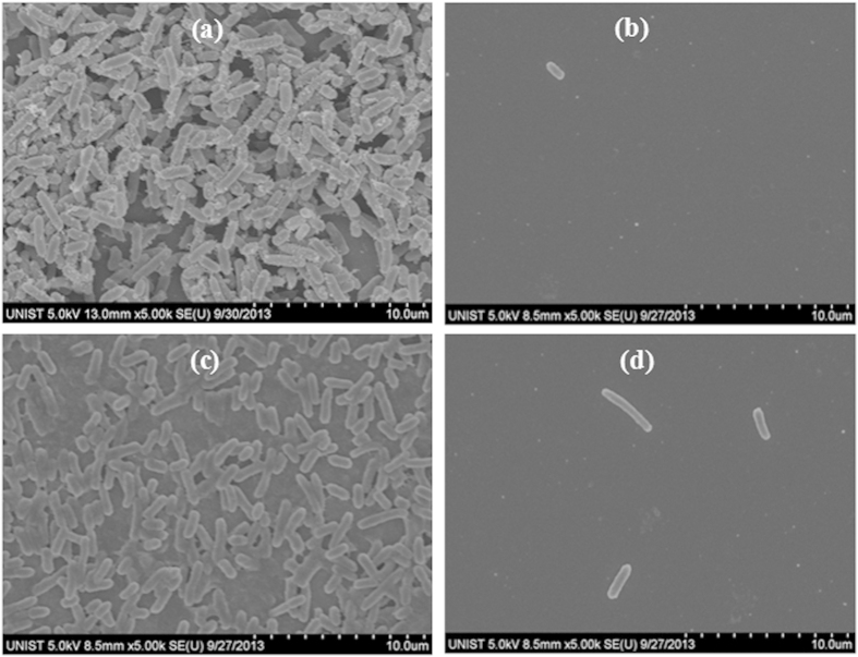
SEM analysis was initially used to analyze E. coli biofilms grown for one day on silicon chips before and after treating them with CO2 aerosols or hydrolytic enzymes (Fig. 1). This figure shows that a uniform growth of E. coli biofilm across the silicon surface was readily apparent for the control chip (Fig. 1a) and that this biofilm was effectively removed after the CO2 aerosol treatment (Fig. 1b). For comparison, an SEM micrograph of the E. coli biofilm after soaking in HEPES buffer is also presented (Fig. 1c) along with an image showing the E. coli biofilm after treatment with both Proteinase K and DNase I (Fig. 1d). As these two enzymes hydrolyze the protein and DNA present within the EPS, treatment of the biofilms with both of these results in a gentle dispersion of the bacteria.
Figure 1. SEM images showing the effects of a treatment with either CO2 aerosols or hydrolytic enzymes on the E. coli biofilms grown for one day.
(a) Untreated control chip showing the presence of an extensive E. coli biofilm on the Si surface. (b) Biofilm after CO2 aerosol treatment. (c) Image of E. coli biofilm after being soaked in 25 mM HEPES buffer for two hours. (d) E. coli biofilm after treatment with HEPES buffer containing both Proteinase K and DNase I.
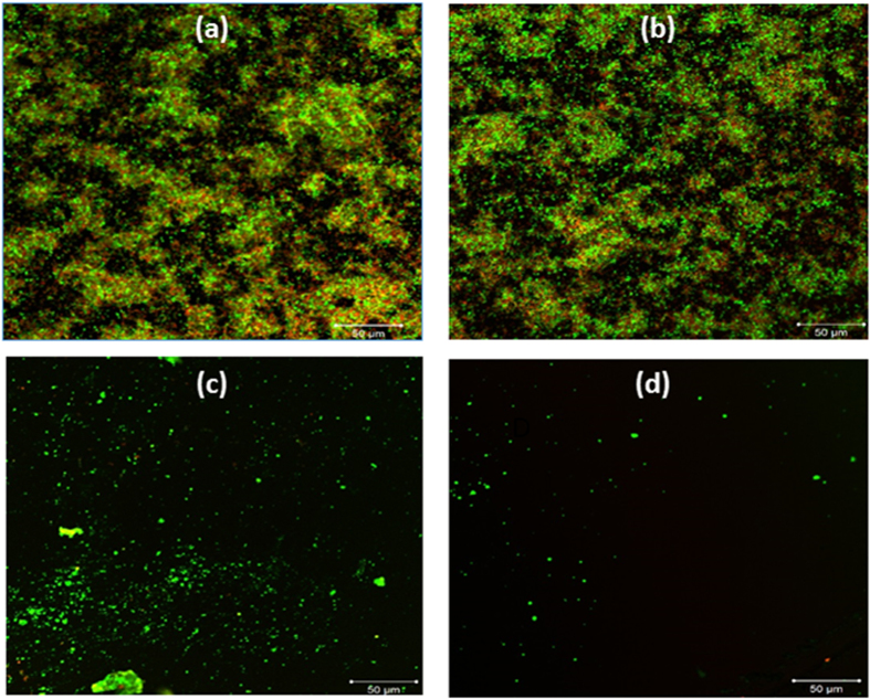
To analyze these results deeper, particularly with regards to biofilm viability, we stained the biofilms with a BacLight stain (Invitrogen, USA) containing both SYTO9 and propidium iodide (PI), which labels the live cells green and the dead cells red, respectively. Figure 2a shows the fluorescent image of the untreated E. coli biofilm. Although not quantitative, the greater prevalence of green fluorescence suggests that the majority of the culture was viable, a finding that also appears true of the HEPES-treated biofilm (Fig. 2b). Treatment of the biofilm with either hydrolytic enzymes (Fig. 2c) or the aerosols (Fig. 2d), however, led to a significant decrease in both fluorescent signals, affirming the findings of Fig. 1 where a significant number of the bacteria cells were removed by both of these treatments.
Figure 2. Confocal microscopic image of one-day grown E. coli biofilms stained with the BacLight stain (SYTO-9 and propidium iodide) after their respective treatments.
(a) Untreated control biofilm. (b) HEPES soaked biofilm. (c) E. coli biofilm after treatment with Proteinase K and DNase I. (d) E. coli biofilm treated with CO2 aerosols. The scale bars are 50 μm.
Removal of biofilms with CO2 aerosols was previously demonstrated by our group19,20. Based upon the above images and a previous report4, it is clear that biofilms harbor a significant number of viable bacteria. However, the effects of CO2 aerosols on the viability of the dispersed bacteria have not been studied to date. To evaluate this, therefore, we collected three groups of samples: enzymatically dispersed bacteria from a biofilm using Proteinase K and DNase I (Control), the bacteria dispersed from the biofilm by CO2 aerosols (Aerosol) and those obtained after treatment of the biofilm first by CO2 aerosols followed by an enzymatic treatment of the biofilm still attached to the silicon chip (Aero + Chip). It should be noted that all the samples collected were treated subsequently with Proteinase K and DNase I to dissociate any bacterial aggregates that were present to ensure that the viable counts in Fig. 3a were correct.
Figure 3. Viable number of E. coli present within the CO2 aerosol treated samples.
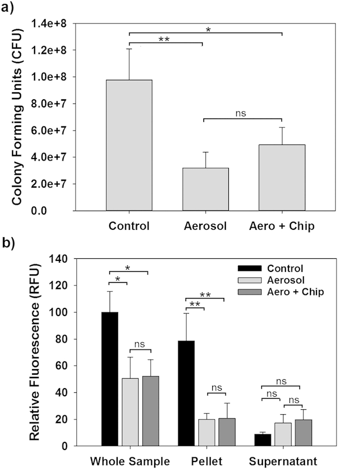
(a) Colony forming units determined by plating out on agar plates. The samples are the Control, which was gently dispersed using a Proteinase K and DNase I treatment, the Aerosol sample, i.e. the bacteria dispersed by CO2 aerosols and captured within the flask, and the Aero + Chip sample, which includes both the flask captured bacteria and the dispersal of any remaining bacteria present on the chip using Proteinase K and DNase I. (b) The relative fluorescence seen in the whole sample or the constituent parts (i.e., cell pellet or supernatant). The presence of nearly half of the fluorescence in the supernatant after CO2 aerosol treatment implies that a significant number of E. coli are being ruptured by this treatment. Statistical analysis was performed using one-way ANOVA followed by the Tukey post hoc test. Statistically significant results are identified with asterisks (*,** =P values < 0.05 or 0.01, respectively; ns – not significant).
For the control sample, which was dispersed using only the enzymes, the total number of viable bacteria per chip was on average 9.8 × 10⁷ CFU. By comparison, the number of viable bacteria in the aerosol-dispersed samples was 3.2 × 10⁷ CFU, or approximately 3-fold less. Inclusion of the bacteria still present on the chip after aerosol treatment increased the viable count to 4.9 × 10⁷ CFU, which is approximately half the original number. Based upon the number still attached to the chip, i.e., 1.7 × 10⁷ CFU, treatment of the biofilms with aerosols removed approximately 80% of the biofilm-associated viable cells from the silicon chip surface. This value is not unexpected as the treatment area did not encompass the entire chip.
As the solid CO2 particles are comparable in size with the bacteria, it was presumed that their momentum and impact may be sufficient to cause damage to the bacteria and their membranes. To study this more in depth, we performed the same experiments with a fluorescent strain of E. coli that expresses the cyan fluorescent protein (CFP). It was hypothesized that if the cell membranes are damaged by the CO2 aerosols, the CFP protein would be released into the collection media, thereby increasing the fluorescence of the cell-free supernatant. This was found to be the case, as shown in Fig. 3b. Addition of Proteinase K to the sample was not a concern since GFP and related fluorescent proteins are known to be resistant to this protease26.
When considering the total fluorescence of each sample as shown in Fig. 3b, approximately half is lost during the aerosol treatments. Of that seen in the captured samples, however, approximately half was associated with cell pellet and the other half was found in the supernatant. Such a significant presence of CFP within the extracellular milieu indirectly confirms that the CO2 aerosols are disrupting the bacterial membrane integrity and helps explain the loss in viability seen in Fig. 3a. This activity of the CO2 aerosols can be attributed to the large momentum (mechanical impact) of the solid CO2 particles and high shear near the silicon surfaces due to the high-speed gas flows, which will be discussed below.
Although the above fluorescence results indicate that a large number of the bacteria are being injured or killed by the aerosol treatment, it still remained uncertain if the decrease in the viable counts (Fig. 3a) is due to cell death or if a portion of the bacterial population was lost due to aerosolization of the biofilm. To address this, we analyzed each of the collections (Control, Aerosol and Aero + Chip) using flow cytometry. As shown in Fig. 4a, when the same volume of sample was analyzed the particle number in the Aerosol sample was approximately 40% lower than that of the Control. However, treatment of the undispersed biofilm still present on the chip with Proteinase K and DNase I (Aero + Chip) increased this to 73%. These values show that a significant number of bacteria are not being captured, and although this contributes to the lower viability seen in Fig. 3a it does not fully account for this discrepancy.
Figure 4. Analysis of the bacteria dispersed during the CO2 aerosol and enzymatic treatments by flow cytometry.
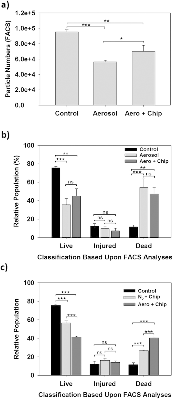
The samples are the same as in Fig. 3. (a) Actual number of particles counted within the FSC gated region when using a fixed time of 10 seconds for each sample. (b) Relative populations of the cells (live, dead and injured) in each sample. (c) Relative populations of the cells (live, dead and injured) were dispersed by enzymatic treatment (control), N2 gas, and CO2 aerosol treatments. In these N2 gas and CO2 aerosol treatments, the dispersed bacteria were added to the bacteria present on the chip surfaces. The classification of bacterial cells into each population was performed by staining the samples with the BacLight stain (SYTO9 and propidium iodide) prior to FACS analysis. A total of 5,000 cells were analyzed for each sample, and the results show the average from three independent tests. Statistical analysis was performed using one-way ANOVA followed by the Tukey post hoc test. Statistically significant results are identified with asterisks (*, **, or *** = P values < 0.05, 0.01 or 0.0001, respectively; ns – not significant).
Flow cytometry combined with Live/Dead staining is also commonly employed to evaluate bacterial viability for the samples derived from various environmental conditions, such as soil and water samples27. Consequently, using live and dead fractions of planktonic bacteria, we were able to gate the live and dead populations according to the FACS analyses (Fig. S1). Subsequently, using a sample number of 5,000 cells for each test condition, we were able to determine the relative live and dead populations (Fig. S2). Interestingly, there were populations that fell between these two categories and, thus, these were serendipitously branded as “injured” bacteria (Fig. 4 and S2).
Figure 4b shows that the majority of the bacteria present within the Control samples are living (76%). This is in stark contrast with the Aerosol and Aero + Chip samples, both of which harbor a significant number of dead and injured bacterial cells. These results, however, are in agreement with the results presented in Fig. 3b since 54% and 47% of the Aerosol and Aero + Chip populations, respectively, are classified as non-viable, i.e., dead. By comparison, in Fig. 3b the supernatant fluorescence intensities for both samples were just below that found in the pellets. The results from both tests indicate that approximately half of the dispersed bacterial population is killed by the aerosols.
From Fig. 3a, the Aerosol sample viability was 33% while for the Aero + Chip sample it was 51%. When the relative number of particles in Fig. 4a, which represents the capture efficiency, is multiplied by the summed percent of live and injured bacteria (Fig. 4b) for each sample, we obtain a relative viability of 30% and 44% for the Aerosol and Aero + Chip samples, respectively, which is slightly lower than but similar to the colony forming units actually found (Fig. 3a). As such, the consistency between the different techniques employed in this study helps to substantiate that the CO2 aerosols inflict a significant amount of damage to the biofilm-associated bacterial population during treatment and that this leads to losing the viability. Moreover, it suggests that the “injured” bacteria are viable and capable of producing colonies.
The aerosols actually consist of two components – the solid CO2 particles and gaseous CO2. As mentioned above, the loss in viability is likely attributed to the large momentum associated with the high-speed solid CO2 particles. To study this further, and in the hope of identifying the component responsible, we also performed tests with only pressurized nitrogen gas whose stagnation pressure was the same as that of CO2 gas. This is based on the fact that CO2 and N2 gases would have almost the same flow patterns and shear stresses on the substrate surfaces if both gases have the same pressures. As shown in Fig. 4c, the viability of the bacteria decreased when the biofilm was treated with nitrogen alone implying viability loss due to the gas component of the aerosols. This loss was more significant when CO2 aerosols were used. A comparison between these two treatments shows that the viability was significantly lowered by the presence of the CO2 aerosols, that is, the solid CO2 particles and gaseous CO2. As such, the activity of the CO2 aerosols on bacterial viability can be attributed to both the large momentum (mechanical impact) of the solid CO2 particles and the high shear stress resulting from the CO2 gas flows. It should also be noted that, although a high-pressure gas treatment only damages many of the bacteria, the removal efficiency was shown previously to be very small19.
Although our group previously found that CO2 aerosols can be used to remove biofilms from various substrate surfaces, this study is the first to show that they also kill a significant portion of the bacteria during the dispersal. Using several different analytical tools and techniques, we found the aerosols significantly impact the biofilm population viability. An analysis of the supernatant fluorescence strongly suggested that the aerosol-treated bacteria experience a loss in membrane integrity, i.e., rupturing, that results in intracellular proteins being released into the surrounding milieu. The net result of this is a dramatic loss in viability, as demonstrated here by the CFU enumeration and flow cytometry analyses, with nearly 50% of the aerosol dispersed E. coli dying as a direct result of this treatment.
Methods
Bacterial growth
E. coli XL1-Blue was obtained from RBC Biosciences, Korea (HIT-Blue Competent Cells, Cat# RH117). This strain was initially grown up on Luria-Bertani (LB) (BD Difco, USA) agar plates (1.6% agar, BD Difco, USA) from which colonies were inoculated into 3 ml LB broth in a 15 ml conical tube (SPL, Korea). These cultures were cultivated at 37 °C with shaking at 250 rpm for 16 hours. Consequently, 25% glycerol stocks were prepared and these were stored at −80 °C. Before preparation of the biofilm, the glycerol stock was streaked on an LB agar plate and incubated overnight at 37 °C to cultivate colonies. From the plate, a single colony was inoculated into 10 ml sterile LB broth in a 50 ml conical tube containing 100 μg/ml ampicillin and incubated with shaking (250 rpm) at 37 °C overnight. To generate a fluorescent variant of XL1-Blue, this strain was transformed with the pAMCyan plasmid (Clontech, USA), which confers resistance to ampicillin.
E. coli biofilm formation on silicon chips
Initially, silicon chips (10 × 10 mm, Shin-Etsu, Japan) were cleaned by piranha solution (hydrogen peroxide (30%):sulfuric acid (96%) =1:1 v/v) for 10 minutes, rinsed with deionized water for 10 minutes, and then dried under pure nitrogen gas. The contact angles of a distilled water droplet on the chips and surface roughness of the chips were 17.0 ± 1.5 degrees and 22.6 ± 0.8 nm, respectively20. As such, this silicon surface is very smooth, which generally facilitates the initial attachment of bacteria28. Moreover, this aerosol technique was quite insensitive to the substrate materials employed with regard to the biofilm removal efficiencies20. Before preparation of the biofilm, the silicon chips were treated with 70% ethanol for 5 seconds, rinsed in autoclaved deionized water for 5 seconds, and then finally rinsed three times in LB broth for 5 seconds each.
To grow the biofilms, an overnight grown culture of E. coli XL1-blue (OD6002.1) was initially diluted 100-fold into fresh LB media, and 180 μl of this diluted culture was further diluted into 5 ml of fresh LB media. This culture contained an average of 240000 (±39600) viable cells per ml and was poured into a 35 mm Petri dish. Two chips were aseptically placed into each dish and incubated for 24 h at 30 °C without shaking.
Biofilm removal via CO2 aerosol treatment
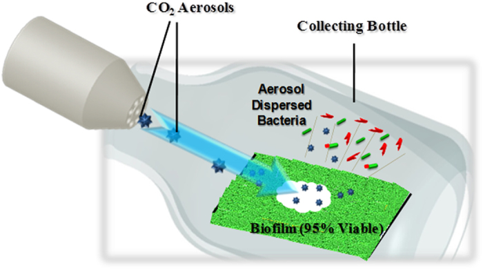
The general experimental procedures employed were published previously19,20. Briefly, the biofilms on the silicon chips were washed gently with 10 mM ammonium acetate buffer (Sigma-Aldrich, USA), and were treated with the aerosols immediately. We placed the E. coli biofilm grown chips horizontally and positioned 2 cm from the nozzle in an aerosol flow. The nozzle axis was maintained at 40° angle relative to the chip surface. The bacteria dispersed by the aerosol treatment were collected using a 200 ml bottle arranged over the chip to minimize loss (Fig. 5). A dual gas unit (K6-10DG; Applied Surface Technologies, NJ, USA) was used for aerosol generation. The N2 gas pressure was 0.7 MPa and the CO2 gas pressure was 5.6 ± 0.2 MPa. The CO2 aerosols were off for 3 seconds during each 8-second cleaning cycle, and the total cleaning time was 5 cycles. The average room temperature was 25.2 (±2.1) °C, and the average relative humidity was 67.5 (±7.3) % during all aerosol treatments.
Figure 5. Schematic illustration of the system used in this study to expose bacterial biofilms to CO2 aerosols and capture dispersed cells29.
Enzymatic disruption of the biofilms
The bacteria collected in 25 mM HEPES buffer (pH 7.2) (Sigma-Aldrich, USA) were subjected to enzymatic treatment with Proteinase K (100 ng/ml, Invitrogen, USA) and DNase I (100 ng/ml, Sigma-Aldrich, USA) for 2 h at 37 °C to disrupt any cell clumps that may be present. Likewise, biofilms still present on the treated chips and those on control chips, i.e., untreated, were also dispersed using the same enzymatic treatment.
Confocal and scanning electron microscopic analysis
Confocal microscopy and SEM were used to visualize the E. coli XL1-Blue biofilms that formed on the silicon chips. The bacterial cells were stained by immersing the chip for 30 min in 25 mM HEPES buffer containing BacLight stain (SYTO9-PI, Invitrogen, USA). The chips were then washed gently to remove any excess dye before imaging using an LSM700 confocal microscope (Carl Zeiss) operated by ZEN 2009 software.
Before SEM imaging, the bacteria were fixed by the chemical fixation procedure described by Dwidar et al.29. The samples were then placed in a critical point dryer (SPI Supplies) for the drying at 35 °C at 1200 psi. Finally, the samples were coated with platinum and imaged with a scanning electron microscope (S-4800, Hitachi).
Enumeration of viable bacteria
For viability measurements, we determined the CFU of three samples separately: 1) from untreated chips using an enzymatic treatment (Control); 2) the bacteria dispersed by the aerosols (Aerosol); and 3) the bacteria dispersed by the aerosols and then an enzymatic treatment of the same chip to obtain any biofilm-associated bacteria still attached to the chip (Aero + Chip). After aerosol treatment, the bacterial samples were collected with 10 ml of buffer as mentioned above and transferred to a 15 ml conical bottom tube (BD Falcon, USA). For the Aero + Chip samples, the treated chip was added to the collected sample in the same tube to disperse the biofilm attached to the surface using Proteinase K and DNase I as described above. The same protocol was used for the Control, so each of the different samples was present within 10 ml of HEPES buffer (25 mM). The samples were centrifuged at 16,000 g for 5 minutes and the supernatant was used for fluorescence determination. The resulting bacterial pellet was re-suspended in 9.8 ml HEPES buffer, and the number of viable bacteria within each sample was determined by spreading serial dilutions out on LB agar plates and growth of the colonies overnight at 37 °C.
Fluorescence measurements
From the 10 ml samples obtained above, a small aliquot was taken for fluorescence measurement. The total fluorescence was determined using 200 μl of the collected samples. In parallel, a 200 μl cell-free supernatant sample was also tested to determine the extracellular fluorescence. These samples were prepared by centrifuging the samples as described above and filtering them through sterile 0.22 micron syringe filters to remove any remaining bacterial cells. Likewise, the resulting cell pellet was re-suspended in the same volume of HEPES buffer (25 mM) and the fluorescence of this sample (pellet) was also determined, representing the cell-associated fluorescence. The fluorescence measurement in each case was performed using a 200 μl sample with 96 well black plates (SPL, South Korea) and a fluorescence plate reader (Infinite® 200 PRO – Tecan, Germany). The excitation and emission wavelengths were set to 410 nm and 495 nm, respectively. The fluorescence from each of the samples was normalized using a standard curve and is shown relative to the whole sample fluorescence obtained from the Control.
Flow cytometry analysis of dispersed bacteria
Each sample was run for 10 seconds using a FACS calibur flow cytometer (BD Biosciences, San Jose, CA), and the number of E. coli cells, both viable and dead, passing through the FSC/SSC gated region were counted. For live/dead discrimination, dead bacteria were prepared by adding 50 μl of disinfectant Extran MA 02 (Merck KGaA, Germany) to 450 μl of an overnight bacterial culture and incubating for 30 min at room temperature. Live cells were directly taken from overnight grown suspension. Both samples were stained with BacLight stain (SYTO9 and PI) prior to FACS analysis. The stain exhibits green fluorescence in all bacterial cells, while PI only stains dead bacteria with a red fluorescence. The different population were gated and gate settings were used for analysis of dispersed bacteria from biofilm.
Similarly, the dispersed biofilm samples were stained and underwent FACS analysis as mentioned above. All the samples were kept at room temperature for 10 minutes before the flow cytometry analysis. Experiments were performed in a BD LSRFortessa Flow Cytometer (BD Biosciences, San Jose, CA) using a blue (488 nm) laser to excite the stained cells. The green fluorescence emission was detected with a 530/30 band pass filter and the red fluorescence emission was detected with a 695/40 band pass filter. The fluorescence emission was acquired for 5,000 cells and displayed in an exponential scale using BD FACS Diva v.6.2 (BD Biosciences San Jose, CA).
Data analysis
Each of the experiments was performed independently in triplicate for error analysis. The average values obtained are shown in the figures with the standard deviations presented as the error bars. Statistical analyses comparing the results amongst the different treatments were performed using a one-way ANOVA test followed by the Tukey post hoc test. Significantly different results are designated with asterisks (*) within the corresponding figures.
Additional Information
How to cite this article: Singh, R. et al. Effects of Carbon Dioxide Aerosols on the Viability of Escherichia coli during Biofilm Dispersal. Sci. Rep. 5, 13766; doi: 10.1038/srep13766 (2015).
Supplementary Material
Acknowledgments
This research was supported by Basic Science Research Program through the National Research Foundation of Korea (NRF) funded by the Ministry of Education, Science and Technology (Grant # 2013R1A1A2008590) and by the Ministry of Science, ICT and future Planning (Grant # 2015R1A2A2A01006446). The authors would also like to acknowledge and thank the staff in the UNIST Central Research Facilities (UCRF) and the UNIST Olympus Biomed Imaging Center (UOBC) for their help in this study.
Footnotes
Author Contributions R.S. and A.K.M. participated in the experiment design, performance and analyzed the data. S.H. handled the CO2 aerosols treatment part. R.J.M. and J.J. supervised the studies and discussed the results. All of the authors reviewed the manuscript and participated in discussions on the results of this research.
References
- Monnappa A. K., Dwidar M., Seo J. K., Hur J. H. & Mitchell R. J. Bdellovibrio bacteriovorus Inhibits Staphylococcus aureus Biofilm Formation and Invasion into Human Epithelial Cells. Sci Rep 4, 3811 (2014). [DOI] [PMC free article] [PubMed] [Google Scholar]
- Simoes M., Simoes L. C. & Vieira M. J. A review of current and emergent biofilm control strategies. Lwt-Food Sci Technol 43, 573–583 (2010a). [Google Scholar]
- Auerbach I. D., Sorensen C., Hansma H. G. & Holden P. A. Physical morphology and surface properties of unsaturated Pseudomonas putida biofilms. J Bacteriol 182, 3809–3815 (2000). [DOI] [PMC free article] [PubMed] [Google Scholar]
- Dwidar M., Leung B. M., Yaguchi T., Takayama S. & Mitchell R. J. Patterning Bacterial Communities on Epithelial Cells. PLoS One 8, e67165 (2013). [DOI] [PMC free article] [PubMed] [Google Scholar]
- Jain A., Gupta Y., Agrawal R., Khare P. & Jain S. K. Biofilms - A microbial life perspective: A critical review. Crit Rev Ther Drug 24, 393–443 (2007). [DOI] [PubMed] [Google Scholar]
- Kim E. H., Dwidar M., Mitchell R. J. & Kwon Y. N. Assessing the effects of bacterial predation on membrane biofouling. Water Res 47, 6024–6032 (2013). [DOI] [PubMed] [Google Scholar]
- Meyer B. Approaches to prevention, removal and killing of biofilms. Int Biodeter Biodegr. 51, 249–253 (2003). [Google Scholar]
- Simoes L. C., Simoes M. & Vieira M. J. Adhesion and biofilm formation on polystyrene by drinking water-isolated bacteria. Anton Leeuw Int J G 98, 317–329 (2010b). [DOI] [PubMed] [Google Scholar]
- Hetrick E. M., Shin J. H., Paul H. S. & Schoenfisch M. H. Anti-biofilm efficacy of nitric oxide-releasing silica nanoparticles. Biomaterials 30, 2782–2789 (2009). [DOI] [PMC free article] [PubMed] [Google Scholar]
- Beloin C., Roux A. & Ghigo J. M. Escherichia coli biofilms. Curr Top Microbiol 322, 249–289 (2008). [DOI] [PMC free article] [PubMed] [Google Scholar]
- Herzberg M. & Elimelech M. Biofouling of reverse osmosis membranes: Role of biofilm-enhanced osmotic pressure. J Membrane Sci 295, 11–20 (2007). [Google Scholar]
- Chen X. & Stewart P. S. Biofilm removal caused by chemical treatments. Water Res 34, 4229–4233 (2000). [Google Scholar]
- Hong S. H. et al. Effect of electric currents on bacterial detachment and inactivation. Biotechnol Bioeng 100, 379–386 (2008). [DOI] [PubMed] [Google Scholar]
- Nandakumar K., Obika H., Utsumi A., Ooie T. & Yano T. In vitro laser ablation of laboratory developed biofilms using an Nd: YAG laser of 532 nm wavelength. Biotechnol Bioeng 86, 729–736 (2004). [DOI] [PubMed] [Google Scholar]
- Bott T. R. Biofouling control with ultrasound. Heat Transfer Eng 21, 43–49 (2000). [Google Scholar]
- Bayoudh S., Ponsonnet L., Ben Ouada H., Bakhrouf A. & Othmane A. Bacterial detachment from hydrophilic and hydrophobic surfaces using a microjet impingement. Colloid Surface A 266, 160–167 (2005). [Google Scholar]
- Gibson H., Taylor J. H., Hall K. E. & Holah J. T. Effectiveness of cleaning techniques used in the food industry in terms of the removal of bacterial biofilms. J Appl Microbiol 87, 41–48 (1999). [DOI] [PubMed] [Google Scholar]
- Sherman R. Carbon dioxide snow cleaning. Particul Sci Technol 25(1), 37–57 (2007). [Google Scholar]
- Kang M. Y., Jeong H. W., Kim J., Lee J. W. & Jang J. Removal of biofilms using carbon dioxide aerosols. J Aerosol Sci 41, 1044–1051 (2010). [Google Scholar]
- Cha M., Hong S., Kang M. Y., Lee J. W. & Jang J. Gas-phase removal of biofilms from various surfaces using carbon dioxide aerosols. Biofouling 28, 681–686 (2012). [DOI] [PubMed] [Google Scholar]
- Singh R., Hong S. & Jang J. Mechanical desorption of immobilized proteins using carbon dioxide aerosols for reusable biosensors. Anal Chim Acta 853, 588–595 (2015). [DOI] [PubMed] [Google Scholar]
- Ausbacher D. et al. Staphylococcus aureus biofilm susceptibility to small and potent beta(2,2)-amino acid derivatives. Biofouling 30, 81–93 (2014). [DOI] [PubMed] [Google Scholar]
- Lee J. T., Jayaraman A. & Wood T. K. Indole is an inter-species biofilm signal mediated by SdiA. BMC Microbiol 7, 42 (2007). [DOI] [PMC free article] [PubMed] [Google Scholar]
- Ren D. C., Sims J. J. & Wood T. K. Inhibition of biofilm formation and swarming of Escherichia coli by (5Z)-4-bromo-5(bromomethylene)-3-butyl-2(5H)-furanone. Environ Microbiol 3, 731–736 (2001). [DOI] [PubMed] [Google Scholar]
- Skogman M. E., Vuorela P. M. & Fallarero A. Combining biofilm matrix measurements with biomass and viability assays in susceptibility assessments of antimicrobials against Staphylococcus aureus biofilms. J Antibiot 65, 453–459 (2012). [DOI] [PubMed] [Google Scholar]
- Yi Z. G., Yuan Z. H., Rice C. M. & MacDonald M. R. Flavivirus Replication Complex Assembly Revealed by DNAJC14 Functional Mapping. J Virol 86, 11815–11832 (2012). [DOI] [PMC free article] [PubMed] [Google Scholar]
- Ferrari B. C., Winsley T. J., Bergquist P. L. & Van Dorst J. Flow cytometry in environmental microbiology: a rapid approach for the isolation of single cells for advanced molecular biology analysis. Methods in molecular biology 881, 3–26 (2012). [DOI] [PubMed] [Google Scholar]
- Crawford R. J., Webb H. K., Truong V. K., Hasan J. & Ivanova E. P. Surface topographical factors influencing bacterial attachment. Adv Colloid Interfac 179, 142–149 (2012). [DOI] [PubMed] [Google Scholar]
- Dwidar M., Hong S., Cha M., Jang J. & Mitchell R. J. Combined application of bacterial predation and carbon dioxide aerosols to effectively remove biofilms. Biofouling 28, 67––80. (2012). [DOI] [PubMed] [Google Scholar]
Associated Data
This section collects any data citations, data availability statements, or supplementary materials included in this article.


