Abstract
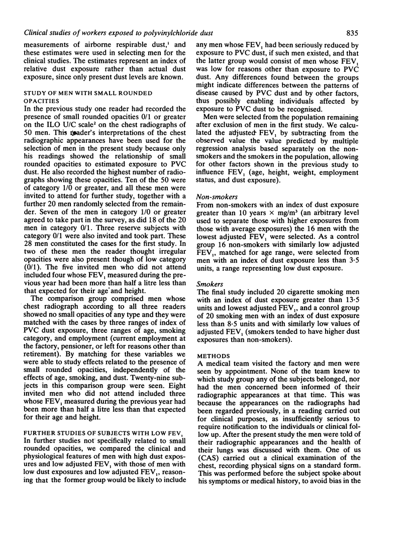
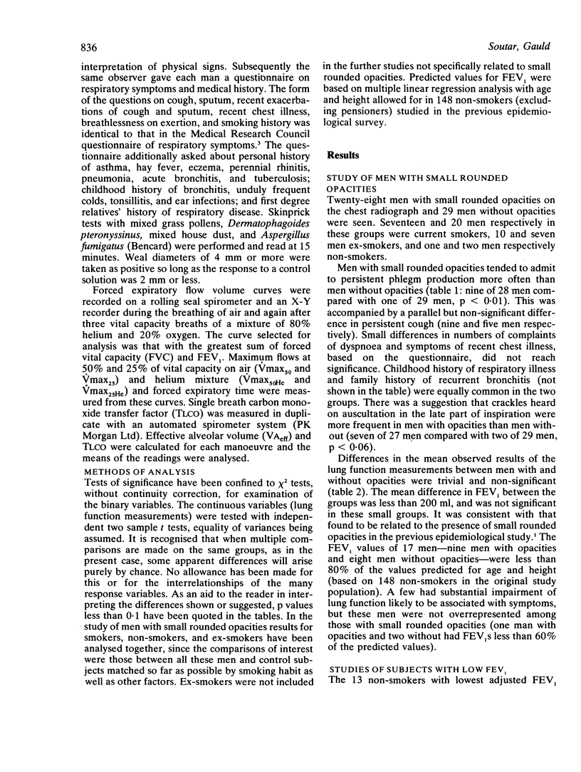
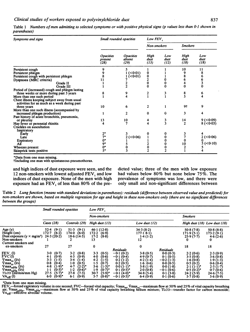
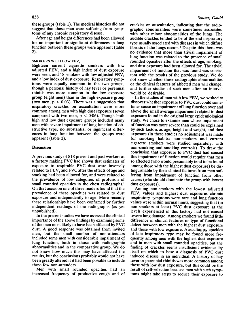
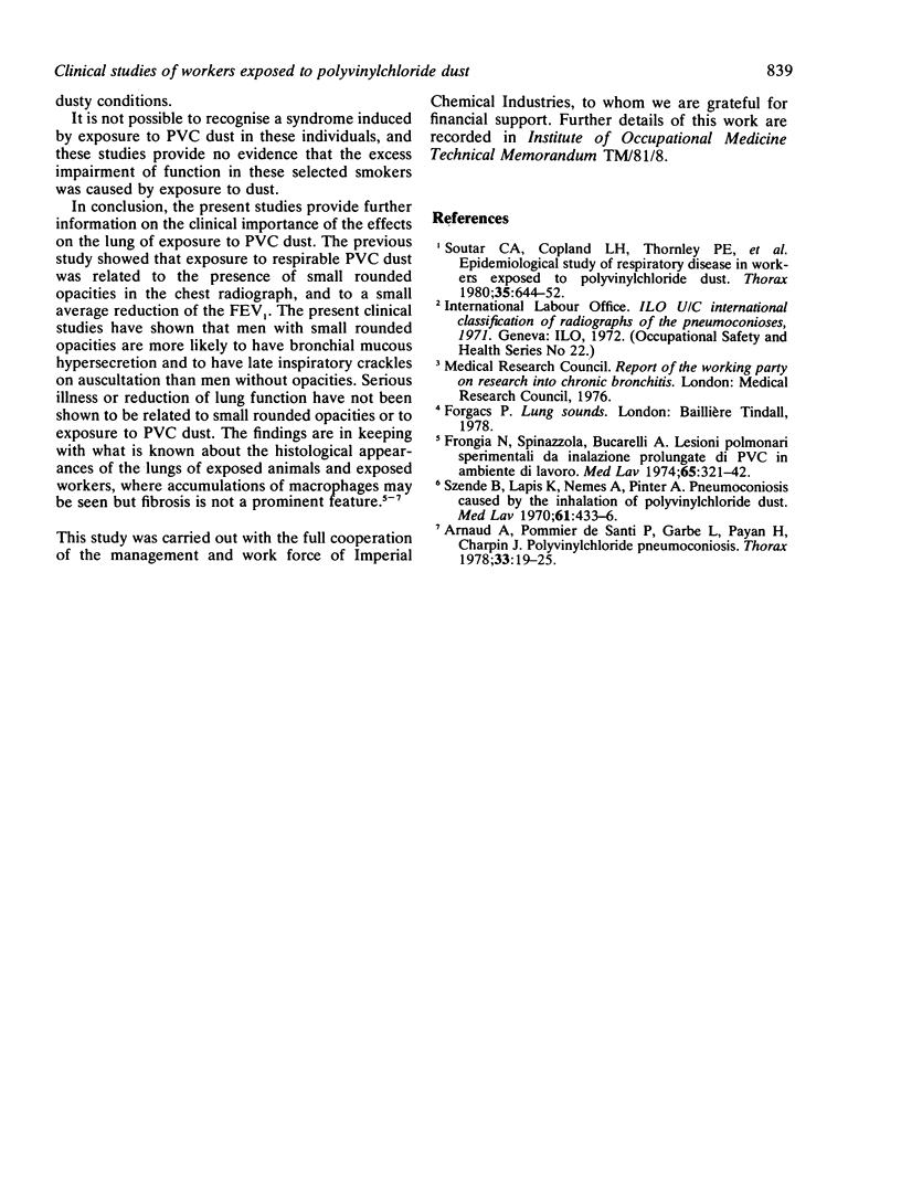
A previous study showed that exposure of workers to polyvinylchloride (PVC) dust was associated with the presence of small rounded opacities in the chest radiograph, and also with a small average reduction of the forced expiratory volume in one second (FEV1). Studies have now been carried out on selected men to determine the clinical importance of the presence of small rounded opacities and also to identify any clinical or physiological features associated with PVC dust exposure. Among 28 men with small rounded opacities, complaints of persistent bronchial mucous hypersecretion were more common (nine men) than amont 29 men of similar age, smoking habits, and dust exposures (one man, p less than 0.01), and there was a suggestion that the physical sign of late inspiratory crackles was more frequent among men with opacities (seven of 27 men) than the other men (two of 28 men, p less than 0.05). The average lung function of men with opacities was only trivially lower than that of the comparative group and the difference between the groups was not significant. In further studies 13 non-smokers with the highest PVC dust exposure and lowest FEV1 (adjusted for age, height, weight, and dust exposure) were examined. None had an observed FEV1 less than 80% of the predicted value for age and height, and these men did not differ in symptoms, signs, or lung function from non-smokers with low dust exposure and similarly low FEV1. In a similar study of 18 smokers with the highest PVC dust exposures and lowest adjusted FEV1, some men had an observed FEV1 well below 80% of predicted values for non-smokers, but with one exception no clinical or functional differences were found between then and 18 smokers with low dust exposure and similarly low FEV1. The exception was again the presence of end inspiratory crackles on auscultation, which were slightly more frequent among men with the highest dust exposures (seven men) than among those with low exposure (two men, p less than 0.06). We conclude that these findings are consistent with the suggestion that the radiographic abnormalities caused by PVC dust exposure are not associated with important functional effects or clinical illness. Further studies are desirable to examine the eventual outcome of the syndrome.
Full text
PDF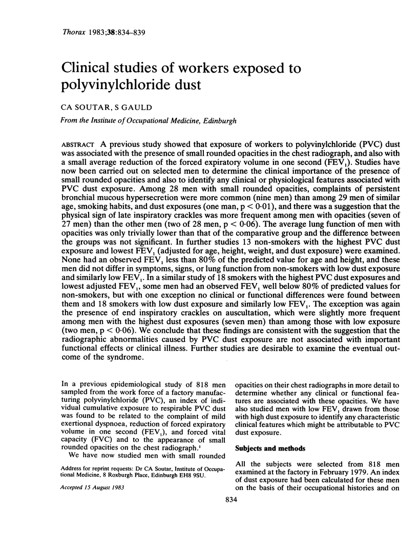
Selected References
These references are in PubMed. This may not be the complete list of references from this article.
- Arnaud A., Pommier de Santi P. P., Garbe L., Payan H., Charpin J. Polyvinyl chloride pneumoconiosis. Thorax. 1978 Feb;33(1):19–25. doi: 10.1136/thx.33.1.19. [DOI] [PMC free article] [PubMed] [Google Scholar]
- Arnaud A., Pommier de Santi P. P., Garbe L., Payan H., Charpin J. Polyvinyl chloride pneumoconiosis. Thorax. 1978 Feb;33(1):19–25. doi: 10.1136/thx.33.1.19. [DOI] [PMC free article] [PubMed] [Google Scholar]
- Frongia N., Spinazzola A., Bucarelli A. Lesioni polmonari sperimentali da inalazione prolungata di polveri di PVC in ambiente di lavoro. Med Lav. 1974 Sep-Oct;65(9-10):321–342. [PubMed] [Google Scholar]
- Soutar C. A., Copland L. H., Thornley P. E., Hurley J. F., Ottery J., Adams W. G., Bennett B. Epidemiological study of respiratory disease in workers exposed to polyvinylchloride dust. Thorax. 1980 Sep;35(9):644–652. doi: 10.1136/thx.35.9.644. [DOI] [PMC free article] [PubMed] [Google Scholar]
- Soutar C. A., Copland L. H., Thornley P. E., Hurley J. F., Ottery J., Adams W. G., Bennett B. Epidemiological study of respiratory disease in workers exposed to polyvinylchloride dust. Thorax. 1980 Sep;35(9):644–652. doi: 10.1136/thx.35.9.644. [DOI] [PMC free article] [PubMed] [Google Scholar]
- Szende B., Lapis K., Nemes A., Pinter A. Pneumoconiosis caused by the inhalation of polyvinylchloride dust. Med Lav. 1970 Aug-Sep;61(8):433–436. [PubMed] [Google Scholar]


