

Abstract
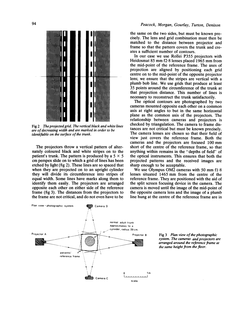
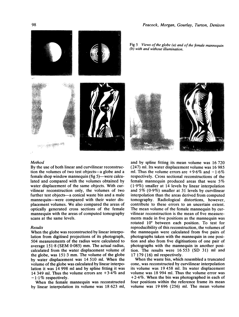
An optical technique has been developed for mapping the size and shape of the thoracoabdominal wall and the change in its shape with breathing. A fixed pattern composed of stripes of light is projected on to both sides of the trunk. These stripes become distorted when viewed from in front and behind, forming contours over the trunk surface. The contours are photographed and then encoded digitally. The digital information can be used to compute automatically the volume of the trunk, the position of any point on its surface, and its cross sectional shape at any level. The technique has been tested on rigid objects (a globe, a cone, and two dummy torsos) that can be measured precisely. With this optical technique linear dimensions can be calculated to within 0.5 mm, cross sectional area to within 5%, and volume to within 1.6-3.7%. These results suggest that this non-invasive technique measures the shape and volume of complex three dimensional surfaces with sufficient accuracy to be tried in clinical practice.
Full text
PDF


Images in this article


Selected References
These references are in PubMed. This may not be the complete list of references from this article.
- Ainsworth H., Joseph M. An assessment of a stereophotogrammetric technique for the study of facial morphology in the child. Ann Hum Biol. 1976 Sep;3(5):475–488. doi: 10.1080/03014467600001741. [DOI] [PubMed] [Google Scholar]
- Britton M. G. Asbestos pleural disease. Br J Dis Chest. 1982 Jan;76(1):1–10. [PubMed] [Google Scholar]
- Burke P. H., Beard F. H. Stereophotogrammetry of the face. A preliminary investigation into the accuracy of a simplified system evolved for contour mapping by photography. Am J Orthod. 1967 Oct;53(10):769–782. doi: 10.1016/0002-9416(67)90121-2. [DOI] [PubMed] [Google Scholar]
- Hök B., Nilsson K., Bjelkhagen H. Imaging of chest motion due to heart action by means of holographic interferometry. Med Biol Eng Comput. 1978 Jul;16(4):363–368. doi: 10.1007/BF02442652. [DOI] [PubMed] [Google Scholar]
- Kovats F., Jr Pléthysmographie optique du tronc: étude du cycle ventilatoire maximal. Bull Physiopathol Respir (Nancy) 1970 Oct-Dec;6(4):833–845. [PubMed] [Google Scholar]
- Lovesey E. J. A simple photographic technique for recording three-dimensional head shape. Med Biol Illus. 1973 Nov;23(4):210–213. [PubMed] [Google Scholar]
- Madden M. C., Karlan M. S. Moiré photography as a means of topographical mapping of the human face. Ann Biomed Eng. 1979;7(2):95–102. doi: 10.1007/BF02363128. [DOI] [PubMed] [Google Scholar]
- Whittle M. W., Herron R. E., Cuzzi J. R. Biostereometric analysis of body form: the second manned Skylab mission. Aviat Space Environ Med. 1976 Apr;47(4):410–412. [PubMed] [Google Scholar]
- van Wijk M. C. Moiré contourgraph--an accuracy analysis. J Biomech. 1980;13(7):605–613. doi: 10.1016/0021-9290(80)90060-3. [DOI] [PubMed] [Google Scholar]