Abstract
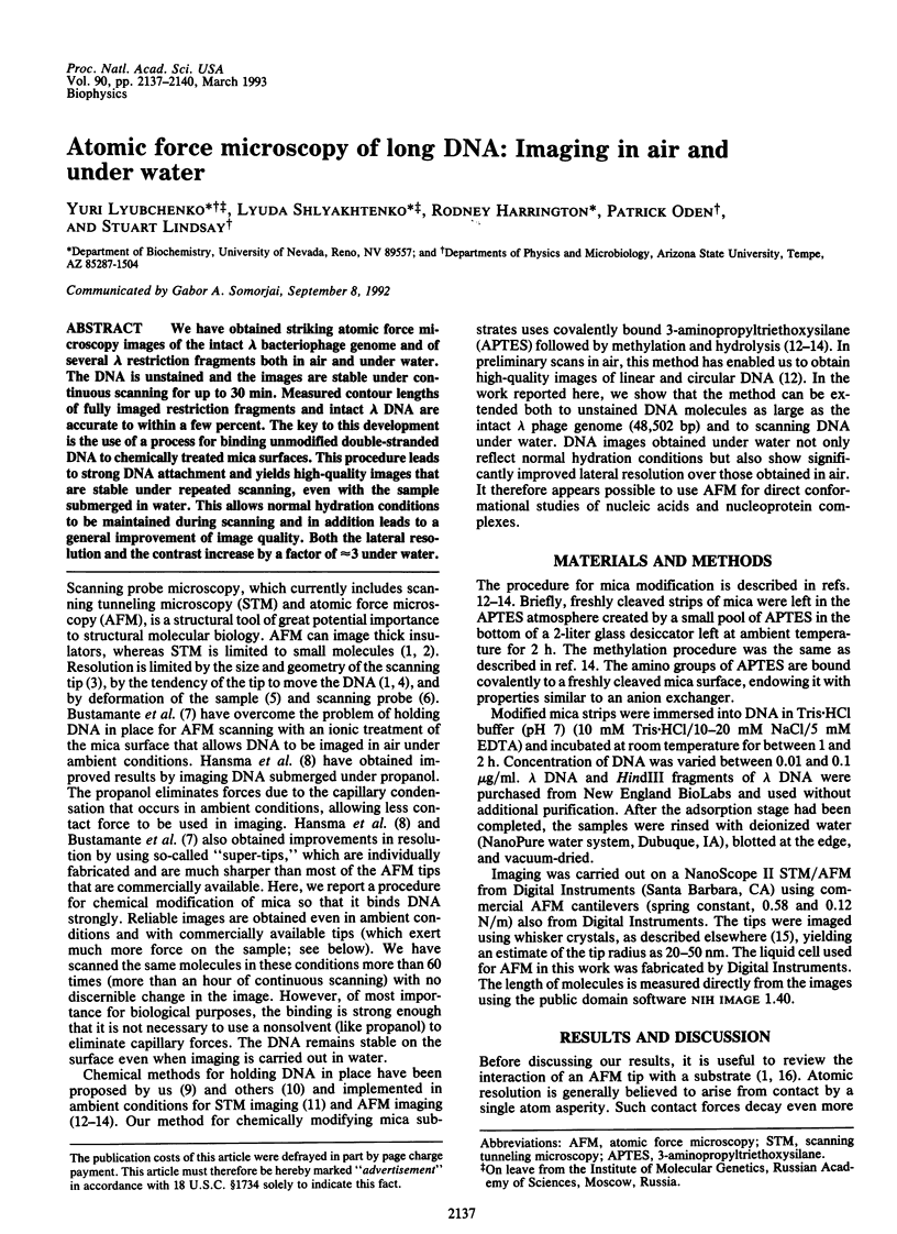
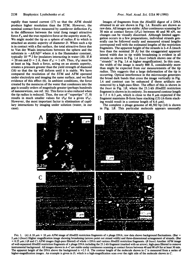
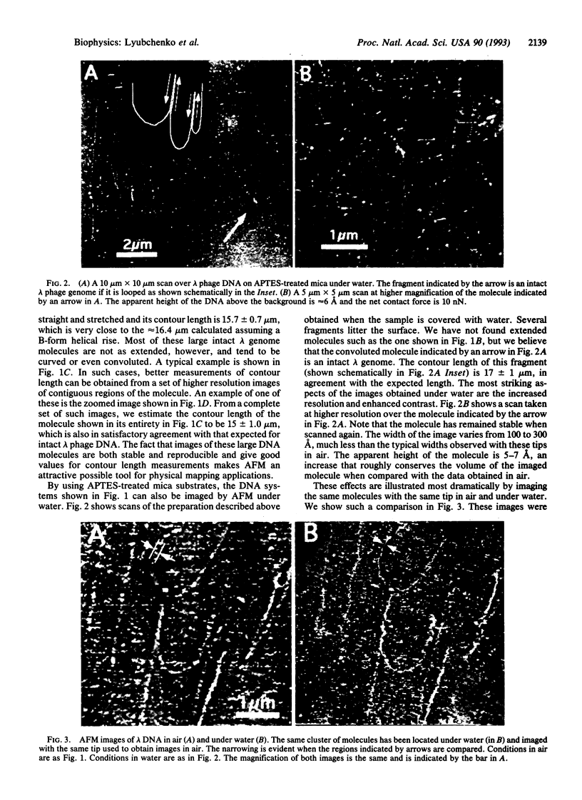
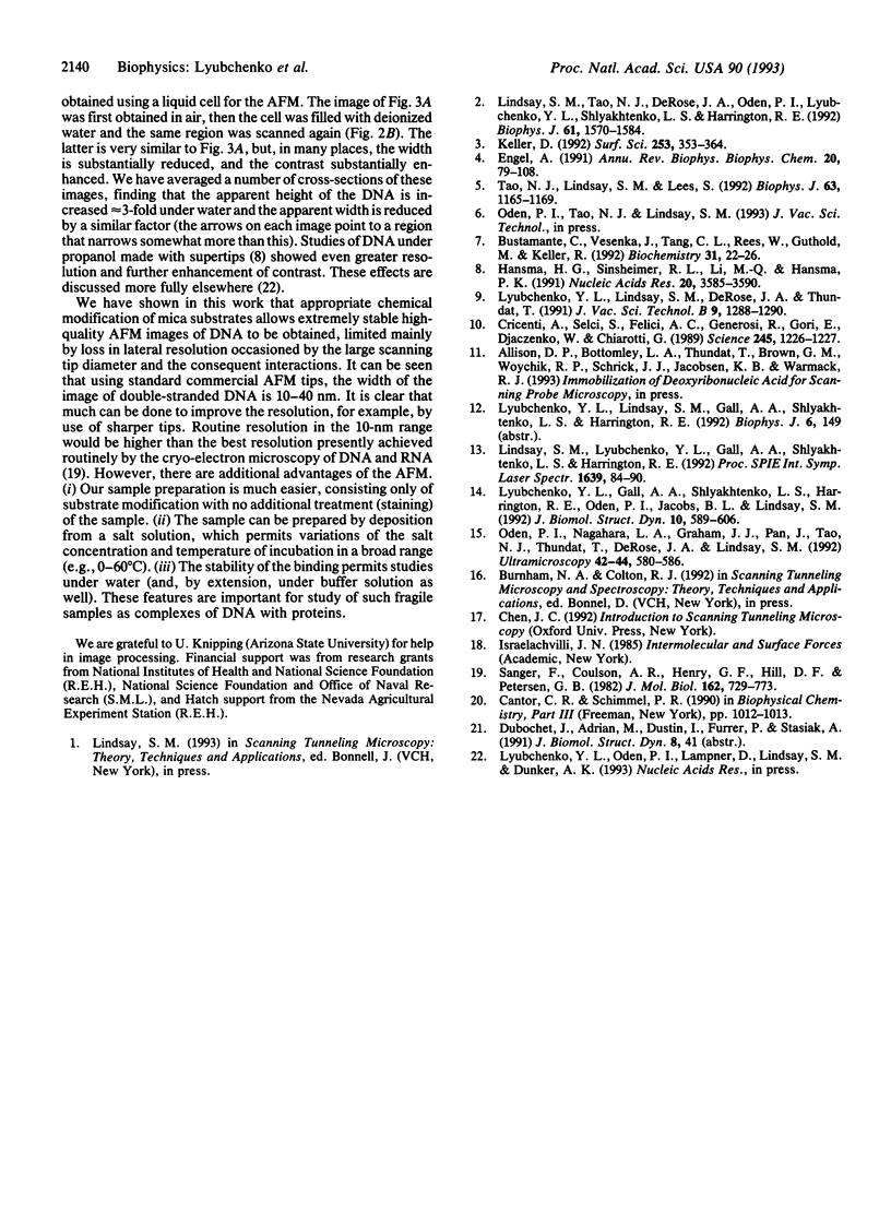
We have obtained striking atomic force microscopy images of the intact lambda bacteriophage genome and of several lambda restriction fragments both in air and under water. The DNA is unstained and the images are stable under continuous scanning for up to 30 min. Measured contour lengths of fully imaged restriction fragments and intact lambda DNA are accurate to within a few percent. The key to this development is the use of a process for binding unmodified double-stranded DNA to chemically treated mica surfaces. This procedure leads to strong DNA attachment and yields high-quality images that are stable under repeated scanning, even with the sample submerged in water. This allows normal hydration conditions to be maintained during scanning and in addition leads to a general improvement of image quality. Both the lateral resolution and the contrast increase by a factor of approximately 3 under water.
Full text
PDF


Images in this article
Selected References
These references are in PubMed. This may not be the complete list of references from this article.
- Bustamante C., Vesenka J., Tang C. L., Rees W., Guthold M., Keller R. Circular DNA molecules imaged in air by scanning force microscopy. Biochemistry. 1992 Jan 14;31(1):22–26. doi: 10.1021/bi00116a005. [DOI] [PubMed] [Google Scholar]
- Cricenti A., Selci S., Felici A. C., Generosi R., Gori E., Djaczenko W., Chiarotti G. Molecular structure of DNA by scanning tunneling microscopy. Science. 1989 Sep 15;245(4923):1226–1227. doi: 10.1126/science.2781279. [DOI] [PubMed] [Google Scholar]
- Engel A. Biological applications of scanning probe microscopes. Annu Rev Biophys Biophys Chem. 1991;20:79–108. doi: 10.1146/annurev.bb.20.060191.000455. [DOI] [PubMed] [Google Scholar]
- Hansma H. G., Sinsheimer R. L., Li M. Q., Hansma P. K. Atomic force microscopy of single- and double-stranded DNA. Nucleic Acids Res. 1992 Jul 25;20(14):3585–3590. doi: 10.1093/nar/20.14.3585. [DOI] [PMC free article] [PubMed] [Google Scholar]
- Lindsay S. M., Tao N. J., DeRose J. A., Oden P. I., Lyubchenko YuL, Harrington R. E., Shlyakhtenko L. Potentiostatic deposition of DNA for scanning probe microscopy. Biophys J. 1992 Jun;61(6):1570–1584. doi: 10.1016/S0006-3495(92)81961-6. [DOI] [PMC free article] [PubMed] [Google Scholar]
- Lyubchenko Y. L., Gall A. A., Shlyakhtenko L. S., Harrington R. E., Jacobs B. L., Oden P. I., Lindsay S. M. Atomic force microscopy imaging of double stranded DNA and RNA. J Biomol Struct Dyn. 1992 Dec;10(3):589–606. doi: 10.1080/07391102.1992.10508670. [DOI] [PubMed] [Google Scholar]
- Sanger F., Coulson A. R., Hong G. F., Hill D. F., Petersen G. B. Nucleotide sequence of bacteriophage lambda DNA. J Mol Biol. 1982 Dec 25;162(4):729–773. doi: 10.1016/0022-2836(82)90546-0. [DOI] [PubMed] [Google Scholar]
- Tao N. J., Lindsay S. M., Lees S. Measuring the microelastic properties of biological material. Biophys J. 1992 Oct;63(4):1165–1169. doi: 10.1016/S0006-3495(92)81692-2. [DOI] [PMC free article] [PubMed] [Google Scholar]


