Abstract
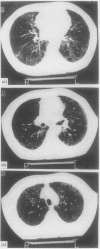
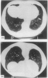
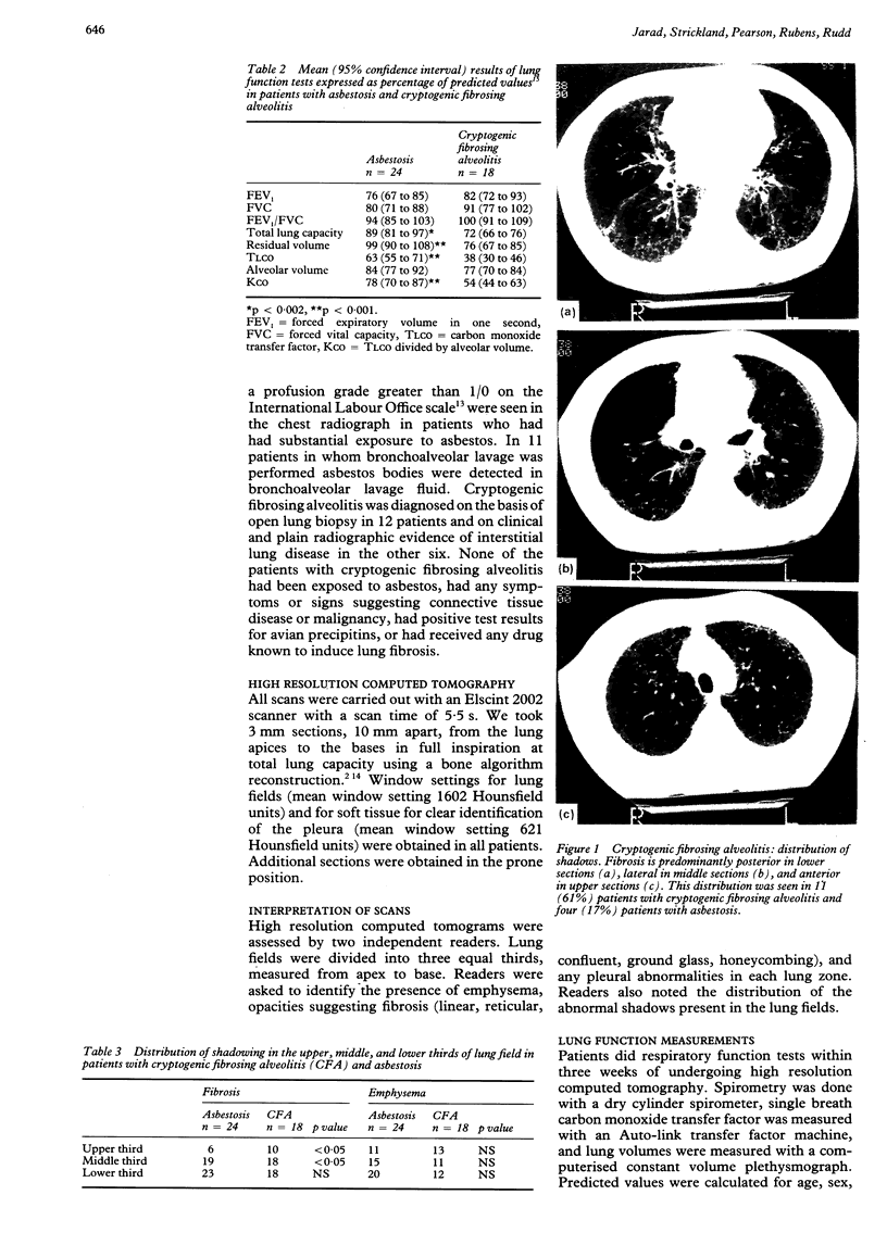
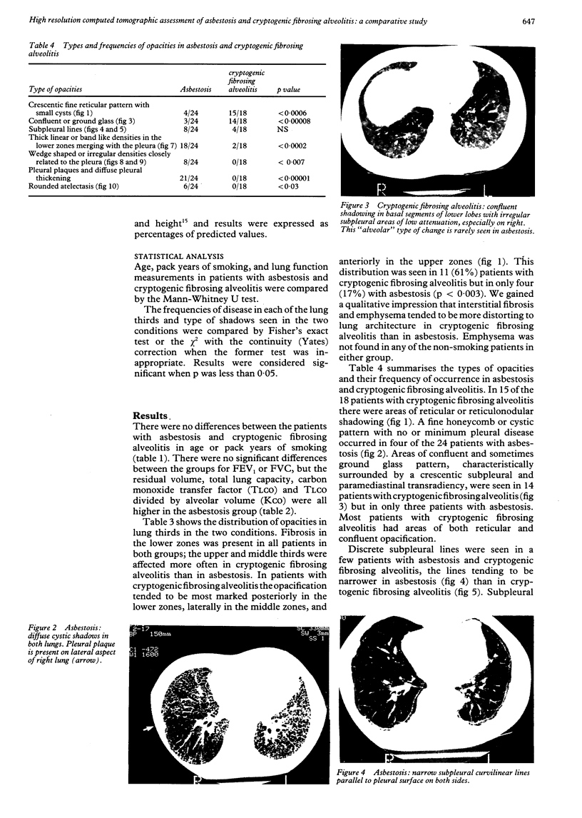
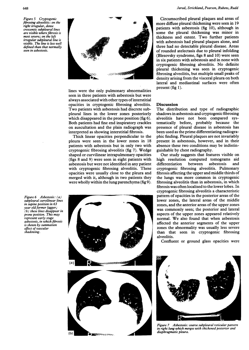
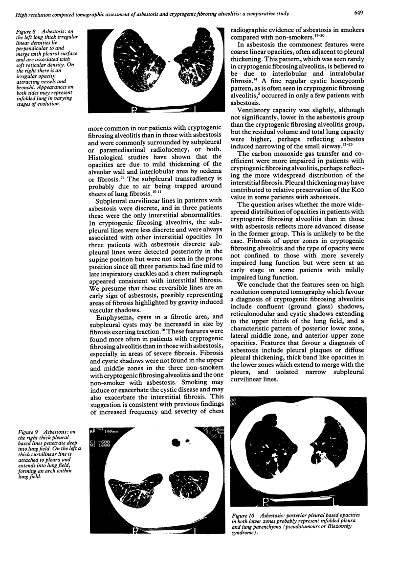
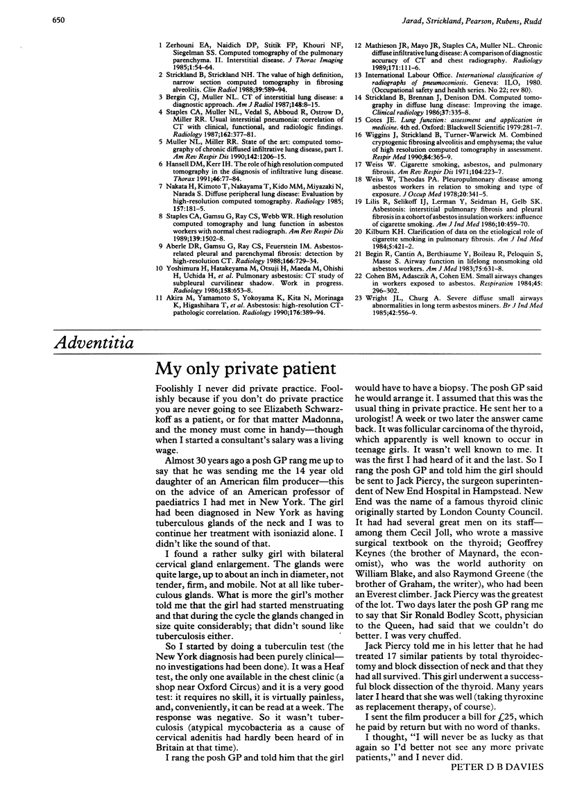
BACKGROUND: The aim of this study was to compare the distribution and configuration of lung opacities in patients with cryptogenic fibrosing alveolitis and asbestosis by high resolution computed tomography. METHODS: Eighteen patients with cryptogenic fibrosing alveolitis and 24 with asbestosis were studied. Two independent observers assessed the type and distributions of opacities in the upper, middle, and lower zones of the computed tomogram. RESULTS: Upper zone fibrosis occurred in 10 of the 18 patients with cryptogenic fibrosing alveolitis and in six of the 24 patients with asbestosis. A specific pattern in which fibrosis was distributed posteriorly in the lower zones, laterally in the middle zones, and anteriorly in the upper zones was seen in 11 patients with cryptogenic fibrosing alveolitis and in four with asbestosis. Band like intrapulmonary opacities, often merging with the pleura, were seen in 19 patients with asbestosis but in only two with cryptogenic fibrosing alveolitis. Areas with a reticular pattern and a confluent or ground glass pattern were the commonest features of cryptogenic fibrosing alveolitis (15 and 14 patients respectively) but were uncommon in asbestosis (four and three patients). Pleural thickening or plaques were seen in 21 patients with asbestosis and in none with cryptogenic fibrosing alveolitis. CONCLUSION: Apart from showing pleural disease high resolution computed tomography showed that confluent (ground glass) opacities are common in cryptogenic fibrosing alveolitis and rare in asbestosis whereas thick, band like opacities are common in asbestosis and rare in cryptogenic fibrosing alveolitis.
Full text
PDF
Images in this article
Selected References
These references are in PubMed. This may not be the complete list of references from this article.
- Aberle D. R., Gamsu G., Ray C. S., Feuerstein I. M. Asbestos-related pleural and parenchymal fibrosis: detection with high-resolution CT. Radiology. 1988 Mar;166(3):729–734. doi: 10.1148/radiology.166.3.3340770. [DOI] [PubMed] [Google Scholar]
- Akira M., Yamamoto S., Yokoyama K., Kita N., Morinaga K., Higashihara T., Kozuka T. Asbestosis: high-resolution CT-pathologic correlation. Radiology. 1990 Aug;176(2):389–394. doi: 10.1148/radiology.176.2.2367652. [DOI] [PubMed] [Google Scholar]
- Bergin C. J., Müller N. L. CT of interstitial lung disease: a diagnostic approach. AJR Am J Roentgenol. 1987 Jan;148(1):9–15. doi: 10.2214/ajr.148.1.9. [DOI] [PubMed] [Google Scholar]
- Bégin R., Cantin A., Berthiaume Y., Boileau R., Péloquin S., Massé S. Airway function in lifetime-nonsmoking older asbestos workers. Am J Med. 1983 Oct;75(4):631–638. doi: 10.1016/0002-9343(83)90449-7. [DOI] [PubMed] [Google Scholar]
- Cohen B. M., Adasczik A., Cohen E. M. Small airways changes in workers exposed to asbestos. Respiration. 1984;45(3):296–302. doi: 10.1159/000194634. [DOI] [PubMed] [Google Scholar]
- Greenberg M. S fibers. Am J Ind Med. 1984;5(5):421–422. doi: 10.1002/ajim.4700050518. [DOI] [PubMed] [Google Scholar]
- Hansell D. M., Kerr I. H. The role of high resolution computed tomography in the diagnosis of interstitial lung disease. Thorax. 1991 Feb;46(2):77–84. doi: 10.1136/thx.46.2.77. [DOI] [PMC free article] [PubMed] [Google Scholar]
- Kneeland J. B., Hyde J. S. High-resolution MR imaging with local coils. Radiology. 1989 Apr;171(1):1–7. doi: 10.1148/radiology.171.1.2648466. [DOI] [PubMed] [Google Scholar]
- Lilis R., Selikoff I. J., Lerman Y., Seidman H., Gelb S. K. Asbestosis: interstitial pulmonary fibrosis and pleural fibrosis in a cohort of asbestos insulation workers: influence of cigarette smoking. Am J Ind Med. 1986;10(5-6):459–470. doi: 10.1002/ajim.4700100504. [DOI] [PubMed] [Google Scholar]
- Müller N. L., Miller R. R. Computed tomography of chronic diffuse infiltrative lung disease. Part 1. Am Rev Respir Dis. 1990 Nov;142(5):1206–1215. doi: 10.1164/ajrccm/142.5.1206. [DOI] [PubMed] [Google Scholar]
- Nakata H., Kimoto T., Nakayama T., Kido M., Miyazaki N., Harada S. Diffuse peripheral lung disease: evaluation by high-resolution computed tomography. Radiology. 1985 Oct;157(1):181–185. doi: 10.1148/radiology.157.1.4034963. [DOI] [PubMed] [Google Scholar]
- Staples C. A., Gamsu G., Ray C. S., Webb W. R. High resolution computed tomography and lung function in asbestos-exposed workers with normal chest radiographs. Am Rev Respir Dis. 1989 Jun;139(6):1502–1508. doi: 10.1164/ajrccm/139.6.1502. [DOI] [PubMed] [Google Scholar]
- Staples C. A., Müller N. L., Vedal S., Abboud R., Ostrow D., Miller R. R. Usual interstitial pneumonia: correlation of CT with clinical, functional, and radiologic findings. Radiology. 1987 Feb;162(2):377–381. doi: 10.1148/radiology.162.2.3797650. [DOI] [PubMed] [Google Scholar]
- Strickland B., Brennan J., Denison D. M. Computed tomography in diffuse lung disease: improving the image. Clin Radiol. 1986 Jul;37(4):335–338. doi: 10.1016/s0009-9260(86)80265-3. [DOI] [PubMed] [Google Scholar]
- Strickland B., Strickland N. H. The value of high definition, narrow section computed tomography in fibrosing alveolitis. Clin Radiol. 1988 Nov;39(6):589–594. doi: 10.1016/s0009-9260(88)80056-4. [DOI] [PubMed] [Google Scholar]
- Weiss W. Cigarette smoking, asbestos, and pulmonary fibrosis. Am Rev Respir Dis. 1971 Aug;104(2):223–227. doi: 10.1164/arrd.1971.104.2.223. [DOI] [PubMed] [Google Scholar]
- Weiss W., Theodos P. A. Pleuropulmonary disease among asbestos workers in relation to smoking and type of exposure. J Occup Med. 1978 May;20(5):341–345. [PubMed] [Google Scholar]
- Wiggins J., Strickland B., Turner-Warwick M. Combined cryptogenic fibrosing alveolitis and emphysema: the value of high resolution computed tomography in assessment. Respir Med. 1990 Sep;84(5):365–369. doi: 10.1016/s0954-6111(08)80070-4. [DOI] [PubMed] [Google Scholar]
- Wright J. L., Churg A. Severe diffuse small airways abnormalities in long term chrysotile asbestos miners. Br J Ind Med. 1985 Aug;42(8):556–559. doi: 10.1136/oem.42.8.556. [DOI] [PMC free article] [PubMed] [Google Scholar]
- Yoshimura H., Hatakeyama M., Otsuji H., Maeda M., Ohishi H., Uchida H., Kasuga H., Katada H., Narita N., Mikami R. Pulmonary asbestosis: CT study of subpleural curvilinear shadow. Work in progress. Radiology. 1986 Mar;158(3):653–658. doi: 10.1148/radiology.158.3.3945733. [DOI] [PubMed] [Google Scholar]
- Zerhouni E. A., Naidich D. P., Stitik F. P., Khouri N. F., Siegelman S. S. Computed tomography of the pulmonary parenchyma. Part 2: Interstitial disease. J Thorac Imaging. 1985 Dec;1(1):54–64. doi: 10.1097/00005382-198512000-00008. [DOI] [PubMed] [Google Scholar]


