Abstract
Aim:
Alpha7-nicotinic acetylcholine receptor (α7 nAChR) is a ligand-gated Ca2+-permeable ion channel implicated in cognition and neuropsychiatric disorders. Activation of α7 nAChR improves learning, memory, and sensory gating in animal models. To identify novel α7 nAChR agonists, we synthesized a series of small molecules and characterized a representative compound, Br-IQ17B, N-[(3R)-1-azabicyclo[2,2,2]oct-3-yl]-5-bromoindolizine-2-carboxamide, which specifically activates α7 nAChR.
Methods:
Two-electrode voltage clamp (TEVC) recordings were primarily used for screening in Xenopus oocytes expressing human α7 nAChR. Assays, including radioisotope ligand binding, Western blots, whole-cell recordings of hippocampal culture neurons, and spontaneous IPSC recordings of brain slices, were also utilized to evaluate and confirm the specific activation of α7 nAChR by Br-IQ17B.
Results:
Br-IQ17B potently activates α7 nAChR with an EC50 of 1.8±0.2 μmol/L. Br-IQ17B is selective over other subtypes such as α4β2 and α3β4, but it blocks 5-HT3A receptors. Br-IQ17B displaced binding of the α7 blocker [3H]-MLA to hippocampal crude membranes with a Ki of 14.9±3.2 nmol/L. In hippocampal neurons, Br-IQ17B evoked α7-like currents that were inhibited by MLA and enhanced in the presence of the α7 PAM PNU-120596. In brain slice recordings, Br-IQ17B enhanced GABAergic synaptic transmission in CA1 neurons. Mechanistically, Br-IQ17B increased ERK1/2 phosphorylation that was MLA-sensitive.
Conclusion:
We identified the novel, potent, and selective α7 agonist Br-IQ17B, which enhances synaptic transmission. Br-IQ17B may be a helpful tool to understand new aspects of α7 nAChR function, and it also has potential for being developed as therapy for schizophrenia and cognitive deficits.
Keywords: α7 nAChR, Br-IQ17B, electrophysiology, ERK1/2 phosphorylation, GABAergic synaptic transmission, CNS diseases
Introduction
The nicotinic acetylcholine receptors (nAChRs) are pentameric integral membrane proteins that belong to a superfamily of ligand-gated ion channels including serotonin 5-HT3 receptor and GABAA receptor. The nAChRs form non-selective cation channels composed of either homomeric α- or heteromeric α/β subunits1. The specific assembly of nAChR subunits determines the biophysical and pharmacological properties of the channel receptor. For instance, homomeric α7 nAChR is characterized by its high calcium permeability (PCa:PNa≈10), its rapid desensitization in the presence of agonists, and its inhibition by the antagonists α-bungarotoxin and methyllycaconitine (MLA)2,3.
The α7 nAChR is a homopentameric receptor in the mammalian central nervous system (CNS) with limited peripheral expression, thus reducing potential side effects from inhibition. The α7 nAChR is highly expressed in the brain regions that are critical for cognition, attention processing, and memory formation, particularly in the hippocampus and frontal cortex2. The localization of α7 nAChR is both presynaptic and postsynaptic, and nAChR is involved in numerous processes, including the modulation of neurotransmitter release4, the regulation of postsynaptic excitability5, and hippocampal synaptic plasticity6.
The disruption of α7 nAChR activity has been implicated in the pathophysiology of neuropsychiatric conditions such as Alzheimer's disease (AD) and schizophrenia. Genetic studies show that the α7 nAChR gene CHRNA7, which is located on chromosome 15q14, is linked with sensory gating deficits in schizophrenic patients7. The analysis for association of promoter variants in the CHRNA7 gene with schizophrenia shows an inhibitory deficit of P50 auditory evoked potential response from schizophrenics and decreased function of transcription from the gene8. In biochemical experiments, the analysis of postmortem tissue reveals a marked reduction of α7 nAChR protein levels in various brain regions of schizophrenics9. In DBA/2 mice, hippocampal α7 nAChR density is closely correlated with sensory gating, and low α-bungarotoin binding is associated with poor gating10. Knocking out α7 nAChR in mice results in a comparable deficit in sustained attention, a characteristic symptom of schizophrenia, when compared to WT mice11. For AD, the levels of α7 nAChR proteins are decreased in the hippocampus of AD patients12. β-Amyloid peptide1-42 (Aβ1–42) binds selectively to neuronal α7 nAChR with picomolar affinity in neuritic plaques13, leading to the intraneuronal accumulation of Aβ1–42 and α7 nAChR complexes14, rapid tau phosphorylation15, the reversible blocking of the α7 nAChR channel16, the disruption of calcium homeostasis, and neuronal cell death17, whereas α7 activation confers neuroprotection against Aβ-induced cellular toxicity18. Therefore, a hypothesis has been proposed that the pharmacological activation of α7 nAChR can serve as potential therapy for the treatment of schizophrenia and AD. A number of distinct α7 agonists have been identified and tested in the early phases of clinical trials. Positive phase II clinical data were previously reported with EVP-6124 ((R)-7-chloro-N-quinuclidin-3-yl)benzo[b]thiophene-2-carboxamide) and TC-5619 ((2S,3R)-N-[2-(pyridin-3-ylmethyl)-1-azabicyclo[2.2.2]oct-3-yl]benzo[b]furan-2-carboxamide) for improving the symptoms of schizophrenia. When combined with atypical antipsychotic treatment, both EVP-6124 and TC-5619 show beneficial effects on cognition and are generally well tolerated in subjects with stable schizophrenia19,20,21. However, there are several α7 agonists that have failed in clinical trials due to adverse events related to cardiovascular and gastrointestinal functions, lack of efficacy, or an insufficient therapeutic index22,23,24, thus indicating the need to identify novel chemicals that can selectively activate α7 nAChR.
In this study, we described a novel, selective α7 nAChR partial agonist, Br-IQ17B, (N-[(3R)-1-azabicyclo[2,2,2]oct-3-yl]-5-bromoindolizine-2-carboxamide) identified from indolizine derivatives using electrophysiology as a primary functional screen. With radioisotope binding assays, brain slice recordings, and Western blotting, we confirmed that Br-IQ17B selectively activates α7 nAChR function with high-affinity binding to α7 nAChR and displays weak interactions with non-α7 nAChR subtypes. The Br-IQ17B activity was further confirmed by its activation of α7 nAChR in native neurons and its improvement of synaptic transmissions recorded from hippocampal brain slices. Our findings demonstrate a selective and novel α7 agonist, Br-IQ17B, that activates α7 currents both in hetero-expression system and neurons. Br-IQ17B can be a helpful pharmacological tool in understanding new aspects of α7 nAChR functionality, and it may lead to the development of therapy for neuropsychiatric disorders such as AD and schizophrenia.
Materials and methods
Chemicals
Br-IQ17B was synthesized in the Medicinal Chemistry Laboratory of the State Key Laboratory of Natural and Biomimetic Drugs, Peking University Health Science Center, China. (−)-Nicotine was obtained from Tocris (Ellisville, MS, USA). [3H]-MLA was provided by American Radiolabeled Chemicals (ARC, St Louis, MO, USA). All other compounds were purchased from Sigma (St Louis, MO, USA).
Animals
Sprague-Dawley rats were obtained from Vital River Laboratories Technology Co, Ltd (Beijing, China). The care and use of all animals were performed in accordance with the guidelines for Animal Use and Care of Peking University Health Science Center.
Two-electrode voltage clamp (TEVC) in Xenopus oocytes
Oocytes were harvested from Xenopus laevis female clawed frogs after anesthesia and washed twice in Ca2+-free OR2 solution (82.5 mmol/L NaCl, 2.5 mmol/L KCl, 1 mmol/L MgCl2, 5 mmol/L HEPES, pH 7.4). Oocytes were transferred to approximately 25 mL tubes and treated with 2 mg/mL collagenase in OR2 solution (Sigma type II, Sigma-Aldrich Inc, St Louis, MO, USA) for 20 min at 20–25 °C with gentle rotation. Stage V or VI oocytes were selected for microinjections.
For two-electrode voltage clamp recordings in oocytes, capped cRNAs were transcribed in vitro using the T3 mMESSAGEmMACHINE Kit (Ambion, Austin, TX, USA) following the linearization of plasmids in pBluescript KSM vectors. The oocytes were injected with 46 nL of cRNA solution containing approximately 20 ng human α7 nAChR cRNA or approximately 1 ng human 5-HT3A cRNA using a microinjector (Drummond Scientific, Broomall, PA, USA). For the expression of heteromeric rat α3β4 and rat α4β2 nAChRs, approximately 2 ng total cRNAs were injected in a 1:1 combination of each subunit. For GABAA receptors, cRNAs encoding α1, 2-, 3- or 5-, β3, and γ2 were mixed in a ratio of 1:1:1 and microinjected into oocytes to a total of approximately 1.5 ng. Oocytes were kept at 17 °C in ND96 solution (96 mmol/L NaCl, 2 mmol/L KCl, 1.8 mmol/L CaCl2, 1 mmol/L MgCl2, 5 mmol/L HEPES, pH 7.4 adjusted with NaOH).
Recordings were made 2–5 days post-injection. Oocytes were impaled with two microelectrodes (0.5–1.0 MΩ) filled with 3 mol/L KCl in a 40-μL recording chamber. The membrane potential was held at −90 mV using standard voltage clamp procedures. Currents were recorded in Ringer's solution (115 mmol/L NaCl, 2.5 mmol/L KCl, 10 mmol/L HEPES, 1.8 mmol/L CaCl2, 1 mmol/L MgCl2, 0.0005 mmol/L atropine) at room temperature (22±1 °C) using a GeneClamp 500B amplifier (Axon Instruments, Union City, CA, USA).
Radioisotope ligand binding assay in crude membranes
Cerebral hippocampi from male Sprague-Dawley rats weighing 230 to 250 g were placed in 0.32 mol/L ice-cold sucrose containing protease inhibitors (Protease Inhibitor Cocktail Tablets; Roche Diagnostics, Indianapolis, IN, USA) (one tablet per 50 mL). For crude membrane preparations, brain tissues were homogenized and centrifuged at 1000×g at 4 °C for 10 min. The supernatant was centrifuged for 10 min at 20 000×g at 4 °C, and the pellet was washed twice by centrifugation at 40 000×g for 10 min. Crude membranes were stored at −80 °C until use.
The saturation binding for α7 nAChR was performed in a final volume of 500 μL binding buffer (120 mmol/L NaCl, 5 mmol/L KCl, 2 mmol/L CaCl2, 2 mmol/L MgCl2, and 50 mmol/L Tris-Cl, 0.1% (w/v) BSA, pH 7.4, 4 °C) containing 200 μg of membrane proteins and different concentrations of the α7 antagonist [3H]-MLA (100 Ci/mmol; ranging from 0.03 to 30 nmol/L) in the presence or absence of 30 μmol/L unlabeled MLA at room temperature for 90 min. The concentration-dependent inhibition of binding was tested by incubating 200 μg of membrane proteins with different concentrations of Br-IQ17B ranging from 3 nmol/L to 3 μmol/L dissolved in buffer with 5 nmol/L [3H]-MLA in 500 μL. The nonspecific binding was determined in the presence of 30 μmol/L MLA. All mixtures were incubated at room temperature for 90 min.
For all experiments, the binding reaction was terminated by vacuum filtration onto Filtermat A filters presoaked with 2% v/v BSA for 2 h. Filters were immediately rinsed twice with 4 mL ice-cold reaction buffer, and the PerkinElmer MicroScint-20 scintillation cocktail was added. The bound [3H]-MLA was measured using a Wallac 1450 MicroBeta TriLux liquid scintillation luminescence counter (PerkinElmer, Waltham, MA, USA).
Western blot analysis
PC12 cells were starved overnight in serum-free medium to reduce basal phosphorylation. After treatment with compounds, reactions were terminated by placing the culture dish on ice and removing the incubation medium by aspiration. Cells were washed twice with D-PBS and treated with ice-cold lysis buffer (150 mmol/L NaCl, 20 mmol/L Tris, 1% Triton X-100, 1% sodium deoxycholate, 0.1% SDS, 10 mmol/L EDTA, pH 8.0), supplemented with protease inhibitor mixtures (Protease Inhibitor Cocktail Tablets; Roche Diagnostics, Indianapolis, IN, USA) on ice for 30 min. Cell lysates were then centrifuged at 13 000×g for 20 min to yield protein extracts in the supernatant.
For Western blots, protein samples were separated on SDS-polyacrylamide gel and transferred electrophoretically onto nitrocellulose membranes (Millipore, Boston, MA, USA). After being blocked with the 5% powdered nonfat milk or 5% BSA in TBS-T (Tris-buffered saline with 0.05% Tween 20), cell membranes were incubated overnight at 4 °C with primary antibodies (1:1000 for anti-ERK from Cell Signaling Technology; 1:2000 for anti-pERK from Cell Signaling Technology, Boston, MA, USA). The membranes were then incubated with their corresponding secondary HRP-conjugated antibodies and detected using an ECL Western blotting detection system (Millipore, Boston, MA, USA). Densitometric analysis was performed using a Bio-Rad image acquisition system (Bio-Rad Laboratories, Hercules, CA, USA).
Culture of hippocampal neurons and whole-cell patch-clamp
Hippocampal explants were isolated from embryos of 18-day-old Sprague-Dawley rats of either sex after decapitation. Hippocampal regions were gently removed and digested with 0.25% trypsin for 30 min at 37 °C followed by trituration with a pipette in plating medium (DMEM with 10% FBS). Dissociated neurons were plated onto poly-D-lysine coated coverslips in 35-mm dishes at a density of 1×10⁶ cells per dish. After 4 h, the medium was displaced by neurobasal medium supplemented with 2% B27 and 0.5 mmol/L GlutaMAX-I (Life Technologies, Carlsbad, CA, USA).
For whole-cell recordings, cells were held at −80 mV and recorded using a HEKA EPC10 amplifier with PatchMaster software (HEKA Elektronik, Pfalz, Germany). Patch pipettes were pulled from borosilicate glass and fire-polished to a resistance of 3–5 MΩ when filled with internal pipette solution composed of the following (in mmol/L): 126 CsCH3SO3, 10 CsCl, 4 NaCl, 1 MgCl2, 0.5 CaCl2, 5 EGTA, 10 HEPES, 3 ATP-Mg, 0.3 GTP-Na, and 4 phosphocreatine, pH 7.2. Cells were perfused with the external solution containing (in mmol/L): 140 NaCl, 5 KCl, 2 CaCl2, 1 MgCl2, 10 HEPES, and 10 glucose, pH 7.4. Atropine (5 μmol/L), CNQX (5 μmol/L), bicuculline (10 μmol/L) and tetrodotoxin (0.5 μmol/L) were included in the bath solution throughout experiments.
Hippocampal brain slice electrophysiology
Male Sprague-Dawley rats (14–21 day-old) were deeply anesthetized with sodium pentobarbital (250 mg/kg) before decapitation. Brains were quickly removed and placed into ice-cold artificial cerebrospinal fluid (ACSF) containing (in mmol/L): 119 NaCl, 2.5 KCl, 2.5 CaCl2, 1.3 MgSO4, 1 NaH2PO4, 26.2 NaHCO3, and 11 glucose, and bubbled with 95% O2/5% CO2. Transverse hippocampal slices (400 μm) were cut with a vibratome (Leica VT1200S, Leica, Wetzlar, Germany). The slices were incubated for 1 h at 34 °C with ACSF and then transferred to a recording chamber on an upright microscope equipped with differential interference contrast (DIC) optics (Nikon FN1, Tokyo, Japan). For IPSC recordings, whole-cell patch clamp configurations were routinely achieved from CA1 hippocampal pyramidal neurons visualized with a DIC microscope. Patch pipette (3–5 MΩ) solution contained (in mmol/L): 136 CsCl, 4 NaCl, 1 MgCl2, 0.5 CaCl2, 5 EGTA, 10 HEPES, 3 Mg-ATP, 0.3 GTP-Na, 4-phosphocreatine (pH adjusted to 7.2 with CsOH and osmolarity adjusted to 290 mOsm). According to the Nernst equation, the reversal potential of Cl− was approximately 3 mV at room temperature. Therefore, when neurons were held at −70 mV, the spontaneous IPSCs were inward. To block fast excitatory synaptic current (EPSC), 5 μmol/L CNQX (in DMSO) and 10 μmol/L APV were added to ACSF. Atropine (5 μmol/L) was used to block muscarinic receptors. Voltage-clamp recordings were conducted with a computer-controlled amplifier (Multiclamp 700B, Molecular Devices, Sunnyvale, CA, USA), and traces were low-pass filtered at 2.6 kHz and digitized at 10 kHz (DigiData 1440A, Molecular Devices, Sunnyvale, CA, USA). Neurons with stable access resistance were included in the analysis.
Data analysis
All data are expressed as the mean±SEM. Statistical significance was assessed by Student's t-test or one-way ANOVA using Prism version 5.0 software. A value of P<0.05 was considered statistically significant. In two-electrode voltage clamp and whole-cell patch clamp recordings, responses were quantified by measuring peak current amplitude, and data were collected and analyzed using PatchMaster and Origin 8.0 software. For hippocampal brain slice electrophysiology, the data were collected and analyzed using Clampfit 10.4 or Clampex 10.4 software. All concentration-response curves were fitted to the Hill equation as follows: Inormalized=Emax/(1+(EC50/C)nH). For radioisotope ligand binding assay, the dissociation constant Ki was determined from the equation Ki=IC50/(1+L/KD).
Results
Screening α7 nAChR agonists by two-electrode voltage clamp
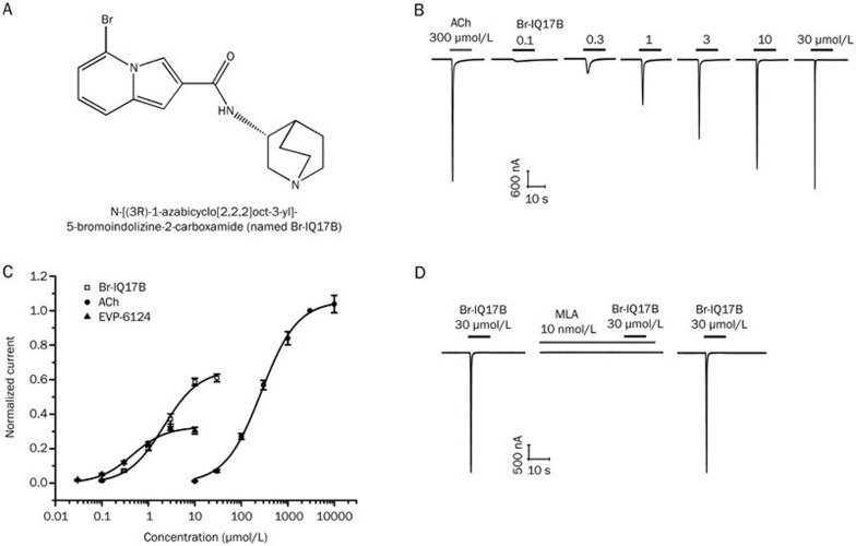
To identify novel α7 agonists, we started with a computer-aided virtual screen and performed structural optimizations using the compound EVP-6124, which is in a phase II clinical trial, as a reference structure21. The substructure search for α7 nAChR agonists from the SureChEMBL database revealed that N-(quinuclidin-3-yl) formamide appeared frequently, and its similarity search (SS) was performed based on the MACCS structural fingerprint25. Approximately 400 compounds were selected, and they clustered into 20 categories. Among the SS-selected compounds, an indolizine-3-carboxylic acid derivative was reported to block the 5-HT3 receptor, which shares significant homology with α7 nAChR26,27. Thus, an indolizine-3-carboxylic acid derivative was used as a hit for structural modifications that were primarily focused on the indolizine moiety by changing the substitution group at different positions. Over 60 compounds were synthesized, and their activities were tested on human α7 nAChR transiently expressed in Xenopus oocytes using two-electrode voltage clamp. Compounds that were able to evoke α7 currents were further assessed for their potency and relative efficacy against acetylcholine. EC50 values of positive compounds were generated by fitting Hill equations, and their maximal effects were normalized to 3 mmol/L ACh from the same cell. Based on the criteria that compounds must have a minimal EC50 and an efficacy of at least 60% that of ACh, one compound, N-[(3R)-1-azabicyclo[2,2,2]oct-3-yl]-5-bromoindolizine-2-carboxamide (Br-IQ17B, MW=348.24) was identified as an ideal lead (Figure 1A). Therefore, Br-IQ17B was further evaluated for its effects on α7 currents.
Figure 1.
Chemical structure of Br-IQ17B and its dose-dependent activation of human α7 nAChR channels expressed in Xenopus oocytes. (A) Chemical structure of N-[(3R)-1-azabicyclo[2,2,2]oct-3-yl]-5-bromoindolizine-2-carboxamide, named Br-IQ17B, molecular weight of 348.24. (B) Representative α7 currents recorded from an oocyte expressing human α7 nAChR in response to 300 μmol/L ACh (a near half-maximal concentration) and increasing concentrations of Br-IQ17B (0.1–30 μmol/L). Oocytes were held at −90 mV, and traces were consecutively acquired with 7-min lapses. (C) Curve with open squares represents concentration-response relationship of inward α7 nAChR currents induced by Br-IQ17B. The maximal response evoked by Br-IQ17B is 64.3%±3.6% of ACh (3 mmol/L) with an EC50 of 1.8±0.2 μmol/L and a Hill coefficient of 1.2±0.1 (n=6 for all data points). Curve with fitted triangles represents concentration-response of EVP-6124 with an EC50 of 0.45±0.08 μmol/L and a Hill coefficient of 1.2±0.2 (n=6 for all data points). The maximal response compared to ACh is 30.6%±1.8%. Curve with fitted circles represents concentration-response of natural agonist ACh, yielding an EC50 of 284.2±14.6 μmol/L and a Hill coefficient of 1.1±0.1 (n=6 for all data points). Peak current amplitudes were measured and normalized to the current evoked by 3 mmol/L ACh. (D) The α7 current evoked by 30 μmol/L Br-IQ17B was inhibited by 10 nmol/L α7 antagonist MLA. The inhibitory effect could be reversed after a 10-min washout.
Dose-dependent activation of α7 currents by Br-IQ17B
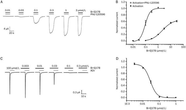
As shown in Figure 1B, the activation of α7 by ACh was first performed as a control before repeated applications of Br-IQ17B. With a 7-min pulse interval between each dose, increasing concentrations of Br-IQ17B dose-dependently activated α7 nAChR with a maximum efficacy of 64.3%±3.6% compared with 3 mmol/L ACh (Figure 1C). The data from dose-dependent activation were fitted with the Hill equation, yielding an EC50 of 1.8±0.2 μmol/L and a Hill coefficient of 1.2±0.1 (n=6) (Figure 1C). The current induced by Br-IQ17B (30 μmol/L) was completely blocked by 10 nmol/L MLA, an α7 nAChR selective antagonist (Figure 1D), indicating that Br-IQ17B mediated its effects through specific α7 activation. To further confirm the agonist activity of Br-IQ17B, we tested Br-IQ17B in the presence of the α7 positive allosteric modulator (PAM) PNU-120596, which is known to enhance the activation of α7 by ACh28. As shown in Figure 2A, co-application of Br-IQ17B with a fixed concentration of PNU-120596 (1 mmol/L) resulted in a dose-dependent activation of α7 currents. The potency of Br-IQ17B was increased from 1.8±0.2 μmol/L to 93.6±6.5 nmol/L (n=6), and the Hill slope was increased from 1.2±0.1 to 2.4±0.1 compared with Br-IQ17B alone, which showed partial activation (Figure 2B).
Figure 2.
Dose-dependent activation of α7 current by Br-IQ17B agonist in the presence of the PAM PNU-120596 and desensitization induced by pre-incubation of Br-IQ17B. (A) Representative traces induced by increasing concentrations of Br-IQ17B co-applied with the selective α7 type II PAM PNU-120596 (1 μmol/L). Oocytes were held at −90 mV, and traces were consecutively acquired with 7-min lapses. (B) Concentration-peak current of α7 induced by Br-IQ17B in the presence of 1 μmol/L PNU-120596 and fitted by the Hill equation (line with open squares), with an EC50 of 93.6±6.5 nmol/L and a Hill coefficient of 2.4±0.1 (n=5 for all data points); partial activation of α7 by Br-IQ17B alone with EC50 of 1.8±0.2 μmol/L (line with filled circles). (C) Representative current traces depicting the response to ACh (100 μmol/L) after 1-min sustained exposure to low concentrations of Br-IQ17B (3–300 nmol/L). Oocytes were held at −90 mV, and consecutive acquisitions of traces were made with 7-min lapses. (D) Fitting of concentration-dependent inhibition of the ACh (100 μmol/L)-evoked response after sustained exposure to Br-IQ17B. Peak current amplitudes were measured and normalized with respect to the amplitude of current elicited by 100 μmol/L ACh alone. Data points were fitted to a Hill equation, yielding an IC50 of 28.5±1.9 nmol/L and a Hill coefficient of 2.8±0.3 (n=6 for all data points).
One of the key properties of α7 current is robust desensitization upon repeated exposures to an agonist29. To test whether Br-IQ17B could cause desensitization, we used a double protocol in which oocytes were exposed to sustained (1 min) increasing concentrations of Br-IQ17B before the addition of a fixed and low concentration of ACh (100 μmol/L). As shown in Figure 2C, 100 μmol/L ACh produced a robust α7 current. In contrast, the incubation of different concentrations of Br-IQ17B decreased the amplitude of α7 current in the presence of the same concentration of ACh (100 μmol/L). Plotting the amplitude of the current evoked by ACh as a function of increasing Br-IQ17B yielded a concentration-dependent inhibition of ACh-induced α7 current with an IC50 at 28.5±1.9 nmol/L and a Hill coefficient at 2.8±0.3 (Figure 2D), which is in good agreement with the Ki of 14.9±3.2 nmol/L for the displacement of [3H]-MLA binding at α7 nAChR. It is noteworthy that the low concentration of Br-IQ17B (3 nmol/L) that had no agonist activity increased the ACh-mediated peak current, whereas the current desensitization became apparent for Br-IQ17B at 10 nmol/L or greater concentration (Figure 2C and 2D).
Displacement of [3H]-MLA binding to α7 nAChR by Br-IQ17B
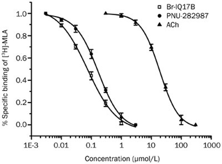
α7 nAChR agonists can displace the binding of the specific competitive antagonist [3H]-MLA to the α7 nAChR extracellular N-terminal domain, which contains the orthosteric binding sites30. To assess whether Br-IQ17B could affect [3H]-MLA binding, we prepared hippocampal crude membranes from rat hippocampi that contain nAChRs and conducted the radioisotope ligand binding assay. As shown in Figure 3, Br-IQ17B displaced the [3H]-MLA binding to crude membranes in a concentration-dependent manner with a Ki value of 14.9±3.2 nmol/L (n=3), which is approximately 300-fold more potent than the natural agonist ACh (Ki=3.9±0.3 μmol/L). PNU-282987, a selective α7 nAChR agonist, is known to bind to α7 nAChR31. As another positive control, the inhibitory effect of PNU-282987 on [3H]-MLA binding was determined. PNU-282987 dose-dependently displaced the binding of [3H]-MLA with a Ki of 34.1±4.3 nmol/L, which is similar to the Ki value as previously described32. These results show that Br-IQ17B can competitively bind to α7 nAChR.
Figure 3.
Displacement of [3H]-MLA ligand binding in crude membranes from rat brain. Displacement of the binding of the radio-ligand α7 antagonist [3H]-MLA to membranes of rat brain hippocampus in the presence of increasing concentrations of Br-IQ17B (line with open squares) and natural agonist ACh (line with filled triangles) with Kis of 14.9±3.2 nmol/L and 3.9±0.3 μmol/L, respectively. The line with filled circles shows the dose-dependent inhibition of [3H]-MLA binding by the orthosteric agonist PNU-282987 as positive control, yielding a Ki of 34.1±4.3 nmol/L.
Selective activation of α7 by Br-IQ17B
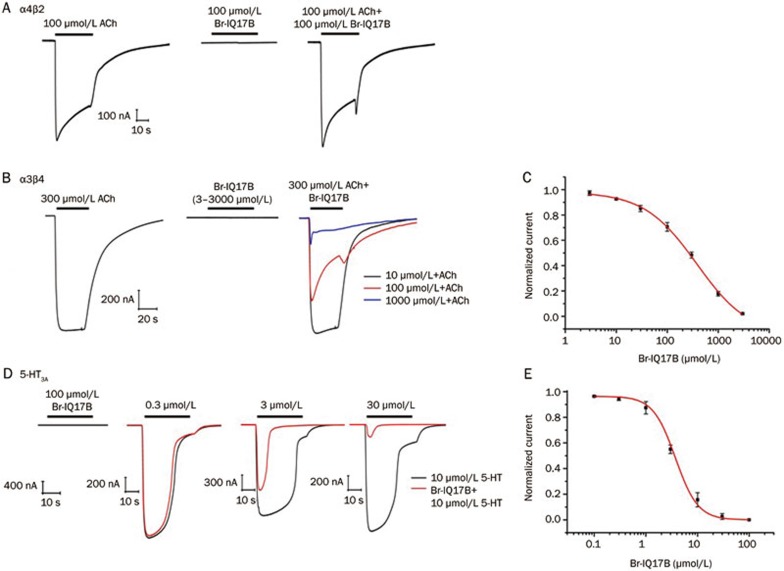
To determine the selectivity of Br-IQ17B, we examined the effects of Br-IQ17B on α4β2 and α3β4, two nAChR subtypes that exhibit distributions that are similar to that of α7 nAChR in the brain33. The expression of rat α4β2 or α3β4 in Xenopus oocytes produced a robust current in response to either 100 or 300 mmol/L ACh (Figure 4A and 4B). The application of Br-IQ17B (100 mmol/L) did not induce any current (Table 1). The lack of effect on α4β2 was further confirmed by either pre-incubation of Br-IQ17B for 1 min or the co-application of Br-IQ17B with ACh, which resulted in no enhancement or inhibition of α4β2 current (Figure 4A). This result indicates that Br-IQ17B had no effect on α4β2. For α3β4, Br-IQ17B showed an inhibitory effect with an IC50 of approximately 381.5±28.6 μmol/L and a Hill coefficient of 0.9±0.1 (n=5) (Figure 4B), thus displaying at least 300-fold selectivity over α3β4 (Figure 4C). The 5-HT3A receptor has significant homology with α7 nAChR and serves as a cross-reactivity target for α7 agonists. Br-IQ17B alone (0.1–100 μmol/L) did not show any agonist activities on human 5-HT3A receptors expressed in oocytes. The co-application of Br-IQ17B with serotonin dose-dependently inhibited the current induced by 5-HT (10 μmol/L) with an IC50 of 3.74±0.64 μmol/L (n=6) and a Hill coefficient of 2.1±0.1 (Figure 4D and 4E, Table 2).
Figure 4.
Selectivity assessments of Br-IQ17B. (A) Representative current traces showing the effects of Br-IQ17B on an oocyte expressing rat α4β2 nAChR in the presence of 100 μmol/L ACh (left), 100 μmol/L Br-IQ17B (middle), and the co-application of ACh and Br-IQ17B (right). (B) Representative current traces showing the effects of Br-IQ17B on an oocyte expressing α3β4 nAChR subunits in the presence of ACh (300 μmol/L) (left), or different concentrations (3–3000 μmol/L) of Br-IQ17B alone (middle), or the co-application of different concentrations of Br-IQ17B ranging from 3–3000 μmol/L (only 3 traces for concentrations of 10, 100 and 1000 μmol/L are shown) with a fixed concentration of ACh (300 μmol/L) (right). (C) Concentration-response curve for inhibition of Br-IQ17B on rat α3β4; the peak current amplitudes were plotted as a function of Br-IQ17B concentration (3–3000 μmol/L) normalized to ACh (300 μmol/L), with an IC50 of 381.5±28.6 μmol/L and a Hill coefficient of 0.9±0.1 (n=5 for all data points). (D) Representative current traces from oocytes expressing 5-HT3A receptors in response to 100 μmol/L Br-IQ17B alone (first trace), 10 μmol/L 5-HT alone, and the co-application of increasing concentrations of Br-IQ17B (0.3–30 μmol/L) with 5-HT (10 μmol/L). (E) Concentration-response relationship for antagonist properties of Br-IQ17B against human 5-HT3A; plot of the peak current as a function of the Hill equation of Br-IQ17B (0.1–100 μmol/L) and normalized to 5-HT (10 μmol/L), with an IC50 of 3.74±0.64 μmol/L and a Hill coefficient of 2.1±0.1 (n=6 for all data points).
Table 1. Selectivity of Br-IQ17B on nAChR subtypes and 5-HT3A receptor determined by TEVC.
| Target | EC50 (μmol/L) | Maximum effect vs 3 mmol/L ACh or 10 μmol/L 5-HT | n |
|---|---|---|---|
| α7, Human | 1.8±0.21 | 64.3%±3.6% | 6 |
| α4β2, Rat | >100 | 0 | 6 |
| α3β4, Rat | >100 | 0 | 6 |
| 5-HT3A, Human | >100 | 0 | 6 |
Table 2. Inhibition of ACh-evoked nAChR subtype and serotonin-evoked 5-HT3A currents by Br-IQ17B in TEVC.
| Target | IC50 (nmol/L) | n |
|---|---|---|
| α7, Human | 28.5±2.5 vs 100 μmol/L ACh | 6 |
| α4β2, Rat | >100 000 vs 100 μmol/L ACh | 6 |
| α3β4, Rat | 381 510±52 251 vs 300 μmol/L ACh | 5 |
| 5-HT3A, Human | 3740±90 vs 10 μmol/L 5-HT | 6 |
Activation of native neuronal α7 nAChR by Br-IQ17B
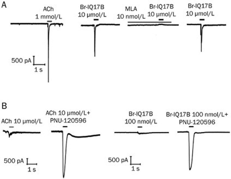
Hippocampal somatic neurons express α7 nAChR or ACh-evoking characteristic α7 currents that can be activated by α7 agonists34. To evaluate whether Br-IQ17B can activate native α7 nAChR, we examined the effects of Br-IQ17B on hippocampal neurons. As shown in Figure 5A, the application of either ACh (1 mmol/L) or Br-IQ17B (10 μmol/L) evoked α7 currents with fast onset and rapid decay. Br-IQ17B evoked currents were sensitive to the α7 antagonist MLA (10 nmol/L), which completely blocked the native α7 current, and the inhibitory effect can be washed out (Figure 5A). We further tested whether the Br-IQ17B-evoked native α7 current can be augmented in the presence of the PAM PNU-120596. A low concentration of ACh (10 mmol/L) evoked an observable α7 current in neurons that could be robustly increased in the presence of PNU-120596 (1 mmol/L) (Figure 5B). Similarly, a low concentration of Br-IQ17B (100 nmol/L) activated a small current that was significantly enhanced by the co-application of PNU-120596 (1 mmol/L), further confirming that the agonist Br-IQ17B can activate native neuronal α7 current (Figure 5B).
Figure 5.
Activation of native α7 nAChR in hippocampal neurons by Br-IQ17B. (A) Representative current traces evoked by agonist ACh (1 mmol/L) and Br-IQ17B (10 μmol/L) in hippocampal neurons. The current elicited by Br-IQ17B was blocked by the inhibitor MLA (10 nmol/L) (n=3). The neurons were held at −80 mV. (B) Representative current traces evoked by a low concentration of ACh (10 μmol/L) or Br-IQ17B (100 nmol/L) in the presence or absence of PNU-120596 (1 μmol/L), respectively (n=3). Current induced by the co-application of Br-IQ17B (100 nmol/L) and PNU-120596 (1 μmol/L) is significantly larger than Br-IQ17B (100 nmol/L) alone. The neurons were held at −80 mV.
Enhanced phosphorylation of ERK signaling by Br-IQ17B in PC12 cells
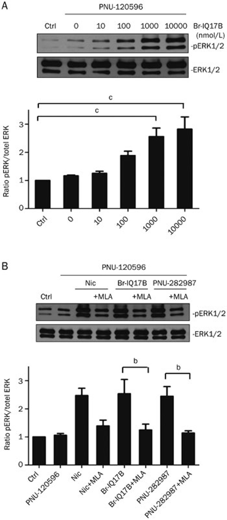
The activation of native α7 nAChR results in enhanced ERK (extracellular signal-regulated kinases) phosphorylation and signaling that is implicated in the regulation of a variety of physiological functions such as cognition enhancement35. To test whether Br-IQ17B had any effect on ERK signaling, we evaluated ERK phosphorylation upon α7 activation in rat PC12 cells, which have intact ERK signaling and robustly expressed pathway components. Because α7 nAChR is rapidly desensitized upon activation, agonist-evoked responses are not easily detectable by conventional methods that are insufficient for the measurement of rapid effects. To overcome this shortcoming, PNU-120596 has successfully been used as a tool to slow the receptor desensitization, thus leading to easy detection of compound response. As shown in Figure 6A, the pre-incubation of PC12 cells with PNU-120596 for 10 min only caused a slight increase of the basal ratio of phosphorylated ERK (pERK1 and pERK2) over total ERK (tERK). In contrast, the pre-incubation of the PAM PNU-120596 with Br-IQ17B (0.01–10 mmol/L) for an additional 7 min significantly increased the pERK/tERK ratio in a concentration-dependent manner (Figure 6A). The maximal increase of the pERK/tERK ratio was approximately 3–4 fold compared with either the blank control or the pre-incubation control without Br-IQ17B. To further confirm whether Br-IQ17B-induced ERK1/2 phosphorylation was mediated by the activation of α7 nAChR, several tool compounds including the selective antagonist MLA (100 nmol/L), the selective α7 agonist PNU-282987 (1 μmol/L), and the traditional non-selective α7 agonist nicotine (10 μmol/L) were utilized to test any specific activation of ERK signaling by Br-IQ17B. As shown in Figure 6B, the incubation of MLA (100 nmol/L) significantly reversed the activation of α7 induced by the agonists, confirming that the enhanced level of pERK is α7 activity dependent.
Figure 6.
Activation of α7 nAChR by Br-IQ17B enhances pERK signaling in PC12 cells. (A) In the upper panel, Western blot analysis of PC12 cells stimulated with different concentrations of Br-IQ17B (0.01–10 μmol/L) for 7 min after pre-incubation of 1 μmol/L PNU-120596 for 10 min. Lower panel, semi-quantitative analysis of pERK/tERK from upper panel data. Data were normalized to that of control without any treatment and expressed as the mean±SEM. Co-application of PNU-120596 (1 μmol/L) and Br-IQ17B significantly increased the ratio of phosphorylated ERK to total ERK (n=3, cP<0.01 vs control, paired t-test). (B) In upper panel, the Br-IQ17B-increased phosphorylation of ERK1/2 was blocked in the presence of the α7 blocker MLA. Western blot analysis of PC12 cells pretreated with or without 100 nmol/L MLA for 10 min followed by the addition of 1 μmol/L PNU-120596 and agonists (10 μmol/L for nicotine; 1 μmol/L for Br-IQ17B, and 1 μmol/L for PNU-282987). Lower panel, semi-quantitative analysis of pERK/tERK from upper panel data. Data were normalized to that of control without any treatment and expressed as the mean±SEM. MLA significantly reversed the increased ratio of phosphorylation ERK caused by either Br-IQ17B or PNU-282987 (n=3, bP<0.05, paired t-test).
Enhancement of GABAergic synaptic transmission by α7 nAChR agonist Br-IQ17B
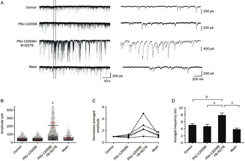
In the hippocampus, α7 nAChR subunits are primarily expressed in GABAergic interneurons, therefore we predicted that the α7 nAChR agonist Br-IQ17B might have an impact on GABAergic transmission. To test this notion, we recorded spontaneously occurring inhibitory postsynaptic current (sIPSC) from CA1 pyramidal neurons that receive inputs from interneurons in response to challenges with Br-IQ17B (10 μmol/L), PNU-120596 (1 μmol/L), or a combination of Br-IQ17B and PNU-120596 (Figure 7A and 7B). To block glutamatergic synaptic transmissions and muscarinic AChR, spontaneous IPSCs were recorded in the presence of glutamate receptor antagonists (CNQX 5 μmol/L, APV 10 μmol/L) and a muscarinic receptor antagonist (atropine 5 μmol/L). When applied alone for 10 min, PNU-120596 produced no detectable changes in the average frequency or amplitude of sIPSC at the population level (Figure 7B and 7C) (baseline activity at 4.5±0.7 Hz vs 4.6±0.7 Hz in the presence of PNU-120596; n=6, P>0.05; amplitude of 30.8±6.8 pA vs 30.6±4.7 pA in the presence of PNU-120596; n=6, P>0.05). In contrast, in the combined presence of Br-IQ17B and PNU120596, the peak amplitudes of IPSC events of individual neurons were significantly increased from 1.3 to 4.9 times in 5 out of 6 neurons recorded (P<0.0001) compared with control or PNU120596 alone (Figure 7B and 7C). After the co-application of PNU-120596 with Br-IQ17B for 1–5 min, the IPSC frequency also increased from 4.6±0.7 Hz to 7.8±0.7 Hz (n=6, P<0.01), and the combined effects of both Br-IQ17B and PNU120596 could be completely washed out (Figure 7D). For one neuron out of six, the co-application of Br-IQ17B and PNU-120596 promoted IPSC frequency without affecting IPSC amplitude (25.3±1.4 pA vs 25.9±1.2 pA). These results demonstrate that α7 nAChR activation by the novel agonist Br-IQ17B enhances synaptic transmissions in hippocampal neurons.
Figure 7.
α7 nAChR agonist Br-IQ17B enhances GABAergic synaptic transmission. (A) Representative raw traces showing that Br-IQ17B (10 μmol/L) reversibly enhanced IPSC (inhibitory postsynaptic current) in the presence of PNU-120596 (1 μmol/L). Superfusion of PNU-120596 alone for 10 min had no detectable effect on spontaneous IPSC (sIPSC); further application of Br-IQ17B (10 μmol/L) increased both the frequency and amplitude of sIPSC. Enhanced sIPSC induced by the co-application of PNU-120596 and Br-IQ17B can be washed out. The area of the dashed rectangle (left panels) is expanded to show a faster timescale at right. The neurons were held at −70 mV. (B) Peak amplitude distributions of all IPSC events detected in the left panel of (A). Co-application of Br-IQ17B and PNU-120596 significantly increased IPSC amplitudes (cP<0.01, compared with control, PNU-120596 and wash, one-way ANOVA). (C) Normalized average IPSC peak amplitudes from all recordings. Co-application of Br-IQ17B and PNU-120596 increased IPSC amplitude of each individual neuron (n=5, record duration for 1 min, one-way ANOVA). (D) Statistical analysis of average IPSC frequency from all neurons recorded (n=6, record duration for 1 min). Co-application of Br-IQ17B and PNU-120596 significantly increased average IPSC frequency (bP<0.05 vs control, cP<0.01 vs PNU-120596 or wash, paired t-test).
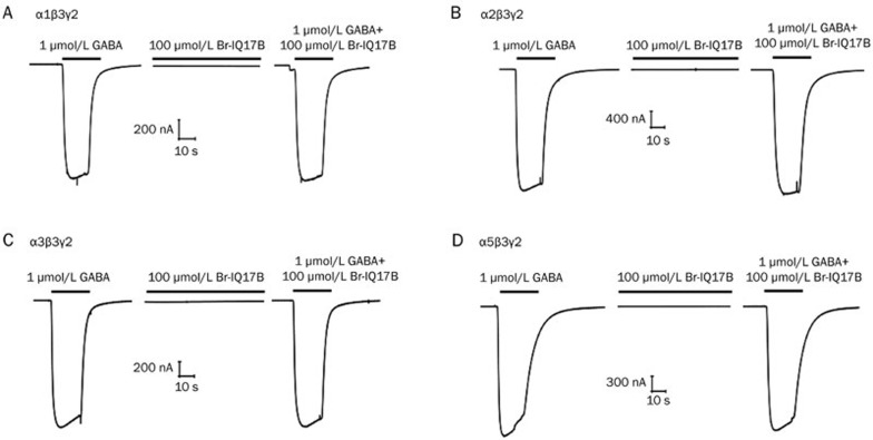
To further examine if the enhancement of IPSC resulted from an increase of GABAergic synaptic transmission, we tested the effect of Br-IQ17B on GABAA receptors with combinations of α1β3γ2, α2β3γ2, and α3β3γ2, which are the major subtypes in the brain36,37. Although the α5 subunit accounts for less than 5% of GABAA receptors in brain, we also tested the selectivity on α5β3γ2 because the α5 subtype is primarily localized in the hippocampus38. As shown in Figure 8, after pre-incubation for 1 min, Br-IQ17B (100 μmol/L) had no observable effects on any of the four subtypes and displayed no allosteric modulation when co-applied with 1 μmol/L GABA. These results show that enhanced GABAergic synaptic transmission likely resulted from the direct activation of α7 nAChR by Br-IQ17B.
Figure 8.
Selectivity assessments of Br-IQ17B on subtypes of GABAA receptors. Representative current traces recorded in oocytes expressing human α1β3γ2 (A), α2β3γ2 (B), α3β3γ2 (C), and α5β3γ2 (D) subtypes showing the lack of effects of Br-IQ17B on GABAA activity in the presence of 1 μmol/L GABA (left), 100 μmol/L Br-IQ17B (middle), or the co-application of GABA and Br-IQ17B (right) (n=3 for all subtypes).
Discussion
The goal of this study was to identify novel potent α7 nAChR agonists. Using electrophysiology as a primary screen, we identified the novel and potent compound Br-IQ17B from a series of indolizine derivatives; this compound selectively activates α7 nAChR and enhances synaptic transmission in vitro. Our α7 agonist Br-IQ17B, N-[(3R)-1-azabicyclo[2,2,2]oct-3-yl]-5-bromoindolizine-2-carboxamide, adds a novel tool in the field that may help understand new aspects of α7 nAChR function and also provides the potential for the development of novel therapy. Br-IQ17B exhibits some typical characteristics of α7 agonists reported in literature. One of the key characteristics is that the sustained exposure of Br-IQ17B progressively prevented ACh from activating α7 nAChR in Xenopus oocytes, whereas the pre-incubation of low concentration of Br-IQ17B potentiated ACh-mediated current. The most plausible explanation is the co-agonistic behavior of agonists binding at low concentrations, as previously reported for tubocurarine and ACh at α3β4 nAChRs39.
In this study, we selected compound EVP-6124 as a reference. In comparison to EVP-6124 from the oocyte assay (Figure 1C), although the EC50 of Br-IQ17B (1.8±0.2 μmol/L) is greater than that of EVP-6124 (0.45±0.08 μmol/L), the maximal response from Br-IQ17B is more than doubled that of EVP-6124 (64.3%±3.6% vs 30.6%±1.8%). Thus, Br-IQ17B may possess better efficacy in the enhancement of α7 activity. Furthermore, the concentration of EVP-6124 needed to reach 30% of the response (maximal response of EVP-6124) of ACh is 3 μmol/L, whereas for Br-IQ17B it is only 1.5 μmol/L. All of these findings suggest that Br-IQ17B may have advantages over EVP-6124.
The identification of selective α7 agonists for CNS therapy is a huge challenge in the field; three different α7 agonists in phase I clinical studies, PHA-543613, PHA-568487, and CP-810123 were recently discontinued due to non-sustained ventricular tachycardia23, and the nonspecific nicotinic ligand varenicline has been associated with serious adverse cardiovascular events24. It is not known whether these cardiovascular events are related to the activation of α7 nAChR or other pharmacological mechanisms, but compounds such as EVP-6124 suggest that the α7 nAChR can be targeted safely in clinical trials21. Because drugs with less selectivity that target multiple unintended receptors may be insufficient to prevent the incidence of side effects, especially gastrointestinal ones, it is necessary to evaluate the selectivity of Br-IQ17B on other nAChR subtypes. In the present study, we were concerned that Br-IQ17B could have activity against α3β4 and α4β2 nAChRs, which have similar distribution patterns as α7 nAChR in CNS33, and the nonspecific nicotinic ligands always have cross reactivity on these receptors (eg, nicotine, varenicline). In oocytes, Br-IQ17B showed no detectable agonist activity at up to 100 μmol/L toward either the α3β4 and α4β2 subtypes and very weak antagonist activity against α3β4 (IC50≈381 μmol/L).
The 5-HT3A belongs to the superfamily of ligand-gated ion channels. Significant sequence homology between 5-HT3A and α7 nAChR including the ligand-binding domain can lead to the cross-reactivity of certain compounds (eg, tropisetron)27. Br-IQ17B inhibited 5-HT-induced current but did not evoke currents when applied alone. The antagonist activity of Br-IQ17B against 5-HT3A receptors is likely a common property of α7 nAChR agonists, such as that reported for AZD032840 and EVP-612441, which are still in clinical development.
Stimulating α7 nAChR increases intracellular Ca2+, either directly or via voltage-gated calcium channels, and leads to the activation of Raf42 and the subsequent activation of the ERK1/2 signaling pathway, thus allowing the phosphorylation of cytoplasmic ERK substrates43. The ERK1/2 pathway is a central component in signal transduction regulating synaptic targets to control plasticity, and it is implicated in the processes of learning and memory44. PC12 cells express native α7 nAChR and are utilized for measuring the activation of native α7 nAChR45,46. Not only the non-specific nAChRs agonist nicotine but also the selective α7 nAChR agonist A-582941 have been proposed to stimulate the phosphorylation of ERK1/2 both in vitro and in vivo47. In our study, we assessed the phosphorylation of ERK1/2 in response to the activation of α7 nAChR by Br-IQ17B within PC12 cells, and a significant concentration-dependent increase in ERK1/2 phosphorylation was observed, indicating that the ERK1/2 pathway is a downstream effector of α7 activation.
It has well been established that within the rat hippocampus, α7 nAChR is located predominantly in interneurons where it modulates GABAergic synaptic transmission involved in sensory gating48. Deficits in auditory sensory gating can lead to an inability to filter out extraneous signals from meaningful sensory inputs, resulting in a state of sensory overload, which contributes to the attentional and cognitive deficits in CNS diseases, most convincingly in schizophrenia49. It has been shown that the α7 nAChR partial agonist GTS-21 [3-(2,4)-dimethoxybenzilidine anabaseine], also known as DMXB-A, appears to be safe and promising for improving P50 gating deficits and attention, and it significantly improves neurocognition in schizophrenia patients with antipsychotic drugs50,51. GABA is an inhibitory synaptic transmitter, and its decreased release can lead to the disinhibition of sensory gating (eg, the deficit of P50 auditory-evoked potential gating is indicated in some schizophrenic patients). To evaluate the ability of Br-IQ17B to modulate GABAergic synaptic transmission, we recorded sIPSC in pyramidal neurons in acutely isolated rat hippocampal slices. The spontaneous GABAergic synaptic events were recorded for 3 to 5 min under baseline conditions, followed by a 10-min pre-incubation of PNU-120596 before the addition of Br-IQ17B for 5 min. In agreement with the previously reported role of α7 nAChR in the hippocampus, there was no detectable change in synaptic activity after the application of PNU-120596 alone, whereas the bath application of 10 μmol/L Br-IQ17B doubled the frequency and increased the amplitude significantly.
In conclusion, we identified Br-IQ17B as a novel and selective α7 nAChR agonist that exhibits favorable potency and efficacy. Br-IQ17B is a prominent candidate that provides opportunities for further investigations in CNS pharmacology. It also serves as a suitable tool for characterizing the role of α7 nAChR in CNS function, and it may have the potential for development into new therapy for CNS disorders.
Author contribution
Jing-shu TANG conducted the majority of biological experiments; Bing-xue XIE and Yu XUE performed the chemical synthesis; Xi-ling BIAN made the recordings of brain slices; Ning-ning WEI, Jing-heng ZHOU, and Yu-chen HAO performed some cell biology experiments; Jing-shu TANG and Xi-ling BIAN analyzed the data; Jing-shu TANG drafted the manuscript; Gang LI, Liang-ren ZHANG, and Ke-wei WANG supervised the project and wrote and finalized the manuscript.
Acknowledgments
We would like to thank Tim G HALES at the University of Dundee for the generous gift of the human 5-HT3A cDNA clone and Lucia G SIVILOTTI at University College London for rat alpha3 and beta4 cDNA clones. We thank laboratory member Yan SONG for technical assistance. We also thank Ming JI and Xiao-guang CHEN for technical assistance in radioisotope ligand binding. Ke-wei WANG wishes to thank JM WANG for her consistent support during this research. This work was supported by research grants to Ke-wei WANG from the Ministry of Science and Technology of China (2013CB531302 and 2014ZX09507003-006-004) and the National Science Foundation of China (31370741 and 81221002), to Liang-ren ZHANG from the National Science Foundation of China (81373272) and to Xi-ling BIAN from Beijing Higher Education Young Elite Teacher Project.
References
- Dani JA, Bertrand D. Nicotinic acetylcholine receptors and nicotinic cholinergic mechanisms of the central nervous system. Annu Rev Pharmacol Toxicol 2007; 47: 699–729. [DOI] [PubMed] [Google Scholar]
- Gotti C, Zoli M, Clementi F. Brain nicotinic acetylcholine receptors: native subtypes and their relevance. Trends Pharmacol Sci 2006; 27: 482–91. [DOI] [PubMed] [Google Scholar]
- Seguela P, Wadiche J, Dineley-Miller K, Dani JA, Patrick JW. Molecular cloning, functional properties, and distribution of rat brain alpha 7: a nicotinic cation channel highly permeable to calcium. J Neurosci 1993; 13: 596–604. [DOI] [PMC free article] [PubMed] [Google Scholar]
- Sher E, Chen Y, Sharples TJ, Broad LM, Benedetti G, Zwart R, et al. Physiological roles of neuronal nicotinic receptor subtypes: new insights on the nicotinic modulation of neurotransmitter release, synaptic transmission and plasticity. Curr Top Med Chem 2004; 4: 283–97. [DOI] [PubMed] [Google Scholar]
- Alkondon M, Braga MF, Pereira EF, Maelicke A, Albuquerque EX. alpha7 nicotinic acetylcholine receptors and modulation of GABAergic synaptic transmission in the hippocampus. Eur J Pharmacol 2000; 393: 59–67. [DOI] [PubMed] [Google Scholar]
- Yakel JL. Nicotinic ACh receptors in the hippocampal circuit; functional expression and role in synaptic plasticity. J Physiol 2014; 592: 4147–53. [DOI] [PMC free article] [PubMed] [Google Scholar]
- Freedman R, Coon H, Myles-Worsley M, Orr-Urtreger A, Olincy A, Davis A, et al. Linkage of a neurophysiological deficit in schizophrenia to a chromosome 15 locus. Proc Natl Acad Sci U S A 1997; 94: 587–92. [DOI] [PMC free article] [PubMed] [Google Scholar]
- Leonard S, Gault J, Hopkins J, Logel J, Vianzon R, Short M, et al. Association of promoter variants in the alpha7 nicotinic acetylcholine receptor subunit gene with an inhibitory deficit found in schizophrenia. Arch Gen Psychiatry 2002; 59: 1085–96. [DOI] [PubMed] [Google Scholar]
- Freedman R, Hall M, Adler LE, Leonard S. Evidence in postmortem brain tissue for decreased numbers of hippocampal nicotinic receptors in schizophrenia. Biol Psychiatry 1995; 38: 22–33. [DOI] [PubMed] [Google Scholar]
- Stevens KE, Freedman R, Collins AC, Hall M, Leonard S, Marks MJ, et al. Genetic correlation of inhibitory gating of hippocampal auditory evoked response and alpha-bungarotoxin-binding nicotinic cholinergic receptors in inbred mouse strains. Neuropsychopharmacology 1996; 15: 152–62. [DOI] [PubMed] [Google Scholar]
- Young JW, Crawford N, Kelly JS, Kerr LE, Marston HM, Spratt C, et al. Impaired attention is central to the cognitive deficits observed in alpha 7 deficient mice. Eur Neuropsychopharmacol 2007; 17: 145–55. [DOI] [PubMed] [Google Scholar]
- Hellstrom-Lindahl E, Mousavi M, Zhang X, Ravid R, Nordberg A. Regional distribution of nicotinic receptor subunit mRNAs in human brain: comparison between Alzheimer and normal brain. Brain Res Mol Brain Res 1999; 66: 94–103. [DOI] [PubMed] [Google Scholar]
- Wang HY, Lee DH, Davis CB, Shank RP. Amyloid peptide Aβ1-42 binds selectively and with picomolar affinity to α7 nicotinic acetylcholine receptors. J Neurochem 2000; 75: 1155–61. [DOI] [PubMed] [Google Scholar]
- Nagele RG, D'Andrea MR, Anderson WJ, Wang HY. Intracellular accumulation of β-amyloid1–42 in neurons is facilitated by the α7 nicotinic acetylcholine receptor in Alzheimer's disease. Neuroscience 2002; 110: 199–211. [DOI] [PubMed] [Google Scholar]
- Wang HY, Li W, Benedetti NJ, Lee DH. Alpha 7 nicotinic acetylcholine receptors mediate beta-amyloid peptide-induced tau protein phosphorylation. J Biol Chem 2003; 278: 31547–53. [DOI] [PubMed] [Google Scholar]
- Liu Q, Kawai H, Berg DK. beta-Amyloid peptide blocks the response of alpha 7-containing nicotinic receptors on hippocampal neurons. Proc Natl Acad Sci U S A 2001; 98: 4734–9. [DOI] [PMC free article] [PubMed] [Google Scholar]
- Lin H, Bhatia R, Lal R. Amyloid beta protein forms ion channels: implications for Alzheimer's disease pathophysiology. FASEB J 2001; 15: 2433–44. [DOI] [PubMed] [Google Scholar]
- Qi XL, Nordberg A, Xiu J, Guan ZZ. The consequences of reducing expression of the alpha7 nicotinic receptor by RNA interference and of stimulating its activity with an alpha7 agonist in SH-SY5Y cells indicate that this receptor plays a neuroprotective role in connection with the pathogenesis of Alzheimer's disease. Neurochem Int 2007; 51: 377–83. [DOI] [PubMed] [Google Scholar]
- Lieberman JA, Dunbar G, Segreti AC, Girgis RR, Seoane F, Beaver JS, et al. A randomized exploratory trial of an alpha-7 nicotinic receptor agonist (TC-5619) for cognitive enhancement in schizophrenia. Neuropsychopharmacology 2013; 38: 968–75. [DOI] [PMC free article] [PubMed] [Google Scholar]
- Meltzer HY, Gawryl M, Ward S, Dgetluck N, Bhuvaneswaran C, Koenig G, et al. EVP-6124, an alpha-7 nicotinic partial agonist, produces positive effects on cognition, clinical function, and negative symptoms in patients with chronic schizophrenia on stable antipsychotic therapy. In: Proceedings of the American College of Neuropsychopharmacology (ACNP) 50th Annual Meeting; December 4–8, 2011; Waikoloa Beach, Hawaii, USA. Neuropsychopharmacology 2011; 36 Suppl 1: S75–197. [Google Scholar]
- Preskorn SH, Gawryl M, Dgetluck N, Palfreyman M, Bauer LO, Hilt DC. Normalizing effects of EVP-6124, an alpha-7 nicotinic partial agonist, on event-related potentials and cognition: a proof of concept, randomized trial in patients with schizophrenia. J Psychiatr Pract 2014; 20: 12–24. [DOI] [PubMed] [Google Scholar]
- Hurst R, Rollema H, Bertrand D. Nicotinic acetylcholine receptors: from basic science to therapeutics. Pharmacol Ther 2013; 137: 22–54. [DOI] [PubMed] [Google Scholar]
- Barrish JC, Carter PH, editors. Accounts in drug discovery: case studies in medicinal chemistrys. London: Royal Society of Chemistry; 2010.
- Singh S, Loke YK, Spangler JG, Furberg CD. Risk of serious adverse cardiovascular events associated with varenicline: a systematic review and meta-analysis. CMAJ 2011; 183: 1359–66. [DOI] [PMC free article] [PubMed] [Google Scholar]
- Willett P. Similarity searching using 2D structural fingerprints. Methods Mol Biol 2011; 672: 133–58. [DOI] [PubMed] [Google Scholar]
- Bermudez J, Fake CS, Joiner GF, Joiner KA, King FD, Miner WD, et al. 5-Hydroxytryptamine (5-HT3) receptor antagonists. 1. Indazole and indolizine-3-carboxylic acid derivatives. J Med Chem 1990; 33: 1924–9. [DOI] [PubMed] [Google Scholar]
- Macor JE, Gurley D, Lanthorn T, Loch J, Mack RA, Mullen G, et al. The 5-HT3 antagonist tropisetron (ICS 205-930) is a potent and selective alpha7 nicotinic receptor partial agonist. Bioorg Med Chem Lett 2001; 11: 319–21. [DOI] [PubMed] [Google Scholar]
- Hurst RS, Hajos M, Raggenbass M, Wall TM, Higdon NR, Lawson JA, et al. A novel positive allosteric modulator of the alpha7 neuronal nicotinic acetylcholine receptor: in vitro and in vivo characterization. J Neurosci 2005; 25: 4396–405. [DOI] [PMC free article] [PubMed] [Google Scholar]
- Briggs CA, McKenna DG. Activation and inhibition of the human alpha7 nicotinic acetylcholine receptor by agonists. Neuropharmacology 1998; 37: 1095–102. [DOI] [PubMed] [Google Scholar]
- Davies AR, Hardick DJ, Blagbrough IS, Potter BV, Wolstenholme AJ, Wonnacott S. Characterisation of the binding of [3H]methyllycaconitine: a new radioligand for labelling alpha 7-type neuronal nicotinic acetylcholine receptors. Neuropharmacology 1999; 38: 679–90. [DOI] [PubMed] [Google Scholar]
- Bodnar AL, Cortes-Burgos LA, Cook KK, Dinh DM, Groppi VE, Hajos M, et al. Discovery and structure-activity relationship of quinuclidine benzamides as agonists of alpha7 nicotinic acetylcholine receptors. J Med Chem 2005; 48: 905–8. [DOI] [PubMed] [Google Scholar]
- Anderson DJ, Bunnelle W, Surber B, Du J, Surowy C, Tribollet E, et al. [3H]A-585539 [(1S,4S)-2,2-dimethyl-5-(6-phenylpyridazin-3-yl)-5-aza-2-azoniabicyclo[2.2.1]hept ane], a novel high-affinity alpha7 neuronal nicotinic receptor agonist: radioligand binding characterization to rat and human brain. J Pharmacol Exp Ther 2008; 324: 179–87. [DOI] [PubMed] [Google Scholar]
- Taly A, Corringer PJ, Guedin D, Lestage P, Changeux JP. Nicotinic receptors: allosteric transitions and therapeutic targets in the nervous system. Nat Rev Drug Discov 2009; 8: 733–50. [DOI] [PubMed] [Google Scholar]
- Jones S, Yakel JL. Functional nicotinic ACh receptors on interneurones in the rat hippocampus. J Physiol 1997; 504 (Pt 3): 603–10. [DOI] [PMC free article] [PubMed] [Google Scholar]
- Dajas-Bailador FA, Soliakov L, Wonnacott S. Nicotine activates the extracellular signal-regulated kinase 1/2 via the alpha7 nicotinic acetylcholine receptor and protein kinase A, in SH-SY5Y cells and hippocampal neurones. J Neurochem 2002; 80: 520–30. [DOI] [PubMed] [Google Scholar]
- Whiting PJ. GABA-A receptor subtypes in the brain: a paradigm for CNS drug discovery? Drug Discov Today 2003; 8: 445–50. [DOI] [PubMed] [Google Scholar]
- McKernan RM, Whiting PJ. Which GABAA-receptor subtypes really occur in the brain? Trends Neurosci 1996; 19: 139–43. [DOI] [PubMed] [Google Scholar]
- Caraiscos VB, Elliott EM, You-Ten KE, Cheng VY, Belelli D, Newell JG, et al. Tonic inhibition in mouse hippocampal CA1 pyramidal neurons is mediated by alpha5 subunit-containing gamma-aminobutyric acid type A receptors. Proc Natl Acad Sci U S A 2004; 101: 3662–7. [DOI] [PMC free article] [PubMed] [Google Scholar]
- Cachelin AB, Rust G. Unusual pharmacology of (+)-tubocurarine with rat neuronal nicotinic acetylcholine receptors containing beta 4 subunits. Mol Pharmacol 1994; 46: 1168–74. [PubMed] [Google Scholar]
- Sydserff S, Sutton EJ, Song D, Quirk MC, Maciag C, Li C, et al. Selective alpha7 nicotinic receptor activation by AZD0328 enhances cortical dopamine release and improves learning and attentional processes. Biochem Pharmacol 2009; 78: 880–8. [DOI] [PubMed] [Google Scholar]
- Prickaerts J, van Goethem NP, Chesworth R, Shapiro G, Boess FG, Methfessel C, et al. EVP-6124, a novel and selective alpha7 nicotinic acetylcholine receptor partial agonist, improves memory performance by potentiating the acetylcholine response of alpha7 nicotinic acetylcholine receptors. Neuropharmacology 2012; 62: 1099–110. [DOI] [PubMed] [Google Scholar]
- Walker SA, Cullen PJ, Taylor JA, Lockyer PJ. Control of Ras cycling by Ca2+. FEBS Lett 2003; 546: 6–10. [DOI] [PubMed] [Google Scholar]
- Davis S, Laroche S. Mitogen-activated protein kinase/extracellular regulated kinase signalling and memory stabilization: a review. Genes Brain Behav 2006; 5 Suppl 2: 61–72. [DOI] [PubMed] [Google Scholar]
- Thomas GM, Huganir RL. MAPK cascade signalling and synaptic plasticity. Nat Rev Neurosci 2004; 5: 173–83. [DOI] [PubMed] [Google Scholar]
- Wang H, Yu M, Ochani M, Amella CA, Tanovic M, Susarla S, et al. Nicotinic acetylcholine receptor alpha7 subunit is an essential regulator of inflammation. Nature 2003; 421: 384–8. [DOI] [PubMed] [Google Scholar]
- Waldburger JM, Boyle DL, Pavlov VA, Tracey KJ, Firestein GS. Acetylcholine regulation of synoviocyte cytokine expression by the alpha7 nicotinic receptor. Arthritis Rheum 2008; 58: 3439–49. [DOI] [PMC free article] [PubMed] [Google Scholar]
- Bitner RS, Bunnelle WH, Anderson DJ, Briggs CA, Buccafusco J, Curzon P, et al. Broad-spectrum efficacy across cognitive domains by alpha7 nicotinic acetylcholine receptor agonism correlates with activation of ERK1/2 and CREB phosphorylation pathways. J Neurosci 2007; 27: 10578–87. [DOI] [PMC free article] [PubMed] [Google Scholar]
- Frazier CJ, Rollins YD, Breese CR, Leonard S, Freedman R, Dunwiddie TV. Acetylcholine activates an alpha-bungarotoxin-sensitive nicotinic current in rat hippocampal interneurons, but not pyramidal cells. J Neurosci 1998; 18: 1187–95. [DOI] [PMC free article] [PubMed] [Google Scholar]
- Leiser SC, Bowlby MR, Comery TA, Dunlop J. A cog in cognition: how the alpha 7 nicotinic acetylcholine receptor is geared towards improving cognitive deficits. Pharmacol Ther 2009; 122: 302–11. [DOI] [PubMed] [Google Scholar]
- Freedman R, Olincy A, Buchanan RW, Harris JG, Gold JM, Johnson L, et al. Initial phase 2 trial of a nicotinic agonist in schizophrenia. Am J Psychiatry 2008; 165: 1040–7. [DOI] [PMC free article] [PubMed] [Google Scholar]
- Olincy A, Harris JG, Johnson LL, Pender V, Kongs S, Allensworth D, et al. Proof-of-concept trial of an alpha7 nicotinic agonist in schizophrenia. Arch Gen Psychiatry 2006; 63: 630–8. [DOI] [PubMed] [Google Scholar]


